


Ehlers-Danlos Syndrome and Your Heart: Valves and Aorta
How Ehlers-Danlos syndrome affects the heart valves and aorta, and how it is monitored.
What is the Ehlers-Danlos Syndrome Heart Connection?
When you or a loved one receives a diagnosis of a connective tissue disorder, understanding the ehlers-danlos syndrome heart connection is vital for your long-term care. Inherited connective tissue conditions: Ehlers-Danlos Syndrome (EDS) is a group of genetic disorders that affect the structural framework holding our cells, organs, and tissues together. Because this tissue is found throughout the body, the cardiovascular system can be deeply affected by these genetic changes.
Impact on the cardiovascular system: Connective tissue provides essential strength and elasticity to blood vessels and the heart. In certain types of EDS, genetic mutations alter collagen production, making these tissues weaker or more fragile than usual. This can directly impact the heart valves and the large arteries attached to it, potentially leading to structural changes that develop slowly over time.
Importance of proactive care: Expert consensus from escardio.org highlights that while not every type of EDS severely impacts the cardiovascular system, the vascular subtypes require careful, specialized attention. Recognizing how your genetic makeup influences your cardiovascular health is the first step toward compassionate, proactive care that protects your heart for the future.
How Connective Tissue Affects Heart Valves
Valve structure and function: The heart contains four valves that open and close to ensure blood flows in the correct direction. These valves are made of strong, flexible flaps of connective tissue. When EDS is present, this tissue may be overly stretchy or fragile, impairing their ability to form a tight seal when they close.
Understanding valve regurgitation: When a valve cannot close properly, blood can leak backward into the previous chamber. This backward flow is known as valve regurgitation. Over time, the heart muscle has to work harder to pump the leaked blood, which can lead to fatigue, shortness of breath, or an irregular heartbeat as the heart struggles to keep up with the body's demands.
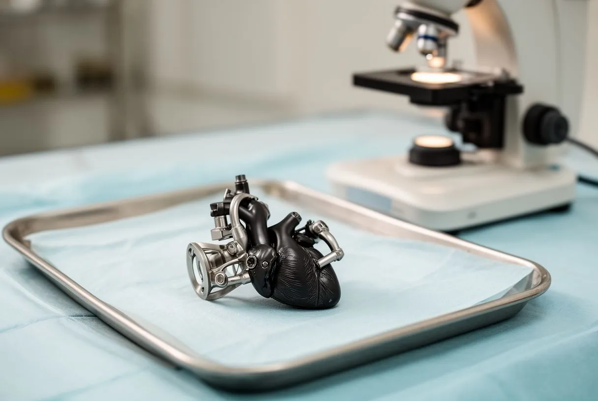
Cellular-level tissue changes: Research into cellular transport, such as the biological properties of extracellular vesicles and their physiological functions, reveals how complex cellular communication is in maintaining healthy connective tissue. While everyday cardiology focuses on the whole organ, these fundamental tissue properties explain why valves stretch abnormally and fail to seal properly in EDS patients.
Understanding Ehlers-Danlos Mitral Valve Prolapse
The mechanics of the mitral valve: The mitral valve sits between the upper and lower chambers on the left side of the heart, acting as a gateway for oxygen-rich blood. In EDS, the connective tissue cords that anchor the valve flaps can stretch out. This causes the valve leaflets to bulge or flop backward into the upper chamber during the heart's contraction, a condition called mitral valve prolapse.
Symptoms and daily impact: Many people with mild prolapse experience no symptoms and require no treatment beyond routine monitoring. However, for others, the stretching can lead to significant leaking. If the regurgitation becomes severe, it may cause an uncomfortable awareness of the heartbeat, dizziness, or decreased stamina during physical activity, which warrants prompt medical attention.
Tracking changes over time: Regular check-ups allow your cardiac team to listen for the characteristic "click-murmur" sound of a prolapsing valve. Tracking these subtle changes early means your surgeon can plan conservative interventions or schedule a safe repair before the heart muscle becomes overly strained or permanent damage occurs.
What are Ehlers-Danlos Aorta Complications?
The role of the aorta: The aorta is the largest artery in the body, responsible for carrying oxygen-rich blood from the heart to the rest of the body. Because it handles high-pressure blood flow, it relies on strong, elastic connective tissue walls to expand and recoil safely with every heartbeat. In EDS, these walls can be weaker and less resilient.
Risks of aortic dilation: Over time, the constant pounding of blood can cause the aorta to stretch beyond its normal limits. This enlargement, known as aortic dilation, is a primary concern for cardiac specialists treating patients with connective tissue disorders. Careful, ongoing monitoring of the aortic diameter is essential to catch this silent stretching early.
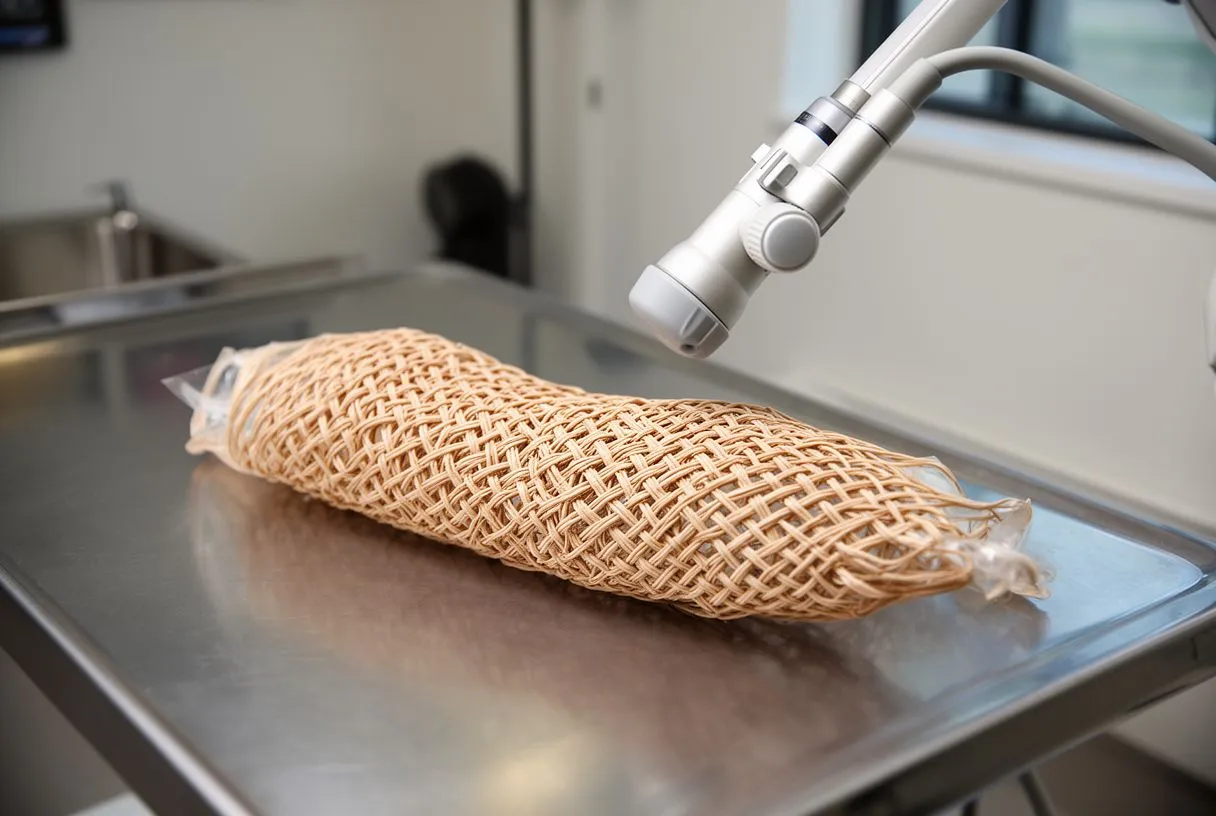
Why the aortic arch matters: The most severe of the ehlers-danlos aorta complications occur when the weakened wall gives way, often near the curved top portion of the vessel called the aortic arch. Because the tissue lacks the normal tensile strength to withstand cardiovascular pressures, a proactive approach to imaging and lifestyle adjustments is the cornerstone of modern cardiac care for EDS patients.
Ehlers-Danlos Aortic Aneurysm Risk Explained
Defining an aneurysm: An aneurysm is a localized, balloon-like bulge in the wall of an artery. For patients with vascular EDS, the risk of developing an aortic aneurysm is higher because the adventitia—the tough outer layer of the blood vessel—may be thinner or more fragile than usual. This anatomical difference requires heightened vigilance from your medical team.
Silent growth and detection: Aortic aneurysms often grow slowly and silently. Without regular imaging, a patient may not realize there is a problem until the aneurysm becomes large enough to press on surrounding structures, causing back pain or a deep ache in the chest, or until it reaches a dangerous size that threatens to rupture.
Planning for surgical safety: The ultimate goal of care is to detect an aneurysm long before it poses a threat. By establishing a baseline scan and tracking the growth rate over time, surgeons can determine the optimal window for a safe, planned aortic aneurysm repair rather than facing a high-risk emergency situation where the fragile tissue has already torn.
Recognizing Ehlers-Danlos Aortic Dissection
What happens during dissection: A dissection is a tear in the inner layer of the aorta. Blood surges through the tear, causing the inner and middle layers of the aortic wall to separate or dissect. This is a life-threatening medical emergency, and patients with connective tissue disorders are at a significantly heightened risk for this catastrophic event.
Recognizing emergency symptoms: The classic symptom of a dissection is sudden, severe, tearing pain in the chest or upper back. However, it is crucial to remember that symptoms can vary widely depending on where the tear occurs. Some patients may experience pain that moves, shortness of breath, or fainting. Immediate emergency medical attention is always necessary if these signs appear.
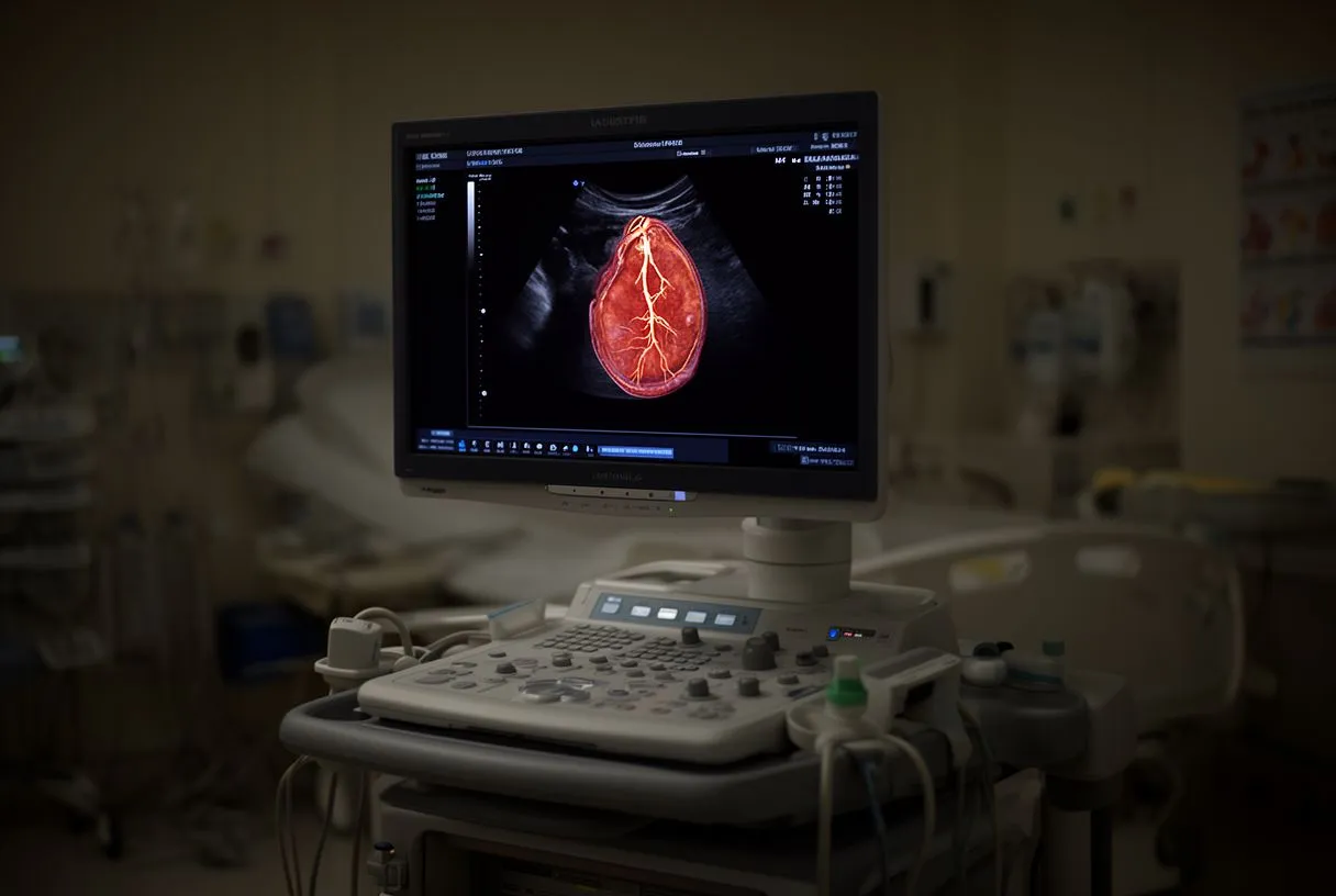
Prevention versus emergency repair: Because the tissue is inherently fragile, prevention is far better than cure. Medications to gently lower blood pressure and reduce the force of the heartbeat are often prescribed to protect the aorta. In some high-risk cases, a surgeon may recommend a prophylactic operation, such as an aortic arch replacement, if the structural weakness is severe and progressive.
Essential Ehlers-Danlos Heart Screening Tests
Establishing a cardiac baseline: Baseline screening is the foundation of a safe care plan. When EDS is diagnosed, a comprehensive cardiac evaluation helps establish the health of the valves and the aorta before any symptoms arise. This involves non-invasive tests that provide a detailed picture of the cardiovascular system to guide future care.
Key diagnostic imaging tools: The most critical screening test is the echocardiogram, an ultrasound of the heart. It allows the cardiologist to watch the valves in motion, measure the size of the aortic annulus (the ring where the aorta meets the heart), and check for any leaking. For a clearer view of the entire chest, a CT scan or MRI may also be ordered.
- Echocardiogram: An ultrasound to evaluate valve function and measure the aortic root.
- CT or MRI Scan: Detailed imaging to check the aortic arch and full aortic diameter.
- Blood Pressure Checks: Routine monitoring to ensure blood pressure stays in a safe, controlled range.
The value of expert interpretation: It is important to have these tests interpreted by a team familiar with connective tissue disorders. They understand the subtle nuances of EDS hearts and can differentiate between normal anatomical variations and early signs of concern, creating a personalized baseline for your ongoing monitoring.
Guidelines for Ehlers-Danlos Cardiac Monitoring
Setting a monitoring schedule: Once baseline screening is complete, ongoing monitoring ensures any changes are caught early. The frequency of these check-ups depends on the specific type of EDS, the size of the aorta, and the presence of any valve issues. An annual echocardiogram is a common and highly recommended standard for many adults with vascular EDS.
Assessing daily heart function: Monitoring is not just about taking images; it is about assessing how your heart is coping on a day-to-day basis. Your doctor will ask about new symptoms, check your blood pressure, and listen to your heart sounds. If you develop anaemia or notice new ankle swelling, these could be subtle signs that the heart is struggling to pump efficiently.

Managing overall cardiovascular risk: Research tracking all-cause mortality in cardiovascular care highlights the importance of managing secondary risk factors. For instance, a landmark study on the effects of lowering LDL cholesterol with simvastatin plus ezetimibe in patients with chronic kidney disease (Study of Heart and Renal Protection): a randomised placebo-controlled trial demonstrates how systemic health improvements benefit the heart. The same principle applies to EDS: controlling blood pressure and cholesterol reduces overall stress on fragile tissues.
Ehlers-Danlos Heart Treatment Options
Conservative medical management: Treatment for cardiac concerns in EDS is highly individualized. For many patients, careful observation and medication are all that is needed for years. Medications like beta-blockers help lower blood pressure and reduce the stress on the aortic walls, while drugs like amiodarone may be used if irregular heartbeats develop. Appropriate antiplatelet therapy may also be considered by your physician to manage other vascular risks safely.
Delicate surgical approaches: When surgery becomes necessary, the approach must be delicate. Because EDS tissues are fragile, standard surgical techniques may require modification. For valve issues, a surgeon might perform an annuloplasty, using an annuloplasty ring to support a stretched valve. Precise annulus sizing is critical to ensure the repair holds securely without tearing the delicate surrounding tissue.
Aortic and valve replacements: For aortic enlargement, surgeons may replace the weakened section of the aorta. During these complex procedures, specialized techniques like antegrade cerebral perfusion are used to protect the brain, and the aortic cross-clamp time is carefully managed to minimize trauma. In certain older or higher-risk patients, transcatheter aortic valve replacement (TAVR) may be considered, though its application in EDS requires highly specialized evaluation due to tissue fragility.
Living With Vascular Ehlers-Danlos Heart Concerns
Building a medical partnership: Living with a connective tissue disorder requires a lifelong partnership with your medical team. Understanding your body, knowing your baseline measurements, and communicating openly with your cardiologist empowers you to live fully while managing your health risks responsibly and safely.
Protecting your cardiovascular system: You should be aware of situations that increase cardiovascular stress. It is often advised to avoid heavy lifting, contact sports, and activities that cause a sudden spike in blood pressure. Simple habits like taking an antimicrobial shower before procedures and maintaining excellent dental hygiene can reduce the need for antibiotic prophylaxis, which is sometimes used to prevent dangerous heart valve infections.
- Avoid Heavy Strain: Refrain from heavy lifting and contact sports to protect fragile blood vessels.
- Medication Adherence: Take prescribed blood pressure medications consistently to reduce stress on the aorta.
- Gentle Tissue Care: Handle skin and tissues gently to prevent tears and monitor healing closely.
Looking forward with confidence: While the anatomy of an EDS heart presents unique challenges, it does not define your future. By combining careful monitoring with expert surgical care when needed, patients lead active, fulfilling lives. We strongly encourage you to discuss your specific symptoms and family history with a qualified cardiac team. Please book a consultation to receive personalized advice rather than self-diagnosing, as every heart and every genetic expression is unique.






