

Fluoroscopy in Heart Procedures: Imaging and Radiation
Fluoroscopy heart procedures use real-time X-ray imaging to guide catheter placement and valve repairs safely.
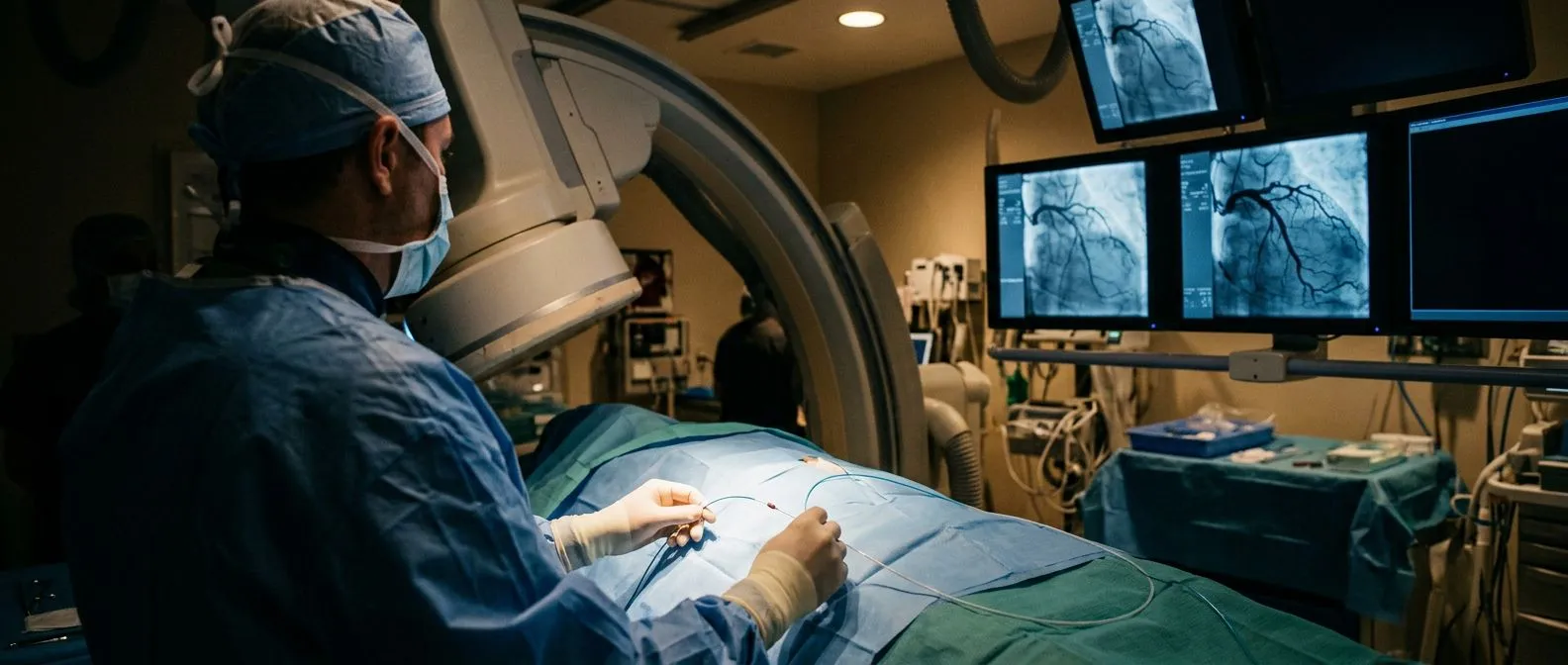
If you or a family member has been told you need a heart catheterization, a stent, or a valve procedure, fluoroscopy in heart procedures is the technology that makes it all possible. Fluoroscopy is a continuous X-ray that shows your heart and moving instruments on a screen in real time, so your medical team can see exactly where they are working without opening your chest. This guide explains how it works, how much radiation is involved, how your team keeps that exposure as low as possible, and what you can expect on the day of the procedure.
What Is Fluoroscopy in Simple Terms?
A continuous X-ray, not a photograph. A standard X-ray is a single still image, like a photo. Fluoroscopy is more like a video: the X-ray machine emits a low, continuous beam and a detector converts it into a live moving picture on a monitor. That real-time imaging is what allows a cardiologist to steer a thin catheter through your blood vessels and watch it travel toward the heart.
Why the heart needs it. The heart is a moving organ inside a moving body. Still images cannot tell a surgeon where a guide wire sits at this exact moment. With fluoroscopy, the interventional team sees the catheter curve around the aortic arch, cross a valve, or release a stent — as it happens.
Contrast dye makes it all visible. Blood vessels and valves do not show up well on X-ray on their own, so a contrast dye (usually iodine-based) is injected through the catheter. The dye lights up on the screen, showing the anatomy of the coronary arteries or the flow of blood through a valve.
Why Fluoroscopy in Heart Procedures Matters
Almost every modern structural heart procedure depends on fluoroscopy in heart procedures being performed precisely and safely. Without it, there would be no way to place a stent, dilate a narrowed valve, or implant a transcatheter aortic valve replacement (TAVR) with confidence.
Precision without open surgery. Fluoroscopy lets the team work through a small puncture in the wrist or groin rather than through a sternotomy (opening the chest). That is the entire reason catheter-based treatment exists: real-time imaging replaces direct vision.
Immediate decisions during the procedure. The live picture allows the team to measure, inject dye, adjust position, and confirm the result before the catheters come out. If a vessel is too narrow or a valve lands poorly, the team knows right away and can correct it in the same session.
In our practice, every catheter-based recommendation is reviewed by the multidisciplinary team against international practice guidelines before anything is proposed, and we explain which imaging studies are genuinely needed so you never undergo unnecessary ones.
What Are the 7 Types of Imaging Used in Heart Care?
Patients often ask how fluoroscopy fits among the many heart tests they hear about. Most centers rely on a core group of imaging technologies, each answering a different question:
- X-ray: still images of the chest, useful for heart size and lung fields.
- Fluoroscopy: live X-ray video used to guide catheters during procedures.
- Echocardiography: ultrasound of the heart, showing valves and pumping function with no radiation.
- MRI (magnetic resonance imaging): detailed soft-tissue images, often for heart muscle and scarring.
- CT scan (computed tomography): detailed cross-sectional images, used to plan TAVR and assess the aorta.
- Nuclear imaging (including PET): shows blood flow and metabolism of the heart muscle; standardized FDG PET/CT protocols for tumour and cardiac work are described in the EANM procedure guidelines.
- Coronary angiography: dye-based X-ray images of the coronary arteries, usually acquired with fluoroscopy during a catheterization.
These tests are not rivals; they are partners. A CT scan may plan the procedure, echocardiography may measure the valve, and fluoroscopy performs the guidance on the day itself.
How Fluoroscopy Cardiac Catheterization Works, Step by Step
Before the procedure. You arrive with nothing to eat for several hours. The team checks your kidney function (because of the contrast dye), your medications, and the access site plan. You receive local anesthetic and light sedation — most patients are relaxed but awake.
During the procedure. A thin tube called a catheter is inserted into an artery, usually at the wrist (radial access) or the groin (femoral access). Under fluoroscopy cardiac catheterization guidance, the catheter is advanced up the aorta to the heart. Dye is injected, and the coronary arteries or heart chambers appear on the monitor in real time.
After the procedure. The catheter is removed and pressure or a small closure device seals the access site. Most diagnostic catheterizations allow going home the same day after a few hours of observation. The fluoroscopy portion itself adds no recovery time — recovery depends on the procedure it guided, not the imaging.
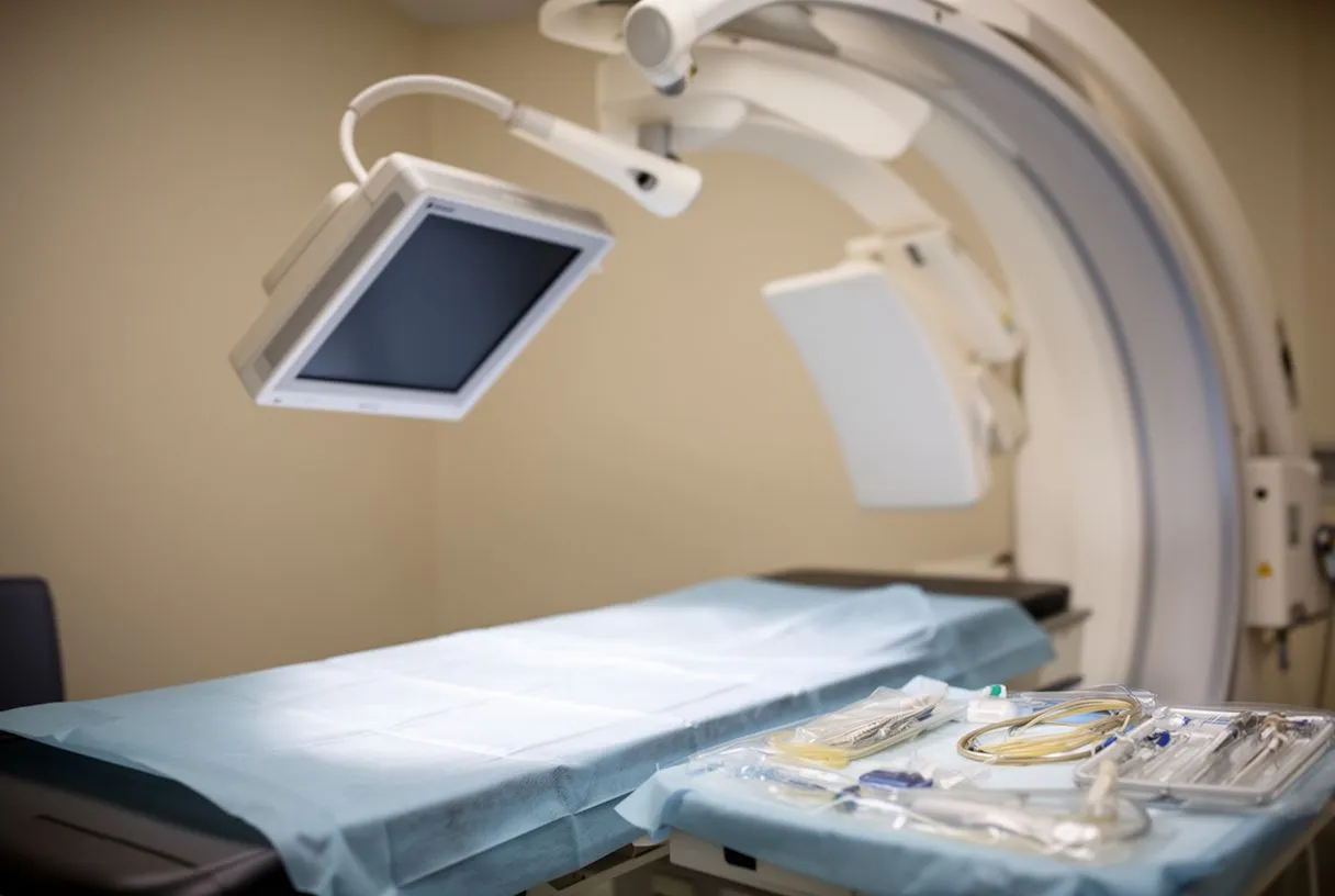
What the images actually show. Beyond the catheter position, fluoroscopy reveals vessel anatomy, valve leaflet motion, and even calcification (calcium deposits) in the aortic valve, which appears bright white on the screen. These visual cues guide decisions within seconds.
Who interprets what you see. This is interventional cardiology imaging in action: a specially trained cardiologist reads the live images continuously and adjusts every movement accordingly. Nothing happens blindly inside the heart.
Fluoroscopy During Stent Placement and Angioplasty
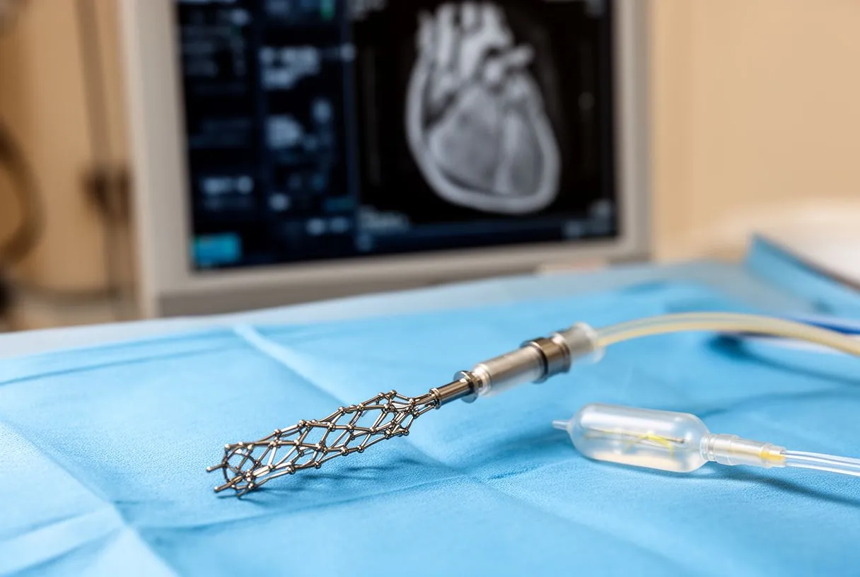
Why fluoroscopy angioplasty radiation dose is measured carefully. Coronary angioplasty — inflating a small balloon to open a blocked artery — and stent placement (inserting a small metal mesh tube to keep the artery open) both rely entirely on fluoroscopy. The procedure can take anywhere from 30 minutes to over an hour, so the team monitors the cumulative radiation dose continuously on a dedicated display.
Step-by-step under the camera. First, angiography maps the blockage. Then a guide wire crosses the narrowing, the balloon inflates at the right spot, and the stent deploys — each step confirmed on the live image. Finally, the team injects dye again to verify the artery is open.
What the patient experiences. You will not feel the imaging itself. The radiation is invisible and painless. What you may notice is a brief warm or flushing sensation when the dye is injected, and occasional instructions from the team, such as holding your breath for a few seconds to sharpen the image.
In our experience, explaining each stage before it happens — what we will see on screen, what the dye will feel like — is one of the most effective ways to calm a nervous patient on the table.
Fluoroscopy Guided Valve Repair and TAVR
The imaging backbone of structural heart procedures. TAVR — transcatheter aortic valve replacement — replaces a narrowed aortic valve through a catheter, usually from the groin, without opening the chest. It is the clearest example of fluoroscopy guided valve repair in modern practice: the new valve is crimped onto a balloon or a self-expanding frame, advanced across the old valve, and released at exactly the right position under live X-ray.
Working together with echocardiography. During TAVR and balloon valvuloplasty (inflating a balloon to widen a narrowed valve), the team almost always combines fluoroscopy with ultrasound inside the heart. The valvuloplasty imaging and deployment are confirmed with both views, because each one shows something the other cannot.
Precision that determines the outcome. A valve placed even a few millimeters too high or too low can leak or block the coronary arteries. The fluoroscopic guidance, paired with pre-procedural CT planning, is what makes the success rate of these procedures possible.
Our team performs conventional, minimally invasive, robotic and transcatheter procedures, and we recommend the less invasive route only where the evidence supports it for that specific patient — the fluoroscopy suite is where those decisions become reality.
Radiation Exposure Heart Catheterization: What You Should Know
How much radiation is involved? Radiation exposure heart catheterization levels vary enormously with the complexity of the procedure. A simple diagnostic angiogram uses far less than a long, complex TAVR. The dose is measured in a unit called the dose-area product, and modern catheterization labs display it in real time. For context, the amounts are generally comparable to or somewhat higher than a set of chest CT scans — meaningful, but managed carefully.
The ALARA principle. Every imaging team follows ALARA — As Low As Reasonably Achievable. This means using the lowest fluoroscopy settings, limiting beam-on time, using pulsed rather than continuous imaging where possible, and shielding the parts of the body not in the field. If you are ever unsure about the dose you received, you can ask for the procedure's radiation report — every modern lab records it.
Reduced radiation fluoroscopy techniques in use today. Modern systems include dose reduction modes, improved detectors, and software that filters the image so the beam can be weaker without losing picture quality. Real-time radiation monitoring tools are highlighted by groups like the Johns Hopkins Medicine system, and standardized imaging protocols — including those set out in the EANM PET procedure guidelines — show how professional bodies codify dose discipline.
Fluoroscopy vs Echocardiography Heart Imaging
Two different tools for two different jobs. When patients compare fluoroscopy vs echocardiography heart imaging, the key difference is radiation. Fluoroscopy uses X-rays; echocardiography (an ultrasound scan of the heart) uses sound waves and involves no radiation at all. Fluoroscopy shows anatomy and instruments with excellent spatial detail; echocardiography shows valve motion, blood flow, and heart function in ways X-ray cannot.
Why echocardiography cannot replace fluoroscopy. Ultrasound cannot see through bone clearly, cannot show a metal stent or catheter, and cannot display the whole vascular pathway at once. It cannot guide a wire through a coronary artery. That is why both are used: echocardiography for diagnosis and function, fluoroscopy for guidance during the procedure.
Minimizing radiation by using both wisely. The best modern practice uses ultrasound wherever possible during a procedure — intracardiac echo (ultrasound from a catheter inside the heart) can partially replace some fluoroscopic views — reserving X-ray for the steps that truly need it. This combination is now standard in high-volume TAVR programs.
What Is IITV in Radiology and What Is a Fluoroscopy Esophagram?
IITV explained simply. IITV stands for Image Intensifier Television — the classic fluoroscopy unit that converts X-rays into a visible signal and displays them on a television monitor. Older labs used image intensifiers; many modern ones use flat-panel detectors, which give sharper images at lower doses. When you hear "IITV" in a radiology report, it simply refers to the fluoroscopy machine used.
The fluoroscopy esophagram. A fluoroscopy esophagram (also called a barium swallow) is a different application of the same technology: you drink a contrast material while the team watches it travel down your esophagus in real time. In heart care, it is sometimes used to investigate swallowing difficulty or esophageal compression from a large aorta or an enlarged left atrium.
Why this matters to heart patients. Symptoms that look digestive — trouble swallowing, chest pressure with meals — can occasionally trace back to cardiac or aortic enlargement. Fluoroscopy in these cases connects the dots between the digestive tract and the heart, without invasive testing.
Heart Procedure Imaging Risks, Recovery, and Questions to Ask
Honest risks worth discussing. Heart procedure imaging risks are generally low, but candid discussion matters. The main considerations are radiation exposure (especially if you may need several procedures over the years), the contrast dye (a small risk to the kidneys and a rare risk of allergic reaction), and the access site (bruising or, rarely, bleeding at the puncture). None of these are reasons to avoid a needed procedure — they are reasons to have them managed by an experienced team.
Fluoroscopy heart procedure recovery. The imaging itself requires no recovery. What determines your recovery timeline is the underlying procedure: same-day discharge for most diagnostic catheterizations and many angioplasties, one to several days in hospital for TAVR, and longer for open surgery. Ask your team specifically what your procedure's timeline looks like, including when you can drive, work, and exercise again.
Questions to bring to your consultation. Come prepared. Useful questions include: What is the expected fluoroscopy time for my procedure? How will you monitor my radiation dose? Do I need a CT scan beforehand, and why? What are the alternatives — medical management, open surgery, or the catheter route? Can I get a second opinion?
Cardiac catheterization imaging costs. Costs vary widely by country, hospital, and procedure. Always request a written estimate covering the procedure, the imaging, the valve or stent device, and the hospital stay — a reputable team will provide one without hesitation. We encourage second opinions and treat seeking one as a normal part of deciding about heart surgery.
In our team, led by Dr Alejandro Molina — trained in cardiothoracic surgery at the Universidad de Guadalajara, in cardiac, minimally invasive, robotic and structural surgery at the University of Montreal, and in structural cardiology and TAVR at CHU Bordeaux — the first contact is a free video consultation: we get to know you, talk through your condition, and tell you which studies are still needed and which are not.
Bringing it all together. Fluoroscopy in heart procedures is the quiet technology behind nearly every modern cardiac intervention — from a simple diagnostic angiogram to a full TAVR. It gives your team real-time vision inside your arteries and valves, and modern techniques keep the radiation as low as reasonably achievable. If you have been advised to have a catheterization, angioplasty, or valve procedure, the most valuable next step is a conversation with a qualified cardiac team who can explain what your specific imaging and treatment plan involves — and who will give you the time to ask every question before you decide. Consider booking a consultation rather than self-diagnosing; your heart deserves that deliberation.






