Contrast Medium in Heart Scans: Safety and Kidneys
Contrast medium heart scans raise important kidney safety questions.
What Is Contrast Medium in Heart Scans?
If your cardiologist has recommended a heart scan that uses contrast medium, you may have questions about what this substance is, how it interacts with your body, and most importantly, whether it is safe for your kidneys. Contrast medium is a special dye injected into the bloodstream to make blood vessels, heart chambers, and surrounding tissues clearly visible on imaging tests. Without it, certain cardiac structures blend together on X-ray, CT, or MRI images, making it difficult to identify blockages, valve problems, or areas of reduced blood flow with precision.
In cardiac imaging, contrast medium serves as a diagnostic tool that helps your medical team see the difference between healthy tissue and areas affected by disease. When the dye travels through the heart and coronary arteries, it highlights narrowing caused by atheromatous plaque — the fatty deposits that build up inside artery walls over years. This visibility is what allows your cardiologist to determine whether you need a lifestyle change, medication, or a procedure such as balloon angioplasty to restore blood flow.
For many patients, the concern is not about the scan itself but about what happens afterward, particularly if kidney function is already reduced. The kidneys are responsible for filtering the contrast medium out of the blood and excreting it in urine, and in some situations this filtering process can place stress on renal tissue. Understanding how contrast medium works, who is most vulnerable, and what safeguards exist will help you approach your scan with realistic expectations rather than fear.
How Contrast Medium Works During Cardiac Imaging
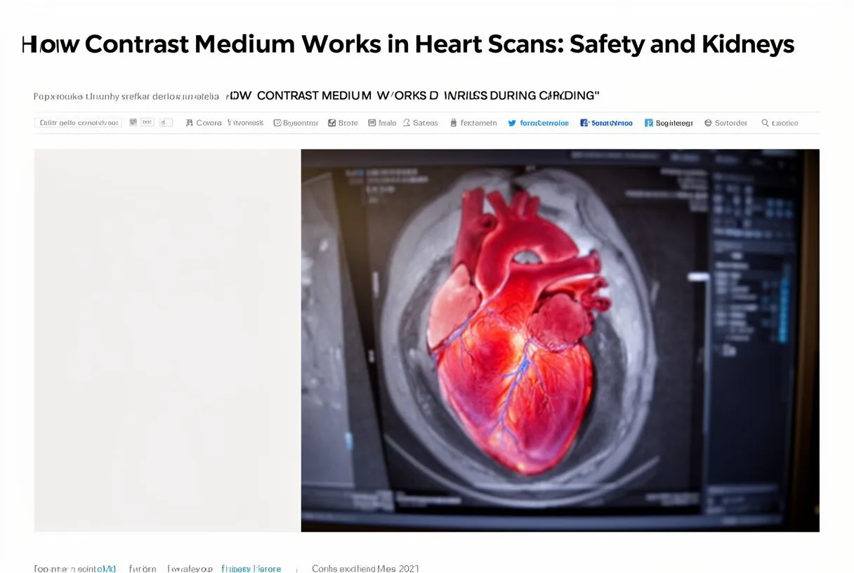
Contrast medium works by changing how X-rays or magnetic fields interact with the blood and tissues they pass through. Most contrast agents used in cardiac CT and catheterization contain iodine, a dense element that blocks X-rays. When the dye fills the heart chambers and coronary arteries, those structures appear bright white on the resulting images, creating a clear silhouette against the darker surrounding tissue. This principle is what makes it possible to see the ascending aorta, coronary artery branches, and even small defects in the heart wall that would otherwise remain invisible.
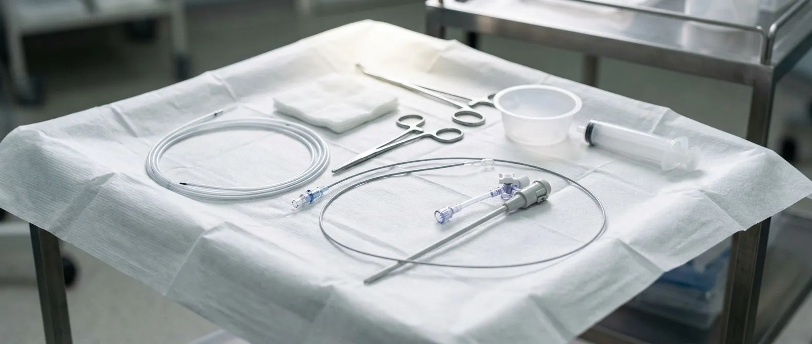
The injection typically happens through an intravenous line in the arm, or in the case of cardiac catheterization, through a thin tube called an arterial line placed in the wrist or groin. Patients often describe a sudden warm sensation spreading through the chest, neck, and arm pain is not expected but a flushed feeling or metallic taste in the mouth is normal and passes within seconds. The imaging itself takes only minutes, though the dye continues circulating through the bloodstream as the kidneys begin filtering it out. According to guidance from the National Heart, Lung, and Blood Institute, the body eliminates most contrast agents within a day, provided kidney function is adequate.
For cardiac MRI, a different type of contrast agent based on gadolinium is used instead of iodine. Gadolinium-based agents interact with magnetic fields rather than X-rays, producing detailed images of heart muscle tissue, scarring, and inflammation. While the mechanism differs, the fundamental principle is the same: the dye enhances the contrast between structures, giving your cardiologist the visual information needed to make an accurate diagnosis and plan the right treatment.
Types of Contrast Agents Used in Heart Scans
Not all contrast dyes are the same, and the type chosen for your scan depends on which imaging test you are having and your individual health profile. The two main families of contrast agents used in heart scans are iodine-based agents for CT and catheterization, and gadolinium-based agents for MRI. Within each family there are newer and older formulations, and the differences between them matter significantly when kidney safety is a concern.
Iodine-based contrast for CT and catheterization: These agents are the most commonly used in cardiac imaging. Modern formulations are called non-ionic, low-osmolar or iso-osmolar contrast media, meaning they have a chemical structure closer to blood plasma than older high-osmolar dyes. This closer match reduces the workload on the kidneys and lowers the risk of adverse reactions. Your cardiologist selects the specific agent based on your kidney function, diabetes status, and whether you take medications such as metformin, which can interact with contrast dye clearance.
Gadolinium-based contrast for cardiac MRI: These agents produce highly detailed images of heart muscle and are particularly useful for evaluating scarring after a heart attack, inflammation of the heart wall, or congenital structural problems. Newer gadolinium agents are designed to stay more tightly bound to their molecular carrier, reducing the amount of free gadolinium the kidneys must process. For patients with significant kidney impairment, however, gadolinium agents carry a rare but serious condition called nephrogenic systemic fibrosis, so alternative imaging approaches may be chosen instead. In all cases, the contrast agent is selected to provide the diagnostic information needed at the lowest risk for your situation.
Contrast Medium Kidney Safety: What Every Patient Should Know
When patients hear that a heart scan involves dye injected into the bloodstream, the most common worry is whether their kidneys can handle it. This concern is legitimate and well-documented in medical literature. The kidneys filter nearly all of the contrast medium from the blood, and in people with normal kidney function this process occurs without difficulty. The dye arrives at the kidneys, passes through the filtering units called nephrons, and exits through urine usually within 24 hours.
Why the kidneys can struggle with contrast dye: Contrast agents are dense, concentrated substances that increase the viscosity or thickness of the blood flowing through the tiny filtering vessels in the kidneys. In a person with healthy renal function, this temporary change is well tolerated. But if the kidneys are already working below capacity — whether from diabetes, longstanding hypertension, or age-related decline — the additional load from the dye can reduce blood flow to the filtering units, causing them to slow down or, in rare cases, temporarily shut down. This is the mechanism behind contrast-induced nephropathy, and it is the reason kidney function is always checked before a contrast scan.
The reassuring news is that modern contrast agents are far gentler on the kidneys than those used decades ago, and the protocols for protecting renal function have improved substantially. Measuring kidney function with a simple blood test called eGFR (estimated glomerular filtration rate) before any contrast scan allows the cardiac team to identify who needs extra precautions and who can proceed without concern. For the vast majority of patients, contrast medium heart scans are completed safely with no lasting impact on kidney health.
What Is Contrast-Induced Nephropathy?
Contrast-induced nephropathy, sometimes called contrast-associated acute kidney injury, is a temporary reduction in kidney function that occurs after exposure to contrast dye. Clinically, it is defined as a rise in serum creatinine — a waste product the kidneys normally clear — by a specific amount within 48 to 72 hours after the scan. It is one of the most discussed complications in cardiac imaging because coronary catheterization and cardiac CT are among the most common procedures using contrast agents.
What actually happens inside the kidney: The contrast medium enters the kidney's filtering system and causes two simultaneous effects. First, it temporarily increases the thickness of blood flowing through the tiny vessels, which slows filtration. Second, it can trigger mild oxidative stress — a chemical imbalance that irritates the cells lining the kidney tubules. Together, these effects reduce the kidney's output of urine and raise creatinine levels in the blood. In most cases, the kidneys recover fully within one to three weeks as the dye is completely cleared and normal blood flow resumes.
It is important to distinguish between mild, temporary changes in kidney blood tests and genuine kidney failure. Most instances of contrast-induced nephropathy are mild and self-limiting, meaning the kidneys recover on their own without dialysis or long-term damage. The patients most likely to experience this are those who already have reduced kidney function, diabetes, or dehydration at the time of the scan. Recognising these risk factors beforehand is the single most effective step in preventing the problem.
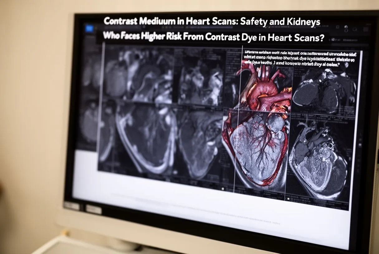
Who Faces Higher Risk From Contrast Dye in Heart Scans?
Not every patient faces the same level of risk when receiving contrast medium for a heart scan. Identifying who is at elevated risk is a critical part of the pre-scan evaluation, and it determines whether extra hydration, medication adjustments, or alternative imaging strategies are needed. The cardiac team assesses risk using a combination of blood tests, medical history, and the planned dose of contrast.
Several factors raise the likelihood of kidney stress after contrast exposure. A study examining postoperative monitoring approaches, referenced in Intraoperative transfontanelle ultrasonography for pediatric patients, illustrates how vigilant assessment before and after procedures involving contrast and surgical intervention improves outcomes through early detection. The same principle applies to adult cardiac imaging: knowing the risk profile beforehand allows the team to intervene preventively rather than reactively.
- Pre-existing kidney disease: An eGFR below 60 mL/min significantly increases vulnerability to contrast-induced nephropathy.
- Diabetes mellitus: Longstanding diabetes can damage the small vessels in the kidneys, reducing their reserve capacity.
- Dehydration: Even mild dehydration at the time of the scan concentrates the dye and worsens its effect on kidney filtration.
- Age over 75: Natural age-related decline in kidney function reduces the organ's ability to clear contrast efficiently.
- Heart failure: Reduced cardiac output means less blood reaches the kidneys, compounding the dye's effect on filtration.
If any of these factors apply to you, it does not mean the scan cannot proceed. It means your cardiac team will take additional protective steps, which may include intravenous hydration before and after the procedure, temporarily stopping certain medications like non-steroidal anti-inflammatory drugs, and choosing the lowest effective dose of a kidney-friendly contrast agent. Openly sharing your full medical history — including over-the-counter medication use — is essential for this risk assessment to be accurate.
How Doctors Protect Your Kidneys During Contrast Heart Scans
Kidney protection during contrast medium heart scans is not an afterthought — it is built into the protocol from the moment the scan is scheduled. The strategies used are well-established and evidence-based, designed to reduce the dye's impact on the kidneys while still obtaining the diagnostic images needed. If you understand these measures, you can participate more actively in your own care and feel more confident going into the procedure.
Pre-scan hydration is the most powerful protective tool: Administering intravenous fluids before the scan dilutes the contrast dye as it enters the bloodstream and helps the kidneys flush it out more efficiently. For high-risk patients, hydration may begin several hours before the procedure and continue afterward. In some centres, a measured protocol using sodium bicarbonate solution is used instead of plain saline, as it helps neutralise the acidic environment in the kidney tubules that contributes to cell stress. Either way, the principle is simple: well-hydrated kidneys handle contrast dye far better than dry ones.
Beyond hydration, the team will review every medication you take and identify any that need to be paused. Metformin, a common diabetes medication, is typically held for 48 hours after contrast exposure because if kidney function is temporarily reduced, metformin can accumulate to dangerous levels. Non-steroidal anti-inflammatory drugs like ibuprofen are paused because they reduce blood flow to the kidneys. Blood pressure medications in the ACE inhibitor or ARS family may also be reviewed, as they can alter kidney filtration dynamics. The contrast agent itself is selected from the newer low-osmolar or iso-osmolar families, and the dose is kept to the minimum that still produces diagnostic-quality images.
Contrast Medium Side Effects and What Patients Experience
Most patients tolerate contrast medium well, and the sensations experienced during injection are brief and expected. Understanding what is normal versus what warrants attention helps reduce anxiety and ensures that genuine concerns are reported promptly. The side effect profile of modern contrast agents is considerably better than older formulations, but reactions can still occur and fall into three categories: expected sensations, mild reactions, and rare allergic responses.
The expected sensation during contrast injection is a sudden wave of warmth that spreads from the injection site through the chest, abdomen, and groin. Many patients also notice a metallic or salty taste in the mouth and a sensation of having urinated, even though this has not actually happened. These feelings are caused by the dye interacting with nerve endings and blood vessels, and they resolve within seconds to minutes. None of these sensations indicate a problem — they simply confirm the dye is circulating as intended.
Mild reactions include nausea, mild hives, or sneezing shortly after injection. These are managed immediately by the imaging team with antihistamines and usually resolve without complications. True allergic reactions to modern non-ionic contrast agents are rare, estimated at less than one per cent of administrations, and severe reactions are even rarer. If you have had a reaction to contrast dye in the past, tell your team — pre-medication with corticosteroids and antihistamines can be given to prevent a repeat response. After the scan, the most common experience is simply increased urination as the kidneys flush out the dye, which is exactly what should happen.
- Warmth and flushing: Normal, brief, and expected during injection.
- Metallic taste: Common, resolves within seconds.
- Increased urination afterward: A sign the kidneys are clearing the dye properly.
- Nausea or mild rash: Report immediately; easily treated by the team.
- Difficulty breathing or swelling: Rare but requires urgent attention; the team is trained for this.
Contrast Dye in Cardiac Catheterization Versus Cardiac CT
The way contrast medium is used differs significantly between cardiac catheterization and cardiac CT, and these differences affect everything from the dose required to the kidney risk involved. Cardiac catheterization is an invasive procedure in which a thin tube is threaded directly into the coronary arteries through an arterial line in the wrist or groin. Contrast dye is injected directly into the coronary arteries themselves, producing real-time X-ray images called angiograms that show blockages with extreme precision. Because the dye is delivered straight to the arteries, relatively small volumes are needed, but the procedure itself carries additional risks related to arterial access and catheter manipulation.
Cardiac CT angiography, by contrast, is non-invasive. The dye is injected through a standard IV in the arm and images are captured by a CT scanner as the dye passes through the heart. This approach requires a somewhat larger volume of contrast to achieve adequate brightness in the coronary arteries, but it avoids the risks of arterial puncture. The choice between these two approaches depends on what your cardiologist needs to learn: CT is excellent for ruling out coronary artery disease in patients with moderate risk, while catheterization is preferred when an intervention like balloon angioplasty or stent placement is likely to be needed during the same procedure.
From a kidney safety standpoint, both approaches use the same modern contrast agents and the same protective protocols. The research summarised in The Specific Features of an Early Period after Minimally Invasive Cardiac Surgery underscores how early post-procedural monitoring — including renal function tracking — contributes to better outcomes, a principle that applies equally to contrast-based imaging. Your kidney function is assessed before either procedure, and if risk is elevated, the lowest effective dose is used along with hydration protocols. In some cases, a non-contrast imaging alternative such as stress echocardiography may be recommended if the diagnostic question can be answered without dye.
Safe Contrast Imaging for Kidneys: Your Follow-Up Plan
What happens after the scan is just as important as what happens during it. The follow-up plan for contrast medium heart scans focuses on ensuring the kidneys have cleared the dye completely and that no delayed effects have developed. For patients with normal kidney function and no risk factors, follow-up may simply mean drinking extra water for the rest of the day and watching for any unusual symptoms. For higher-risk patients, a follow-up blood test to check creatinine levels 48 to 72 hours after the scan is standard practice.
Knowing when to seek help is part of being an informed patient. In the days following your scan, pay attention to changes in urination — a significant decrease in output, dark or concentrated urine, or swelling in the legs or ankles could indicate the kidneys are not clearing the contrast as expected. Mild fatigue is common after any procedure, but persistent nausea, confusion, or breathlessness warrant a call to your cardiac team. These symptoms are uncommon, but reporting them early allows for prompt evaluation and intervention if needed.
If you are facing a heart scan and have concerns about contrast medium and kidney safety, the most productive step is to have a direct conversation with your cardiac team. Bring your complete medication list, share any history of kidney problems or diabetes, and ask what specific protective measures will be used for your situation. The team at Cirugía de Corazón, led by Dr Alejandro Molina, provides individualised assessments for patients in Guadalajara, Querétaro, and Hermosillo, and can explain in plain language which imaging approach offers the lowest risk for your specific anatomy and medical history. Booking a consultation is the right next step — self-diagnosing or postponing necessary imaging out of fear carries its own risks that a qualified team can help you weigh accurately.