Heart Tests and Diagnosis: Which Test You Need
Heart tests and diagnosis which test you need depends on your symptoms.
What Are Heart Tests and Diagnosis? Which Test You Need for Your Symptoms
Heart tests and diagnosis which test you need for your symptoms is one of the most common questions we hear in consultation. The answer depends on what your body is telling you: chest pain, breathlessness, palpitations, dizziness, or swollen ankles each point toward a different family of tests. The good news is that modern cardiology has an orderly way of working through this, starting with simple, painless tests and moving toward more detailed studies only when they are truly needed.
Types of heart diagnostic tests. Broadly, the tests fall into four groups: electrical tests that record your heart's rhythm, imaging tests that look at the heart's structure and pumping, stress tests that show how the heart behaves under exertion, and blood tests that measure substances the heart releases when it is strained. In most cases your cardiologist will start in the first group and work forward. No single test tells the whole story on its own.
Early detection matters. Heart disease early detection tests are not reserved for people who already feel unwell. Adults with risk factors — high blood pressure, diabetes, smoking, or a family history of early heart disease — often benefit from a baseline evaluation before symptoms ever appear. If you are deciding where to begin, a first conversation with a cardiac team can save you from ordering studies you do not need. Our team reviews every case together against international practice guidelines before recommending anything, and the patient receives a clear proposal with the alternatives.
What Is an Electrocardiogram (ECG) and When Is It Ordered?
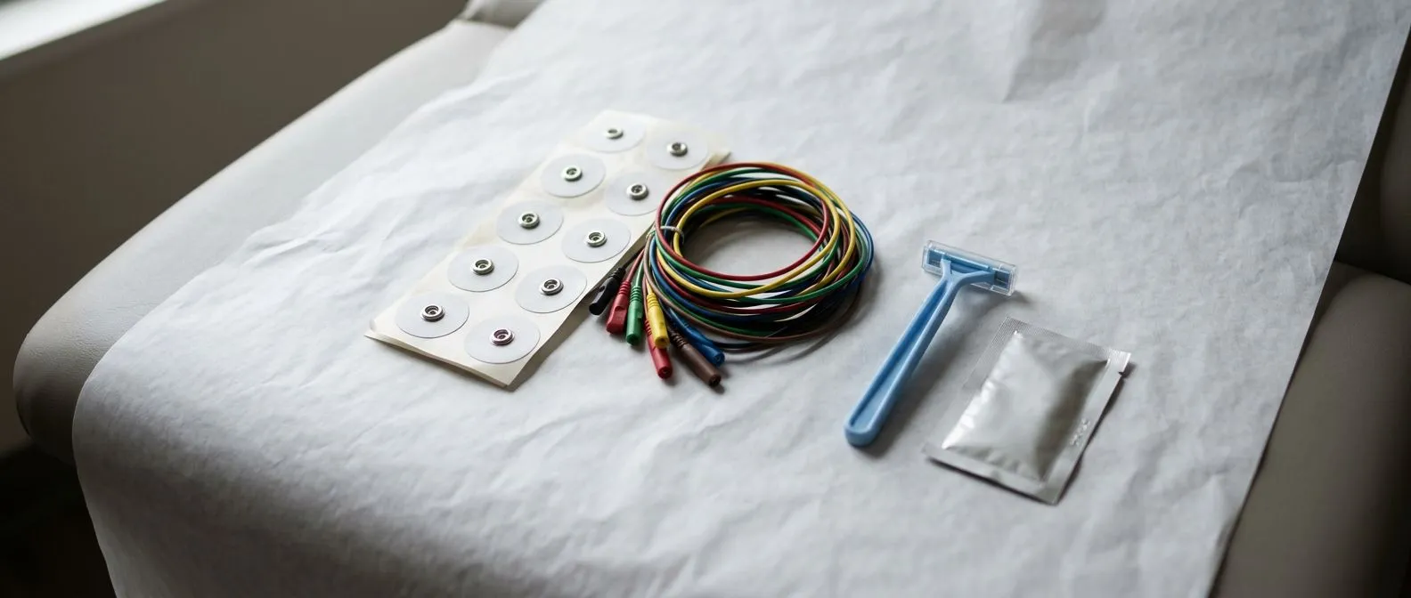
An electrocardiogram (ECG) is the most familiar heart test of all. Small sticky pads are placed on your chest, arms, and legs, and wires record the tiny electrical impulses that make your heart muscle contract. The whole thing takes about ten minutes, involves no needles, and produces a printed tracing of your heartbeat. It is usually the very first test ordered for palpitations, chest discomfort, or fainting spells.
Electrocardiogram ECG interpretation. A trained cardiologist reads the tracing for many things: whether the rhythm is regular, whether the electrical signals travel through the heart in the normal sequence, whether there is evidence of a previous heart attack, and whether the heart muscle walls appear thickened. Some findings are clear-cut; others are subtle and need comparison with older tracings. Johns Hopkins Medicine notes that an ECG is a standard first step in evaluating chest pain and suspected heart rhythm disorders.
What an ECG cannot tell you. The main limitation is timing. An ECG records a single snapshot lasting seconds, so a rhythm problem that comes and goes — or coronary narrowing that only shows itself under exertion — can be completely absent from the tracing. A normal ECG is reassuring, but it rarely closes the investigation on its own. When we explain results in consultation, we take the time to say in plain words what the tracing shows and, just as importantly, what it does not show, before any decision is made.
Echocardiogram Heart Ultrasound: What It Shows About Your Valves
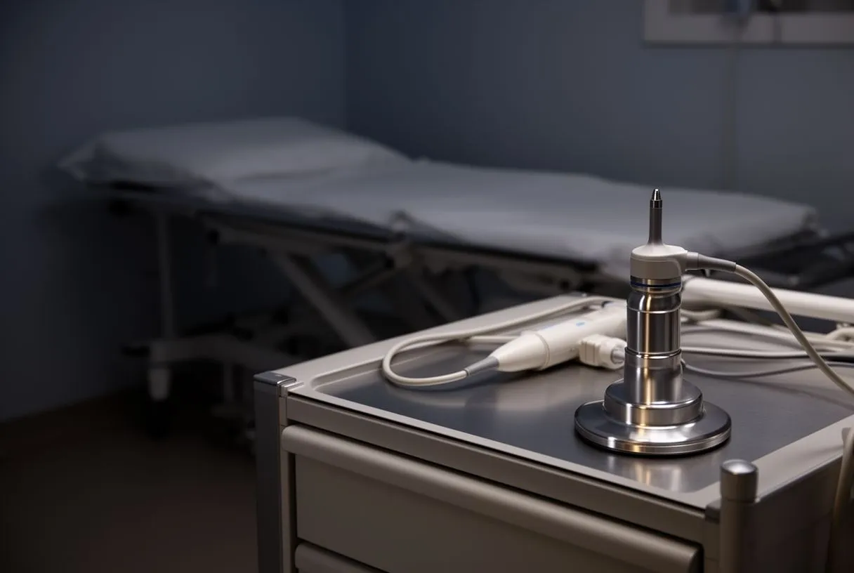
An echocardiogram is an ultrasound of the heart, sometimes called an "echo." It uses sound waves — the same technology used to look at a baby during pregnancy — to build a moving picture of your heart beating in real time. There is no radiation, no preparation, and no discomfort. It is the workhorse test for questions about the heart's structure rather than its electrical activity.
Echocardiogram heart ultrasound findings. An echo answers questions an ECG cannot. It shows the four chambers of the heart and how well they squeeze, the four valves and whether they leak or become narrowed, and the pumping function of the left ventricle, expressed as a percentage called the ejection fraction. In patients with heart failure, the 2016 European Society of Cardiology guidelines place echocardiography at the centre of diagnosis precisely because it distinguishes between different causes of breathlessness.
When valves are the problem. If you have a murmur — a sound your doctor hears through a stethoscope — an echocardiogram will show which valve is responsible and how severe it is. This matters enormously, because decisions about valve surgery or catheter-based repair depend on precise measurements that only imaging can give. In our practice, covering valve disease, coronary disease, the aorta and structural heart conditions, the echocardiogram is the study that most often shapes the treatment proposal we put in front of the patient.
For many people, an echo and an ECG together answer the whole question. For others, especially those whose symptoms appear only with exercise, the next step is a stress test — which brings us to a comparison patients ask about constantly.
Heart Stress Test vs Echocardiogram: Which One Do You Need?
The question of heart stress test vs echocardiogram comes up in almost every consultation, and the honest answer is that they answer different questions. An echocardiogram looks at the heart at rest. A stress test looks at the heart at work. If your symptoms only appear when you climb stairs or walk uphill, a resting study may look perfectly normal — which is exactly why the exertion component matters.
What each test is for. Here is a simple way to keep them straight:
- Echocardiogram: best for valve problems, murmurs, heart muscle function, and causes of breathlessness at rest.
- Stress test: best for chest pain on exertion, exercise tolerance before surgery, and suspected coronary artery narrowing.
- Both together: a stress echocardiogram combines the two, imaging the heart immediately before and after exertion.
Nuclear perfusion heart test. One variant of the stress test adds a tiny amount of radioactive tracer to show blood flow through the heart muscle. This nuclear perfusion study is useful when the ECG changes during exercise are hard to interpret, or in patients whose anatomy or previous surgery makes standard exercise testing less reliable. It involves a mild radiation dose, so we reserve it for cases where it genuinely adds information rather than ordering it by default.
Treadmill Stress Test Preparation: How to Get Ready
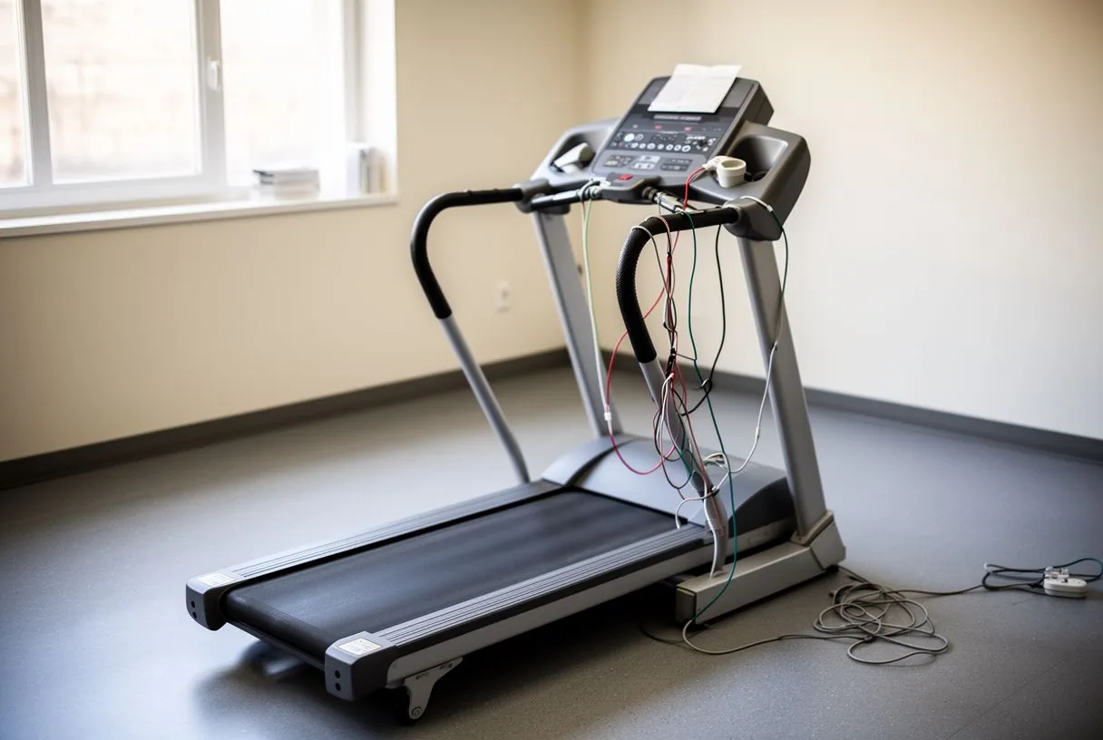
A standard exercise stress test, sometimes called a Treadmill Test, walks you on a belt that gradually gets faster and steeper while your ECG, blood pressure, and symptoms are monitored. The test stops when you reach a target level of exertion, when symptoms appear, or when the tracing shows changes that need attention. It is not dangerous for most patients, and the staff are trained to stop it safely at any point.
Treadmill stress test preparation. Good preparation makes the result more reliable, and the steps are simple:
- Before the day: ask your doctor which heart medications to hold — some, like beta-blockers, can blunt the heart rate response and must be paused temporarily.
- On the day: avoid heavy meals and caffeine for several hours beforehand, and wear comfortable shoes and clothing you can exercise in.
- Bring information: a list of your current medications and any previous ECGs, so the doctor can compare tracings.
What to expect during the test. You will feel the effort — that is the point. Most people walk for six to twelve minutes. Tell the staff exactly what you feel: chest tightness, breathlessness, leg fatigue, or dizziness. The team monitoring you is there to make the test safe, and stopping early does not mean the test failed. In our practice, the first contact is a free video consultation, where we talk through the condition and say which studies are still needed — so patients arrive at testing day with a clear reason for each test rather than a long list of unnecessary ones.
Holter Monitor Heart Test: Catching Rhythm Problems
Palpitations that come and go are the classic reason a standard ECG comes back normal. The rhythm disturbance simply was not happening during those ten minutes. A heart rhythm monitoring test solves this by recording continuously over a longer period, and the most common version is the Holter monitor.
Holter monitor heart test. A Holter is a small portable ECG device worn for 24 to 72 hours, with patches on your chest and a lightweight recorder on a belt or strap. You go about your normal day — work, walking, sleeping — and press an event button whenever you feel a symptom. Later, the cardiologist lines up your symptoms against the recording, minute by minute. Extended monitors can be worn for two weeks or more when the episodes are infrequent.
What the recording can reveal. Holter monitoring can identify atrial fibrillation, an irregular rhythm that raises the risk of stroke and often goes unnoticed; runs of abnormally fast rhythms; and dangerous pauses in the heartbeat. It also settles a common worry: many people who feel palpitations turn out to have perfectly benign extra beats, and seeing that on paper is a genuine relief. For an anxious patient, simply confirming that the rhythm is normal often changes the whole conversation.
Cardiac Biomarker Blood Test: What Your Blood Says About Your Heart
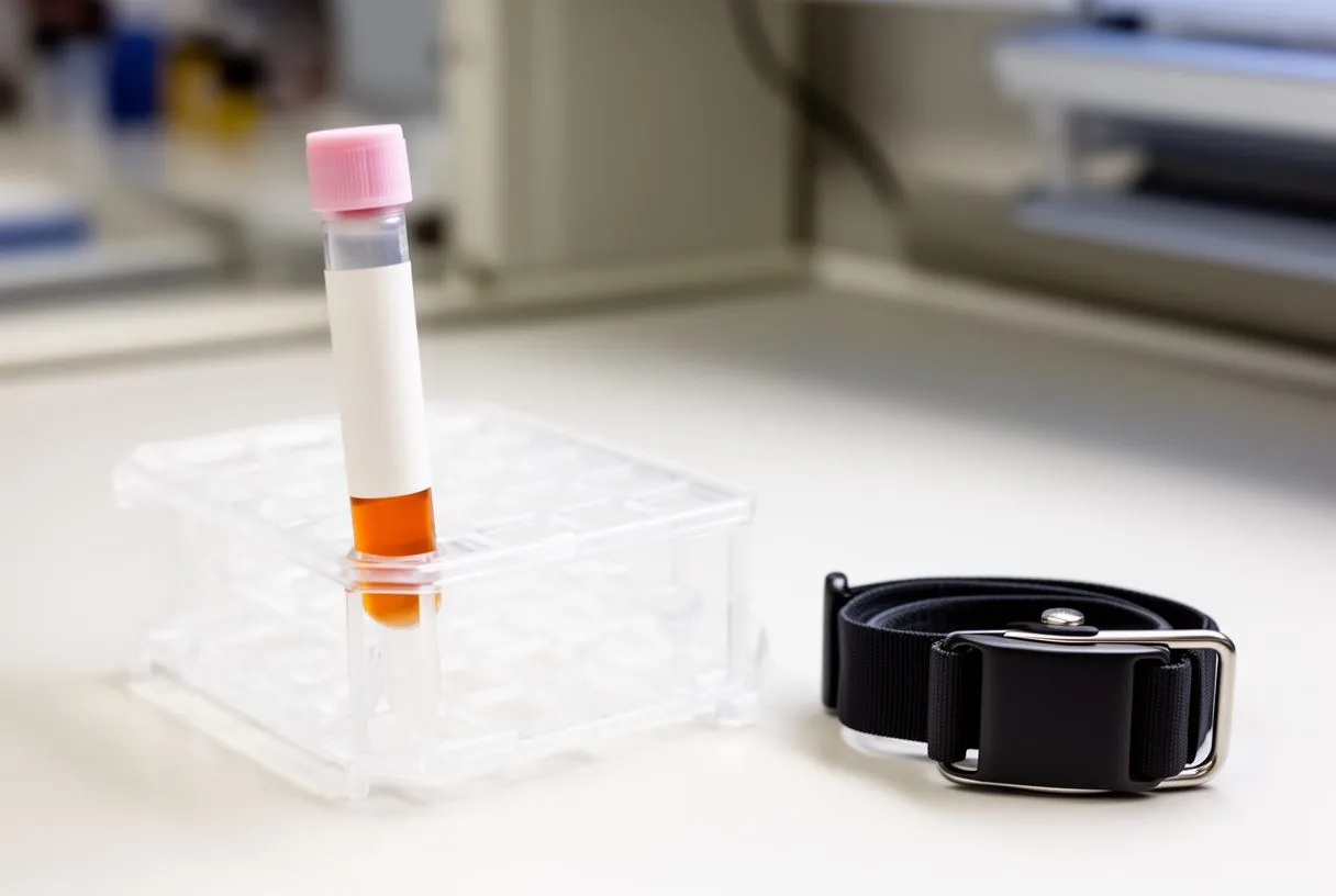
Not every heart test involves a machine with wires or cameras. A cardiac biomarker blood test measures proteins the heart muscle releases into the bloodstream when it is injured or under severe strain. The most important one is troponin, which rises within hours of heart muscle damage and is the cornerstone of diagnosing a heart attack in the emergency department.
Other blood tests that matter. Beyond troponin, several blood tests shape cardiac diagnosis and long-term risk. These include:
- B-type natriuretic peptide (BNP or NT-proBNP): rises when the heart is stretched by heart failure and helps distinguish cardiac from lung-related breathlessness.
- Lipid panel: cholesterol and related fats that drive the gradual narrowing of coronary arteries.
- Blood sugar and kidney function: diabetes and kidney disease are major cardiac risk factors and influence which treatments are appropriate.
- Liver Function Tests: often checked before starting certain heart medications and before major procedures, since several cardiac drugs are processed by the liver.
How blood results fit the picture. Blood tests rarely point to a diagnosis on their own; they confirm, exclude, or quantify what imaging and the clinical examination suggest. The Pulmonary Function Tests serve a similar complementary role — when breathlessness is the complaint, they help show whether the lungs rather than the heart are the source. Good diagnosis is about assembling the right tests for the right question, not about running every available study.
Heart CT Calcium Score Test: Should You Get One?
Computed tomography (CT) has become one of the most useful tools in modern cardiology, and two of its applications are worth knowing about. A heart CT calcium score test uses a fast scanner to measure the amount of calcium deposited in the walls of the coronary arteries — the vessels that feed the heart muscle. Calcium is a marker of atherosclerosis, the gradual build-up of plaque that narrows arteries over decades.
What the calcium score means. The result is a number. A score of zero means no detectable coronary calcium, which carries a very low risk of a cardiac event in the near term and can spare a patient years of unnecessary worry or further testing. Higher scores indicate increasing plaque burden and prompt a conversation about aggressive risk-factor control and, in some cases, further investigation. The scan is quick, requires no contrast injection, and involves a modest radiation dose.
Coronary CT angiography. A related scan, coronary CT angiography, uses an iodine contrast injection to image the actual channel of the coronary arteries and can show narrowings directly, without the invasiveness of a catheter study. It is increasingly the test of choice in intermediate-risk patients with chest pain, because it can rule out significant coronary disease without a catheterization. The decision between these options is exactly the kind of question our multidisciplinary team reviews together, because the right answer depends on the patient's whole picture, not on any single number.
Heart Catheterization and Coronary Angiography Diagnosis
When non-invasive tests suggest a significant coronary artery problem — or when the picture remains unclear — the definitive study is cardiac catheterization. This is the test that shows the coronary arteries in fine detail, and it remains the gold standard for planning interventions such as stents or bypass surgery.
Coronary angiography diagnosis, step by step. A thin tube, called a catheter, is introduced through an artery, usually at the wrist, and guided to the heart. Contrast dye is injected through the catheter, and X-ray imaging shows the dye flowing through the coronary arteries, revealing any narrowings or blockages. The procedure is done under local anaesthetic and mild sedation, and patients are usually awake, watching the images on screen. Most go home the same day.
Heart catheterization test and pressure measurements. Catheterization is not only about the arteries. It also measures pressures inside the heart chambers, which is essential for assessing valve disease severity, and it can be combined with the Treadmill Test concept by doing exercise or pharmacological stress during the study. In patients being considered for aortic valve replacement or repair, the catheterization findings often decide which approach — surgical or transcatheter — offers the lowest risk for that specific patient. In our practice, every catheterization report is reviewed by the full multidisciplinary team before a recommendation is made, and the final decision is always the patient's.
Risks and recovery. Serious complications of catheterization are uncommon, but they exist — bleeding at the puncture site, arrhythmia, and, rarely, damage to the artery or kidney injury from contrast. These risks are higher in patients with kidney disease or advanced valve disease, which is why the test is ordered only when the information it provides will change the plan of care. Discussing this openly is part of every consultation we hold.
Cardiac MRI Scan Test and Transesophageal Echocardiogram: Advanced Imaging
Two advanced imaging studies complete the diagnostic toolkit, and they are reserved for questions the standard tests cannot answer. A cardiac MRI scan test uses a powerful magnetic field — no radiation — to produce exceptionally detailed images of the heart muscle, and it is the best test for identifying scarring, inflammation, and certain rare muscle diseases.
Cardiac MRI and transesophageal echocardiogram TEE: when each is used. Here is how cardiologists decide between them:
- Cardiac MRI: for heart muscle scarring after a heart attack, suspected myocarditis (inflammation of the heart muscle), and characterizing cardiomyopathies that an echo cannot fully define.
- Transesophageal echocardiogram TEE: for a close-up view of the heart valves and for looking for blood clots or infection on the valves, since the probe sits in the oesophagus directly behind the heart.
- Both: in complex structural heart disease, the two studies are often complementary before a major intervention.
What the experience is like. A cardiac MRI means lying still inside the scanner for thirty to sixty minutes, sometimes with breath-hold instructions; claustrophobia is manageable in most cases and should be mentioned beforehand. A TEE involves numbing the throat and passing a slim, flexible probe to the oesophagus under sedation — briefly uncomfortable, then forgotten once you wake. Both are outpatient procedures.
Putting the tests together. No patient needs every test on this page, and a good cardiac team will say plainly which ones apply to you and why. In our consultation rooms in Guadalajara, Querétaro and Hermosillo, we explain what each recommended study actually shows, in plain words, and we answer every question before any decision — and we actively encourage a second opinion as a normal part of the process. If you have symptoms that concern you, start with a conversation rather than a list of tests: a free video consultation with a cardiac team is the most efficient first step, and booking one is far safer than trying to self-diagnose. Your heart deserves a clear answer, and now you know how cardiologists find it.