Osler Nodes: A Skin Sign of Heart Valve Infection
Osler nodes endocarditis causes painful finger nodules as a warning sign of heart valve infection.
What Are Osler Nodes Endocarditis Skin Signs?
Osler nodes endocarditis represents one of the most distinctive dermatological clues that a heart valve infection is underway. When bacteria settle on the valves of the heart — a condition called infective endocarditis — the immune system reacts in ways that can produce small, tender bumps on the pads of fingers and toes. These bumps, known as Osler nodes, are not the infection itself travelling to the skin; rather, they are the body's response to tiny fragments of immune complexes lodging in small blood vessels. For patients and families first encountering this sign, the sudden appearance of painful nodules can be alarming, especially when the connection to the heart is not immediately obvious.
Why the skin reflects what happens inside the heart: The heart's valves sit at the junction of chambers — the atrium above and the ventricle below — and when bacteria colonise these structures, fragments of the bacterial cluster can break free. These fragments travel through the bloodstream and deposit in distant capillaries, particularly in the fingertips. The immune system responds with localised inflammation, producing the tender, pea-sized nodules that Sir William Osler first described in the late nineteenth century. Unlike a rash or an allergic reaction, these nodes carry a clear temporal pattern: they appear, remain tender for hours to days, and then fade, sometimes leaving a faint discolouration.
Who typically develops this sign: Osler nodes do not appear in every patient with endocarditis. They are more commonly seen in cases of subacute bacterial endocarditis, where the infection develops gradually over weeks rather than striking suddenly. Patients with pre-existing valve damage — whether from rheumatic fever, a congenital defect, or a prosthetic valve — carry a higher baseline risk. It is worth noting that the absence of Osler nodes does not rule out endocarditis; many confirmed infections never produce any visible skin signs at all. This is why cardiology teams rely on a combination of clinical examination, blood cultures, and echocardiography rather than any single physical finding. The American Heart Association publishes detailed guidance on recognising and managing these presentations, and clinicians refer to resources from ahajournals.org for current diagnostic criteria.
What Causes Osler Nodes in Bacterial Endocarditis?
The underlying cause of Osler nodes is infective endocarditis itself, but the chain of events leading from a heart valve infection to a painful nodule on a fingertip involves several steps. Bacteria — most commonly streptococci or staphylococci — enter the bloodstream through a portal such as the mouth during dental work, an intravenous line, or a skin wound. Once circulating, they adhere to the endocardial surface, particularly on valves that have rough or damaged areas where an atheromatous plaque or previous injury has disrupted the smooth endothelial lining. There, the bacteria multiply within a protective matrix called a vegetation.
The role of immune complex deposition: As the infection persists, the immune system produces antibodies against the bacteria. These antibodies bind to bacterial antigens, forming immune complexes that circulate in the blood. Some of these complexes become trapped in the small vessels of the finger pads, toe pads, and sometimes the thenar eminence of the palm. The trapped complexes trigger complement activation and local inflammation, which is what produces the pain, redness, and slight swelling characteristic of an Osler node. The process is immunological rather than infectious — meaning the nodule itself does not contain live bacteria. This distinction matters because it explains why treating the skin alone would be futile; the root problem lies within the heart.
Common bacterial culprits: The organisms most frequently responsible include viridans group streptococci, which originate in the oral cavity, and Staphylococcus aureus, which can enter through skin breaches or indwelling catheters. Less common but clinically significant organisms include enterococci and the so-called HACEK group of bacteria. Each organism tends to produce a slightly different clinical picture, with viridans streptococci more often associated with the subacute presentation that produces Osler nodes, while Staphylococcus aureus tends to cause a more acute, destructive infection. Research into cellular mechanisms behind these inflammatory cascades continues to evolve, and studies such as those examining autophagy in major human diseases help scientists understand how cells respond to these immune-driven injury patterns.
Osler Nodes Symptoms: How to Recognize Painful Finger Nodules
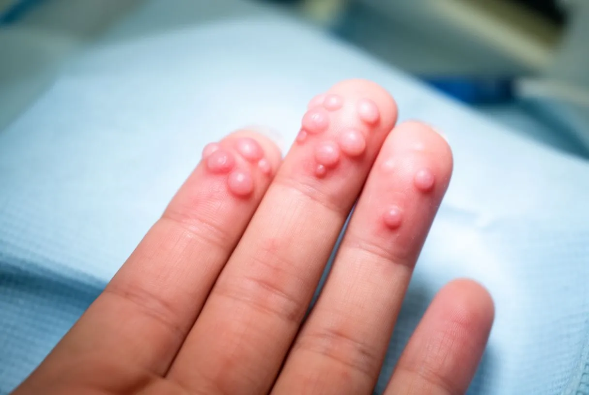
Recognising Osler nodes symptoms early can be the first step toward diagnosing a heart valve infection that might otherwise remain hidden until it causes serious damage. The classic presentation is a small — typically two to five millimetres — tender nodule on the fleshy pad of a finger or toe. Patients often describe the sensation as a deep, aching pain rather than a surface soreness, and the tenderness is usually disproportionate to the visible size of the lesion. The nodes commonly appear on multiple digits simultaneously and may recur in crops over the course of the illness.
What the nodules look and feel like: Osler nodes are usually slightly raised, with a reddish or purplish hue that reflects the underlying inflammation in the dermal capillaries. When pressed, they are distinctly tender — patients often withdraw the hand quickly. Unlike insect bites or minor traumas, these nodules are not associated with a break in the skin; the surface remains intact. They tend to resolve over a few days without scarring, though a faint area of pigmentation may persist for weeks. Because they come and go, patients sometimes dismiss them, especially if they are also experiencing the low-grade fever, fatigue, and weight loss that often accompany subacute endocarditis.
Accompanying symptoms that complete the picture: Osler nodes rarely appear in isolation. Patients with infective endocarditis frequently describe a constellation of symptoms that includes intermittent fever, night sweats, muscle aches, and a general sense of being unwell. Some develop arm pain if embolic material lodges in a peripheral vessel, and others notice small splinter haemorrhages under the fingernails — linear dark streaks that also reflect microembolic events. When clinicians listen to the heart through a stethoscope — a procedure called auscultation — they may detect a new or changing heart murmur, which is the audible signature of turbulent flow across an infected valve. These findings together form the diagnostic puzzle that points toward endocarditis, even before blood cultures or imaging confirm it.
Osler Nodes vs Janeway Lesions: Key Differences
Patients and clinicians sometimes confuse Osler nodes with Janeway lesions, but the distinction is clinically meaningful. Both are dermatological manifestations of infective endocarditis, yet they differ in appearance, location, and underlying mechanism. Understanding the difference helps families know what they are looking at and helps doctors narrow the diagnostic approach.
How each lesion looks and feels: Janeway lesions are flat, painless, macular spots — usually red or maroon — found most often on the palms and soles. They result from septic microemboli, meaning tiny infected clumps actually lodge in the skin's capillaries. Osler nodes, by contrast, are raised, tender, nodular, and found on the finger and toe pads. The pain of Osler nodes stems from immune complex deposition rather than direct infection, which is why the node itself is sterile. In practical terms, a patient who notices a painful lump on a fingertip should be aware that this is more consistent with Osler nodes, while a painless flat spot on the palm leans toward Janeway lesions. Both signs warrant urgent medical evaluation.
Why the difference matters diagnostically: The presence of either sign suggests that the heart valve infection is active and producing systemic effects, but the specific pattern can hint at the type of organism and the tempo of the illness. Janeway lesions are more frequently associated with Staphylococcus aureus and a more acute presentation, while Osler nodes tend to accompany the subacute form of the disease. For a surgical team evaluating a patient, the combination of skin signs, blood culture results, and echocardiographic findings determines whether antibiotics alone will suffice or whether surgical valve replacement becomes necessary. The European pathology community has contributed to understanding these patterns, and detailed post-mortem studies such as the guidelines for autopsy investigation of sudden cardiac death reinforce how systemic infections leave traces across multiple organs.
- Osler nodes: Raised, tender nodules on finger and toe pads — immune complex mediated, sterile.
- Janeway lesions: Flat, painless macules on palms and soles — septic microemboli, may contain bacteria.
- Diagnostic value: Both strongly suggest infective endocarditis; their pattern helps predict organism type and disease tempo.
How Doctors Diagnose Osler Nodes and Heart Valve Infection
The diagnosis of infective endocarditis rests on a combination of clinical findings, laboratory tests, and imaging — no single piece of evidence is sufficient on its own. When a patient presents with painful nodules on the fingertips, a cardiologist will take a careful history asking about recent dental procedures, intravenous drug use, congenital heart conditions, and prosthetic valves. The physical examination focuses on the heart — listening for murmurs, checking for signs of heart failure — and on the skin, eyes, and nails for peripheral manifestations.

The Modified Duke Criteria explained: Cardiologists worldwide use the Modified Duke Criteria to organise diagnostic evidence into major and minor categories. Major criteria include positive blood cultures for typical endocarditis organisms and echocardiographic evidence of valve involvement — such as a vegetation, abscess, or new partial dehiscence of a prosthetic valve. Minor criteria include predisposing heart conditions, fever above 38°C, vascular phenomena like Janeway lesions, and immunological phenomena — which is where Osler nodes, glomerulonephritis, and other immune-mediated signs fit. Meeting two major criteria, one major plus three minor, or five minor criteria classifies the case as definite endocarditis. This structured approach reduces the risk of over-diagnosing based on a single skin finding or under-diagnosing when the presentation is subtle.
Imaging and laboratory confirmation: Blood cultures remain the cornerstone — typically three sets drawn at different times before antibiotics begin. Transthoracic echocardiography (TTE) provides a non-invasive first look at the valves, and if suspicion remains high but TTE is inconclusive, transoesophageal echocardiography (TEE) offers much higher resolution, particularly for the mitral valve and for small vegetations on prosthetic material. In some centres, advanced imaging such as positron emission tomography (PET) can detect abnormal metabolic activity around prosthetic valves. Additional blood tests look for elevated inflammatory markers, anaemia, and renal impairment — all of which can accompany endocarditis. A patient being evaluated for suspected endocarditis should expect multiple blood draws, at least one echocardiogram, and close monitoring while results are pending.
Infective Endocarditis Skin Signs Beyond Osler Nodes
While Osler nodes endocarditis is a well-known cutaneous manifestation, the skin can display several other signs that point toward a heart valve infection. Each sign reflects a different pathological mechanism — some are embolic, some immunological, and some a combination of both. Families who recognise these patterns can communicate more effectively with their medical team and understand the rationale behind specific tests.
Splinter haemorrhages and petechiae: Splinter haemorrhages appear as thin, dark red or brown lines under the fingernails or toenails, running parallel to the nail's long axis. Though they can also result from minor trauma — a common occurrence in manual workers — when they appear in multiple nails without a clear cause, especially alongside fever, they raise the possibility of microemboli from an infected valve. Petechiae are pinhead-sized red spots that do not blanch when pressed; they can appear on the palate, conjunctiva, or skin. Both signs are non-specific, meaning they occur in many conditions, but in the right clinical context they add weight to the endocarditis diagnosis.
Clubbing and other peripheral stigmata: In chronic endocarditis, some patients develop finger clubbing — a widening and rounding of the fingertips with a loss of the normal angle between the nail and the nail fold. Clubbing reflects chronic hypoxia or inflammatory change and develops slowly over months. Other signs include Roth spots — retinal haemorrhages with pale centres visible on fundoscopic examination — and tender nodules on the thenar eminence. It is important to recognise that some patients with endocarditis never develop any of these signs; an atypical infarction or an unexplained stroke may be the first indication of valve infection, especially in older patients or those with prosthetic valves where the presentation can be muted.
Osler Nodes Treatment: Addressing the Underlying Heart Infection
There is no specific treatment for Osler nodes themselves — they resolve as the underlying endocarditis is treated. The therapeutic focus is entirely on eradicating the heart valve infection, which requires targeted antibiotic therapy and, in a significant proportion of cases, surgical intervention. Treatment decisions depend on the organism identified, the extent of valve destruction, the presence of complications, and the patient's overall health.
Antibiotic therapy: duration and choice: Once blood cultures identify the responsible organism, clinicians tailor antibiotic therapy to that specific bacterium and its resistance profile. Treatment typically lasts four to six weeks intravenously, beginning in hospital and sometimes continuing at home with close monitoring. The initial regimen, before culture results return, covers the most likely organisms empirically. For streptococcal infections, penicillin or ceftriaxone is often effective. For staphylococcal infections, particularly methicillin-sensitive Staphylococcus aureus, anti-staphylococcal penicillins such as flucloxacillin are standard. Patients sometimes ask whether aspirin or antiplatelet therapy has a role; current guidelines do not recommend routine antiplatelet treatment for endocarditis, though some patients may be on these medications for other cardiac conditions.
When surgery becomes necessary: Approximately one third to one half of patients with infective endocarditis require surgery during or shortly after the acute episode. Surgical indications include uncontrolled infection despite antibiotics, large mobile vegetations at risk of embolisation, heart failure from valve destruction, abscess formation extending into surrounding tissue, and infections caused by organisms resistant to medical therapy. Depending on the valve involved and the extent of damage, the surgeon may repair or replace the valve. Replacement options include mechanical valves — durable but requiring lifelong anticoagulation — and biological valves, which may be derived from animal tissue or, in some approaches, fashioned from a patient's own tissue (an autograft). When the aortic valve is diseased, surgery may involve the ascending aorta if infection has extended beyond the valve itself. After surgery, patients receive a full course of antibiotics to complete the eradication of infection.
Subacute Bacterial Endocarditis: The Chronic Infection Pattern
Subacute bacterial endocarditis is the form of valve infection most commonly associated with Osler nodes. Unlike the acute variant, which can destroy a previously normal valve within days, the subacute form develops gradually over weeks to months. This slower tempo allows the immune system to mount the responses that produce peripheral signs like Osler nodes, splinter haemorrhages, and glomerulonephritis. Patients often feel unwell for a prolonged period before seeking care, attributing their fatigue, low-grade fever, and weight loss to a lingering virus or the stress of daily life.
Why the indolent course matters: The gradual progression of subacute endocarditis can make diagnosis more difficult. The patient may not appear acutely ill, the fever may be intermittent and low, and the murmur may be subtle, especially if the pre-existing valve abnormality already produced a murmur before infection set in. By the time Osler nodes appear, the infection has often been present for weeks. This is why clinicians take a careful history of any change in a known murmur — a new murmur or a change in the character of an existing one is a significant red flag. Patients with prosthetic valves require especially vigilant monitoring, as infection on prosthetic material can present atypically and progress silently.
Organisms and risk factors: The bacteria most often responsible for the subacute pattern are viridans group streptococci, which normally live in the mouth and enter the bloodstream during dental procedures, vigorous brushing, or chewing. Other organisms include Enterococcus species, which originate in the gastrointestinal and genitourinary tracts, and the HACEK group — a set of fastidious bacteria that require special culture conditions. Patients at highest risk include those with prosthetic valves, previous endocarditis, certain congenital heart defects, and those with indwelling cardiac devices. Preventive antibiotic prophylaxis before dental procedures is recommended for the highest-risk groups, though this remains a decision made individually with the cardiac team rather than a blanket recommendation for all patients.
- Subacute tempo: Weeks to months of low-grade fever, fatigue, weight loss — easy to miss initially.
- Typical organisms: Viridans streptococci (oral), enterococci (GI/GU), HACEK group.
- Skin signs: Osler nodes, splinter haemorrhages, petechiae — reflect immune-mediated and embolic activity.
Infective Endocarditis Complications: What Patients Should Know
Infective endocarditis complications extend well beyond the heart valve itself. The infection can produce embolic events, immune-mediated damage, and structural cardiac problems that may emerge during or after the acute illness. Understanding these potential complications helps families anticipate what the medical team is monitoring for and why certain tests are repeated throughout treatment.
Embolic events and their consequences: Fragments of the infected vegetation can break off and travel through the arterial circulation. The most feared destination is the brain, where an embolus can cause a stroke. Other common sites include the spleen, kidneys, and mesenteric arteries. Peripheral emboli may cause sudden limb pain or ischaemia — and in rare cases, an arterial graft or prosthetic material elsewhere in the body can become seeded. The risk of embolism is highest in the first days of treatment and diminishes significantly once effective antibiotic therapy begins. Large, mobile vegetations — particularly those on the mitral valve — carry a higher embolic risk, and this is one situation where early surgery may be recommended even before the full antibiotic course is complete.

Cardiac and systemic complications: Beyond emboli, endocarditis can cause heart failure when valve destruction leads to regurgitation — blood leaking backward because the valve can no longer close properly. Infection can extend into the conduction system, causing rhythm disturbances such as atrial flutter, or into the surrounding myocardium, producing an abscess. Renal complications include glomerulonephritis from immune complex deposition and, less commonly, renal infarction from emboli. In patients with an arterial line or central venous catheter in place, these devices require careful management to prevent secondary infection. Some patients may need advanced support — for example, an automated external defibrillator for life-threatening arrhythmias, or more complex circulatory support in refractory cases — though these scenarios represent the severe end of the spectrum and are not the experience of most treated patients.
Recovery and Long-Term Outlook After Endocarditis
Recovery from infective endocarditis is a marathon, not a sprint. Even after the infection is eradicated and any surgical intervention is complete, patients face a period of physical rehabilitation, ongoing surveillance, and attention to long-term valve health. The duration and complexity of recovery depend on whether the valve was repaired or replaced, the organism involved, the presence of complications, and the patient's overall cardiovascular health.
The first weeks and months after treatment: Patients completing antibiotic therapy can expect a gradual return of energy, though fatigue may persist for weeks. Those who underwent valve surgery will have a recovery timeline shaped by the surgical approach — minimally invasive techniques generally allow faster return to activity than full sternotomy, though the specific valve and extent of repair matter more than the incision alone. Regular echocardiograms monitor valve function. Blood tests track inflammatory markers to confirm the infection has fully resolved. Patients with prosthetic valves learn about anticoagulation management if applicable, and all patients receive guidance on dental hygiene and the importance of communicating their endocarditis history to any future healthcare providers.
Long-term considerations and recurrence prevention: Patients who have had one episode of endocarditis carry a higher risk of recurrence, which makes preventive care particularly important. This includes meticulous dental hygiene, prompt treatment of skin infections, careful management of any indwelling devices, and — for high-risk patients — antibiotic prophylaxis before certain procedures. Some patients may need lifelong echocardiographic surveillance, especially if a prosthetic valve or residual valve damage is present. The good news is that with modern antibiotic therapy and surgical techniques, most patients recover fully and return to meaningful, active lives. The key is early recognition — and signs like Osler nodes, though small and sometimes dismissed, can be the first clue that the heart needs attention. If you or a family member have noticed painful nodules on the fingertips, unexplained fever, or a new heart murmur, we encourage you to discuss these symptoms with a qualified cardiac team. Dr Alejandro Molina and the team at Cirugía de Corazón in Guadalajara, Querétaro, and Hermosillo are available to evaluate your situation and provide a clear, evidence-based plan. Booking a consultation is the most reliable way to move from concern to clarity — please do not attempt to self-diagnose based on skin signs alone.