Patent Foramen Ovale Closure: Who Needs It
Patent foramen ovale closure explained—learn who needs this procedure, symptoms, diagnosis, and recovery.
What Is Patent Foramen Ovale Closure?
Before you were born, your heart had a small flap-like opening between its two upper chambers — the left and right atrium (the receiving chambers at the top of the heart). This opening, called the foramen ovale, allowed blood to bypass your lungs, which were not yet in use. In most people, this flap seals shut within the first few months after birth. But in roughly one in four adults, it never fully closes. When that flap remains open, the condition is called a patent foramen ovale, or PFO. Patent foramen ovale closure is the medical procedure used to seal that opening when it causes problems.
Is a PFO the same as a heart defect? Not in the way most people think. A PFO is extremely common — about 25% of the adult population has one — and the vast majority never know it is there. It is a remnant of normal fetal circulation rather than a structural malformation. Many people live full, healthy lives with a PFO and never require treatment. The flap typically stays closed under normal pressure because the left side of the heart pushes at higher pressure than the right, keeping the opening pressed shut in most everyday situations.
When does a PFO become a problem? The issue arises when pressure changes in the chest — during coughing, straining, or certain physical manoeuvres — allow the flap to open momentarily. Blood can then slip from the right atrium to the left without passing through the lungs, which normally filter out small blood clots. If a clot crosses through the PFO and travels to the brain, it can cause a stroke. This is why patent foramen ovale closure is recommended for select patients, particularly those who have already experienced a cryptogenic stroke — a stroke with no other identifiable cause.
How Does a PFO Affect Your Heart?
Under normal circumstances, a PFO has almost no effect on day-to-day heart function. The heart continues to pump efficiently, and oxygen delivery to the body is not compromised. The opening is a one-way flap, not a constant hole, so in most resting states it remains functionally closed. This is why so many people with a PFO are entirely asymptomatic and the finding is discovered incidentally during testing for something else.
Can a PFO change how blood flows through the heart? Yes, but only under specific conditions. When intra-thoracic pressure rises — for instance, during heavy lifting, a Valsalva manoeuvre (bearing down), or even vigorous coughing — the pressure gradient between the right and left atrium can briefly reverse. In that moment, a small volume of venous blood can shunt from right to left. This is called a right-to-left shunt. Most of the time the amount is tiny and harmless. However, if that blood carries debris or micro-clots, the consequences can be serious. According to WebMD, PFOs are most clinically significant when they serve as a pathway for paradoxical embolism — a clot that originates in the veins but ends up in the arterial circulation, bypassing the lung's filtering bed.
Does a PFO strain the heart over time? Unlike larger atrial septal defects (which are true structural holes present from birth), a PFO does not typically cause volume overload or enlarge the heart chambers. The flap does not let enough blood through under normal pressures to create the kind of chronic strain that leads to pulmonary hypertension or right-heart failure. This distinction matters because it explains why most PFOs are left alone unless a specific event — usually a stroke or a severe migraine pattern — draws clinical attention to them. The heart itself is not "working harder" because of a PFO; the danger is the potential for clots to slip through.
What Are the Symptoms of a Patent Foramen Ovale?
Most people with a PFO have no symptoms whatsoever. The condition is silent. It does not cause chest pain, shortness of breath, or palpitations in the way that valve disease or coronary artery disease might. A doctor listening to the chest with auscultation (the clinical term for using a stethoscope to listen to heart sounds) will not hear a distinctive murmur that points to a PFO, because the opening is a flap rather than a turbulent gap. This means the diagnosis almost always comes as a surprise.

What symptoms prompt doctors to look for a PFO? The first clue is often neurological rather than cardiac. A young or middle-aged person who experiences a stroke with no obvious risk factors — no severe hypertension, no atrial fibrillation, no significant atherosclerosis — may be investigated for a PFO. Migraine with aura is another pattern that has drawn research interest. Some studies suggest a higher prevalence of PFOs in migraine sufferers, and a subset of patients report fewer migraines after closure, though the evidence is mixed and closure is not routinely recommended for migraines alone.
Could a PFO cause shortness of breath during exercise? In rare cases, a PFO can contribute to oxygen desaturation during exertion, particularly in divers or people at high altitude. When pulmonary pressure rises during intense exercise, more right-to-left shunting can occur, meaning deoxygenated blood mixes with oxygenated blood and slightly reduces arterial oxygen levels. This is uncommon and usually only noticeable in elite endurance athletes or professional divers whose oxygen demands are extreme. For the average person, a PFO alone does not explain exercise intolerance, and other cardiac causes should be explored first.
Who Needs Patent Foramen Ovale Closure Surgery?
The decision to close a PFO is not made lightly. Because so many people carry one without consequence, patent foramen ovale closure is reserved for those in whom the PFO has already caused a measurable problem or poses a clearly elevated risk. The strongest indication is a cryptogenic stroke — a stroke in which thorough investigation (including imaging of the brain, carotid arteries, and heart rhythm monitoring) fails to find another explanation. In these patients, closing the PFO can significantly reduce the risk of a recurrent stroke.
What guidelines drive the decision? Major cardiology guidelines recommend PFO closure in adults under 60 who have had a cryptogenic stroke and in whom the PFO is confirmed with contrast echocardiography (bubble study). The closure is typically performed at least a few months after the stroke, once the patient has stabilised and a full workup has ruled out alternative causes. Patients are also usually prescribed Aspirin or other antiplatelet medication as part of their long-term management. Importantly, closure is not automatically recommended for every stroke survivor with a PFO — the clinical picture must support that the PFO was the likely pathway for the clot.
- Cryptogenic stroke survivors under 60: the primary candidate group, supported by multiple randomised trials showing reduced recurrence.
- Severe migraine with aura (select cases): considered only after standard treatments have failed and a right-to-left shunt is confirmed; not a routine indication.
- Professional divers with decompression illness: rare, but closure may be discussed when recurrent neurological symptoms are linked to venous bubbles crossing the PFO.
How Is a Patent Foramen Ovale Diagnosed?
Diagnosis begins with a high index of suspicion. Because a PFO produces no characteristic murmur and no obvious symptoms, doctors look for it specifically when a younger patient has a stroke that lacks a conventional explanation. The diagnostic pathway usually starts with a transthoracic echocardiogram — an ultrasound of the heart performed through the chest wall — often enhanced with a "bubble study," in which agitated saline is injected into a vein and tracked to see if micro-bubbles appear on the left side of the heart within a few heartbeats.
What if the transthoracic echo is inconclusive? A transoesophageal echocardiogram (TOE or TEE) offers a much clearer view. A probe is passed into the oesophagus, which sits directly behind the heart, providing high-resolution images of the interatrial septum. This study can confirm the PFO, measure its size, and document the degree of right-to-left shunting. In some centres, a transcranial Doppler (TCD) study is used to detect bubbles in the cerebral circulation, adding another layer of confirmation. These tests together help the cardiac team decide whether the PFO is clinically meaningful and whether closure is warranted.
Are other tests needed before deciding on closure? Yes. Before recommending patent foramen ovale closure, the team must exclude other stroke mechanisms. This typically includes brain MRI, carotid ultrasound, Holter or extended rhythm monitoring (to check for paroxysmal atrial fibrillation or atrial flutter), and blood tests for clotting disorders. If any of these reveal an alternative cause, that cause is treated first. The goal is to ensure the PFO is not just present but plausibly implicated in the event. Closure is a targeted intervention, not a blanket fix for every patient with a flap that never sealed.
What Happens During the PFO Closure Procedure?
Patent foramen ovale closure is a minimally invasive catheter-based procedure, not open-heart surgery. It is performed in a cardiac catheterisation laboratory under general anaesthesia or deep sedation, typically guided by transoesophageal echocardiography and fluoroscopy (live X-ray imaging). The entire procedure usually takes 30 to 60 minutes, and most patients are discharged the following morning.
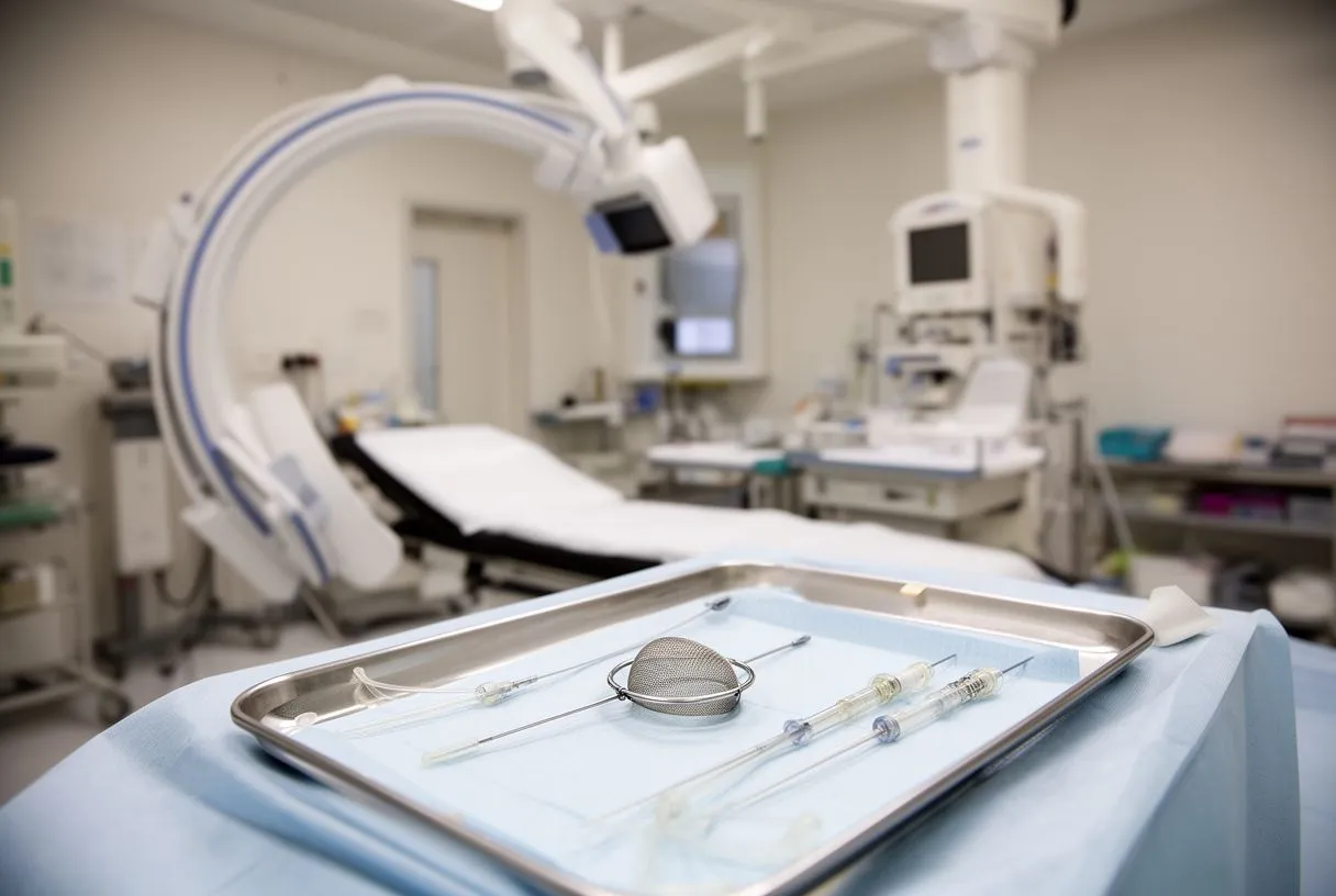
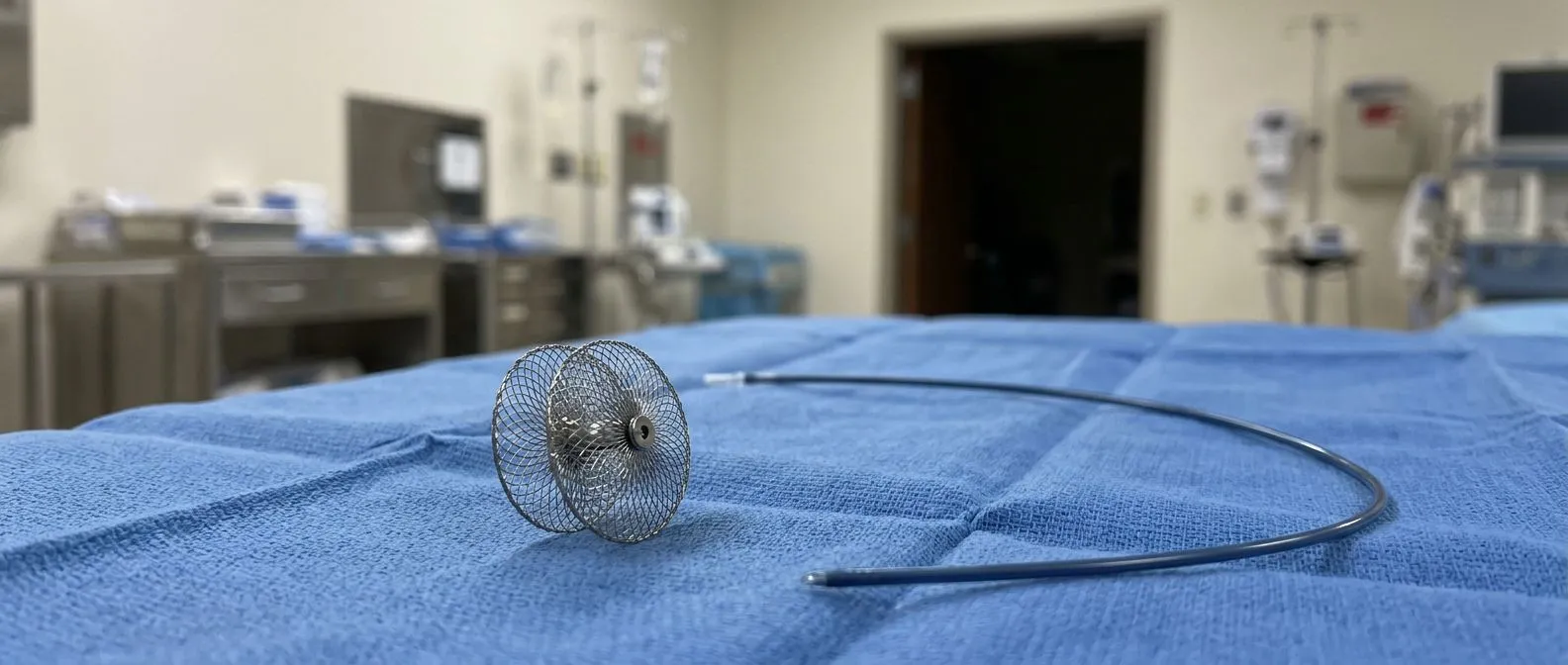
How is the device placed? The cardiologist inserts a thin tube (catheter) into a vein in the groin and threads it up through the right atrium to the PFO. A closure device — typically a small, collapsible umbrella- or disc-shaped implant made of a nitinol wire mesh and a fabric patch — is delivered through the catheter and deployed across the opening. The device sits on both sides of the flap, sandwiching the septum, and over the following weeks the body's own tissue grows over it, sealing the defect permanently. No large incision is made, and the chest is never opened. In some cases, a balloon is briefly inflated across the PFO to measure its true size — a technique related to balloon angioplasty but used here for sizing rather than widening a vessel.
Is it the same technology used for larger septal defects? The principle is similar, but the devices and approach differ. A PFO closure device is designed for a flap that is functionally closed most of the time, whereas an atrial septal defect closure device must span a true, fixed hole. The sizing, imaging requirements, and follow-up protocols vary accordingly. For a PFO, the procedure is generally faster and carries a lower complication rate than closure of a true septal defect, because the anatomy is more forgiving and the defect is smaller.
PFO Closure Recovery: What to Expect
Recovery from PFO closure is usually straightforward. Most patients spend one night in hospital for observation and go home the next day. The groin catheter site may be tender or bruised for a few days, but significant pain is uncommon. Patients are typically advised to avoid strenuous activity, heavy lifting, and contact sports for about a month to allow the device to settle and early tissue ingrowth to begin. Normal daily activities — walking, light household tasks, desk work — can resume within days.
What medications are prescribed after closure? Antiplatelet therapy — usually Aspirin alone or Aspirin combined with clopidogrel for the first one to three months — is standard. This prevents clots from forming on the device surface before the body's tissue fully covers it. Some patients also receive short-term anticoagulation depending on their individual stroke risk profile. After three to six months, once follow-up imaging confirms the device is sealed and no residual shunt remains, medications may be simplified. The decision is individualised; a blanket protocol does not suit every patient.
- First 24–48 hours: hospital observation, monitor for arrhythmia or access-site bleeding, light walking encouraged.
- Weeks 1–4: avoid heavy lifting and strenuous exercise; routine daily activities are fine; take prescribed antiplatelets exactly as directed.
- Months 1–6: follow-up echocardiogram to confirm device position and complete sealing; medication review; gradual return to full activity.

Are There Risks or Complications of PFO Closure?
No medical procedure is risk-free, and PFO closure is no exception. However, the procedure has been performed for over two decades, and in experienced hands the complication rate is low. The most common issues are minor and self-limiting: transient arrhythmias (particularly atrial flutter, a rapid beating of the atrium that may feel like a fluttering in the chest), bruising or minor bleeding at the catheter site, and mild chest discomfort for a few days as the device settles against the septum. Serious complications are rare but real.
What are the serious risks? Device embolisation — the implant migrating from its intended position — occurs in a very small fraction of cases and can usually be retrieved and repositioned during the same procedure. Pericardial effusion (fluid around the heart) is another recognised but uncommon risk. There is also a small long-term risk of device erosion or thrombus formation on the device surface, which is why follow-up echocardiography is essential. These events are rare enough that large registries and trials consistently show a favourable risk–benefit profile in properly selected patients. A review of Wnt/β-catenin signalling pathways notes that endothelial and tissue-remodelling responses — the biological processes that drive device sealing — are generally robust, though individual healing responses vary (see Wnt/β-catenin signalling: function, biological mechanisms, and therapeutic opportunities).
Can the device ever need to be removed? Removal after the tissue has grown over the device is extremely difficult and rarely attempted. This is why patient selection is so important — the decision to close a PFO is essentially a permanent one. If a patient develops a new arrhythmia months or years later, it is managed with medication or ablation rather than device removal. In the rare event of device-related infection (endocarditis), antibiotic therapy is first-line; surgical removal is reserved for cases that do not respond. Patients should also understand that dental and surgical procedures require antibiotic prophylaxis guidelines to be reviewed with their cardiologist, though routine PFO closure devices do not always require the same precautions as prosthetic valves.
Patent Foramen Ovale and Stroke: Understanding the Connection
The link between PFO and stroke is the single most important reason closure is performed. The mechanism is called paradoxical embolism: a clot that forms in the venous system (often a deep vein thrombosis in the leg) travels to the right heart, and instead of being trapped by the lungs, it slips through the PFO into the left heart and then up to the brain, causing an ischaemic stroke. In younger stroke patients where no carotid disease, atrial fibrillation, or atheromatous plaque in the ascending aorta is found, the PFO becomes the leading suspect.
How strong is the evidence that closure prevents recurrent strokes? Several landmark randomised trials — including RESPECT, PC, and CLOSE — initially produced mixed results, but longer follow-up and pooled analyses have clarified that closure does reduce recurrent stroke risk in well-selected patients, particularly those under 60 with a confirmed shunt and no competing stroke mechanism. The benefit is most pronounced when the PFO is large or associated with a substantial right-to-left shunt on bubble study. Research has also explored overlapping pathologies; for example, a study on the Prevalence and Outcomes of Concomitant Aortic Stenosis and Cardiac Amyloidosis illustrates how multiple cardiac conditions can coexist and complicate risk assessment, underscoring why a thorough workup matters before attributing a stroke solely to a PFO.
Does closure eliminate the need for blood thinners after a stroke? Not necessarily. In some patients, closure replaces long-term anticoagulation with antiplatelet therapy alone, but in others — particularly those with additional clotting risk factors — anticoagulation may continue. The decision is individualised based on whether the PFO was the sole mechanism, whether residual shunting is present after closure, and whether the patient has venous thromboembolism risk. Patients should not assume that the device means they can stop all stroke-prevention medications; the medication plan is tailored to the full clinical picture and reviewed at each follow-up.
When Should You Talk to a Cardiac Specialist About PFO?
If you have had a stroke or transient ischaemic attack (TIA, sometimes called a ministroke) and no clear cause was found, ask your neurologist or physician whether a PFO evaluation has been performed. If you are under 60, have a confirmed PFO with a right-to-left shunt, and your stroke workup has excluded other mechanisms, you may be a candidate for patent foramen ovale closure. The evaluation should be done by a team experienced in structural heart disease and congenital cardiology — not every cardiologist performs PFO closure, and the decision requires careful multi-specialty input.
What if you have a PFO but have never had a stroke? In the absence of a clinical event, closure is almost never recommended. The risks of the procedure, however small, outweigh the hypothetical benefit in someone who has never had a stroke. The same applies to patients whose migraines are well controlled with standard medication. A PFO found incidentally on an echocardiogram done for another reason should be noted in your medical record but not treated unless circumstances change. The old adage holds: treat the patient, not the echo finding.
If you or a family member has been told you have a PFO after a stroke, or if you are considering a second opinion on whether closure is right for you, we encourage you to speak with a qualified cardiac team. At Cirugía de Corazón, Dr Alejandro Molina and his team in Guadalajara, Querétaro, and Hermosillo offer thorough PFO evaluation and personalised guidance on whether closure is the right step for your individual situation. We do not rush to close every PFO — we take the time to understand your full clinical picture, explain the anatomy and the options in plain language, and help you make a decision grounded in evidence rather than anxiety. Book a consultation to discuss your case in detail rather than self-diagnosing from online information alone.