


Pulmonary Regurgitation: When a Leak Needs Treatment
Pulmonary regurgitation treatment guide from Guadalajara heart surgeons.
Pulmonary regurgitation treatment becomes a real question when a valve on the right side of the heart stops closing tightly and blood leaks backward with every heartbeat. Many people live with a small leak for years and never feel it. Others — often adults who were born with a heart defect — reach a point where the leak starts to tire out the right ventricle, the chamber that pumps blood to the lungs. This guide explains what the condition is, what symptoms to watch for, how doctors measure it, and when repair or replacement becomes the safer path.
What Is Pulmonary Regurgitation?
The valve and its job. The pulmonary valve sits between the right ventricle and the pulmonary artery, the large vessel that carries blood from the heart to the lungs to pick up oxygen. Like all heart valves, it opens to let blood forward and snaps shut to stop it flowing backward. When its three thin leaflets do not meet cleanly, blood washes back into the right ventricle — that backflow is pulmonary regurgitation, sometimes called pulmonary insufficiency.
Why the right side feels it differently. The right ventricle is built for a low-pressure circuit, so it tolerates a leaking valve better than the left side would. This is why a mild leak often causes no symptoms at all. But the heart is not designed to accept that extra volume forever, and over years the chamber slowly enlarges as it handles the same blood twice.
Mild versus significant leaks. Cardiologists grade pulmonary regurgitation as trivial, mild, moderate or severe. Trivial and mild leaks are common findings on routine scans and usually need nothing more than periodic check-ups. Moderate and severe leaks are the ones that raise the question of intervention, because the volume of backflow begins to change the shape and function of the right ventricle.
How Serious Is Pulmonic Regurgitation?
The honest answer: it depends on degree and duration. A trace leak discovered on an echocardiogram is one of the most common incidental findings in adult cardiology and rarely matters. Severe pulmonary regurgitation, left untreated for years, is a different story: the right ventricle enlarges, its pumping efficiency falls, and patients can develop rhythm problems, exercise intolerance and eventually right heart failure.
Volume overload and the heart muscle. Any chronically leaking valve forces its receiving chamber to handle extra blood, and research models of valvular regurgitation show how this overload drives enlargement and remodeling of the ventricle over time (PubMed). The right ventricle is more forgiving than the left, which is precisely why the warning signs arrive late — and why regular surveillance matters even when you feel well.
Why timing matters more than panic. The goal of good care is to intervene in the window where the right ventricle is still recovering well, before irreversible damage sets in. Intervene too early and you take on the risks of surgery without clear benefit; too late and the heart muscle may not fully bounce back. That balance — not fear — is what should drive the decision.
What Causes a Leaky Pulmonary Valve?
Congenital heart defect and pulmonary valve problems. The most common cause by far in adults is repaired congenital heart disease — conditions people were born with. In a congenital heart defect involving the pulmonary valve, the valve may be malformed from birth, and surgeries performed in childhood often leave it insufficient years later. Tetralogy of Fallot repair is the classic example: pulmonary regurgitation after tetralogy of Fallot is one of the most frequent reasons adults with congenital heart disease eventually need a new valve.
Pulmonary valve stenosis and regurgitation together. Some patients grew up with pulmonary valve stenosis — a narrowed valve that had to be stretched open, either with a balloon catheter or surgically. Pulmonary valve stenosis and regurgitation frequently coexist after these procedures, because the same maneuvers that relieve narrowing can stretch or scar the leaflets so they no longer close completely.
Other causes. Less commonly, the leak develops from infection of the valve (endocarditis), from high pressure in the pulmonary arteries, from prior surgery that damaged the valve directly, or from rare conditions affecting connective tissue. A structurally normal valve can also become insufficient when the pulmonary artery widens and pulls the leaflets apart.
- Repaired tetralogy of Fallot: the patch and surgical techniques used decades ago often leave the valve wide open to backflow.
- Balloon dilation or stenting: effective for stenosis, but a common trade-off is a degree of regurgitation afterwards.
- Infection (endocarditis): can perforate or destroy leaflets and turn a stable leak into an urgent one.
Pulmonary Regurgitation Symptoms: When Surgery Is Considered
Early on, there may be nothing to feel. Because the right side of the heart operates at low pressure, pulmonary regurgitation can progress silently for many years. When symptoms do appear, they tend to be gradual: declining exercise tolerance, breathlessness on activity that used to be easy, fatigue, or a sensation of fullness or palpitations. In advanced cases, fluid retention shows up as swelling in the legs or abdomen.
Symptoms worth reporting promptly. Fainting or near-fainting during exertion, palpitations that feel rapid or irregular, and breathlessness that wakes you at night are all signals to be evaluated quickly. These can indicate that the enlarged right ventricle is starting to affect heart rhythm — a complication seen in long-standing, severe leaks, especially after tetralogy repair.
The contrast with other valve leaks. Patients sometimes ask how this compares with mitral regurgitation, the better-known leak on the left side of the heart, where symptoms and the drive to intervene typically appear earlier. The right-sided leak hides longer, which is why pulmonary regurgitation symptoms and surgery decisions rely heavily on imaging measurements rather than how you feel alone.

Pulmonary Regurgitation Echocardiogram Findings
What the echo shows. An echocardiogram — an ultrasound scan of the heart — is the first and main tool. Typical pulmonary regurgitation echocardiogram findings include a Doppler jet of blood flowing back across the valve, a rapid equalization of pressure between the pulmonary artery and the right ventricle, and indirect clues such as a dilated pulmonary artery and a right ventricle working against no obstruction.
Complementary tests. Cardiac MRI is the gold standard for measuring how much blood actually regurgitates — expressed as a regurgitant fraction — and for tracking right ventricular size and pumping function precisely. If questions remain about the pulmonary arteries or prior surgical anatomy, a CT scan may be added. Each test answers a different question, and together they decide whether observation or intervention is right.
How we read these studies with patients. In our consultation we take the time to explain what the echocardiogram and MRI actually show, in plain words, and to answer every question before any decision is made. Understanding your own numbers — the regurgitant fraction, the ventricular volumes, the pressures — makes the treatment conversation far easier.
When to Treat Pulmonary Regurgitation
Guideline signals for intervention. International guidelines for adult congenital heart disease suggest considering pulmonary valve intervention when the regurgitation is severe and there are clear markers of harm: a significantly enlarged right ventricle, falling pumping function, worsening symptoms, or new rhythm problems. Some teams also act earlier in repaired tetralogy patients when the right ventricle is steadily enlarging on serial scans, even without symptoms yet.
Observation is also a treatment. If the leak is mild or moderate with a stable right ventricle, the right plan is usually surveillance — echocardiograms or MRI every one to three years, depending on severity. Jumping to surgery before the criteria are met exposes you to risk without proven benefit. Knowing when to treat pulmonary regurgitation is as much about restraint as it is about action.
Every recommendation reviewed by the whole team. Every case is reviewed by our multidisciplinary team against international practice guidelines before a recommendation is given; you receive a clear proposal with the alternatives, and the final decision is always yours. We also encourage second opinions and treat seeking one as a normal part of deciding about heart surgery.
Pulmonary Valve Replacement Options
Surgical valve replacement. The established treatment for severe pulmonary regurgitation is replacing the valve, either with a biological tissue valve or with a valved conduit — a tube containing a valve — when the original outflow tract needs reconstruction. For children and adults with complex anatomy from prior congenital surgery, customized valved conduit reconstruction has been an important advance, with design approaches increasingly guided by intelligent modeling of each patient's anatomy (IEEE Xplore).
Transcatheter valve-in-valve. For patients who already have a surgical conduit or bioprosthetic valve in place, a new valve can sometimes be delivered through a catheter — a tube threaded from a blood vessel in the groin — and expanded inside the old one. This avoids open-heart surgery, though it requires the existing structure to be a suitable frame. Not every patient's anatomy permits it.
Valve-sparing and repair approaches. Where the anatomy allows, some surgeons attempt valve-sparing pulmonary repair, reconstructing the patient's own valve or narrowing an enlarged pulmonary artery so the leaflets can meet again. When it works, it avoids a prosthesis, but it is only appropriate for selected anatomies and demands careful case selection.
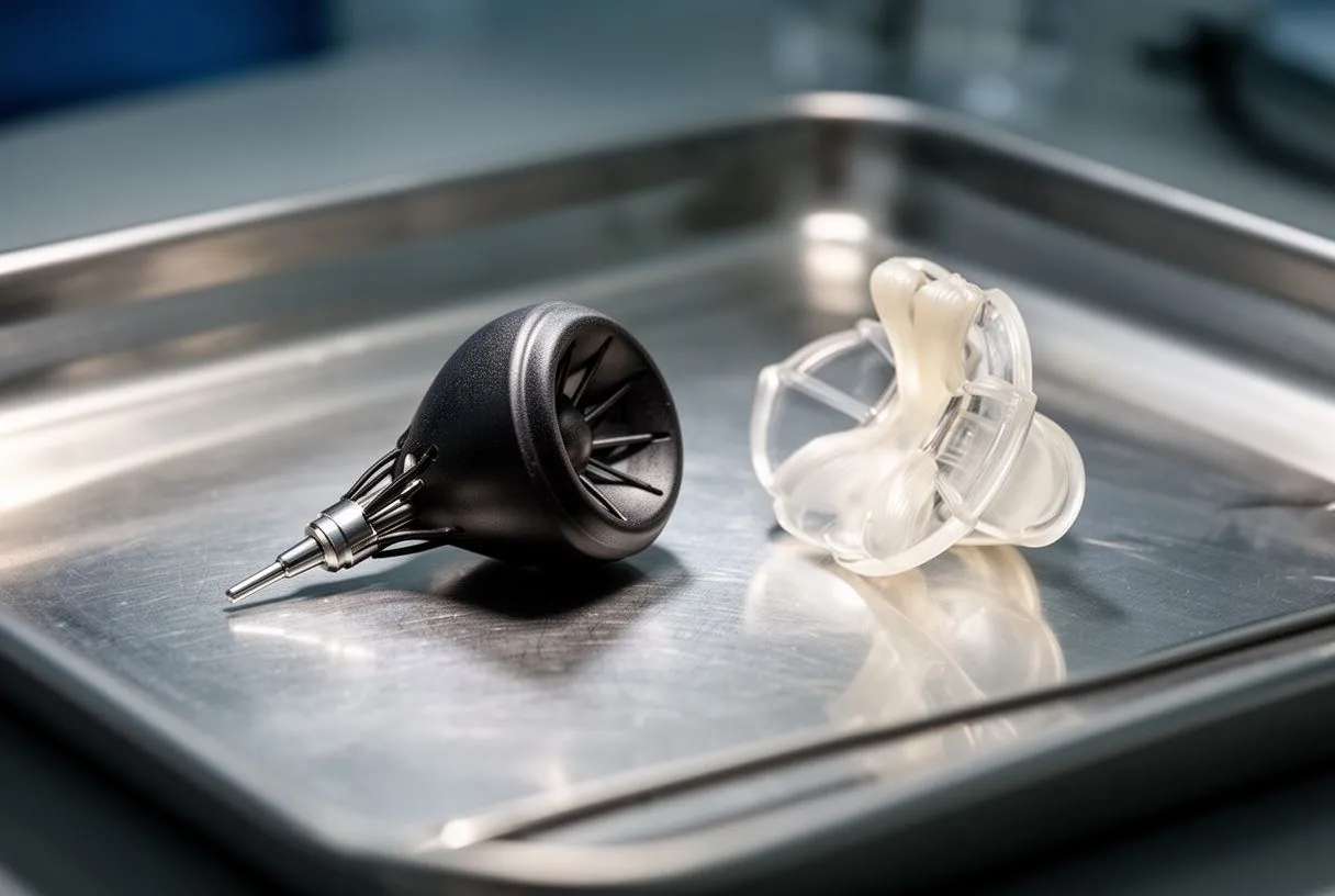
Minimally Invasive Pulmonary Valve Surgery
What "minimally invasive" really means here. Minimally invasive pulmonary valve surgery replaces the valve through a small incision — often between the ribs on the right side of the chest — rather than a full sternotomy through the breastbone. The valve work is the same; the recovery difference comes from the smaller access. For suitable patients this can mean less pain, a shorter stay and a faster return to activity.
Candid trade-offs. Smaller incisions can limit visibility and access for the surgeon, and not every anatomy — especially after multiple prior operations — is suitable. Some patients are better served by a conventional approach. The right question is not "which technique is newest" but "which technique offers the lowest risk for my situation."
Our experience across the spectrum. We perform conventional, minimally invasive, robotic and transcatheter procedures, and we recommend the less invasive route only where the evidence supports it for that patient. Dr Molina trained in cardiac, minimally invasive, robotic and structural surgery at the University of Montreal and in structural cardiology and TAVR at CHU Bordeaux, and performed the first robotic mitral valve repair in the Ángeles Health System and the first robotic heart surgery in Guadalajara.
Pulmonary Valve Replacement Recovery Time
The usual milestones. After open pulmonary valve replacement, most patients spend one to three days in hospital (sometimes longer after complex reoperations), walk within the first day or two, and return to desk work in roughly four to six weeks. Full recovery — including unrestricted exercise — typically takes around three months, when the breastbone or chest incision has fully healed. With a minimally invasive or catheter-based approach, these timelines are often shorter.
What recovery actually feels like. Expect fatigue for the first weeks; this is normal and improves steadily. You will be given breathing exercises, a walking plan, and rules about lifting. Most patients notice improved exercise tolerance within the first months as the right ventricle unloads — although if the ventricle was enlarged for a long time before surgery, some improvement may take longer and be partial.
Follow-up and lifelong surveillance. A replacement valve — tissue or catheter-based — is not a once-and-forget solution. Biological valves can degenerate over years, so echocardiogram surveillance continues for life. Imaging follow-up at every stage means the next valve decision — often years away — need not be a surprise.

Practical questions international patients ask. For patients considering heart valve leakage treatment in Mexico, cost is a legitimate part of the decision, alongside surgical quality. Pulmonary valve disease treatment cost varies with the approach chosen — surgical versus transcatheter — the type of valve or conduit used, the complexity of prior operations, and the length of hospital stay. A candid, itemized estimate should be part of any serious consultation, and we encourage you to ask for one.
How our process works. The first contact is a free video consultation: we get to know the patient, talk through the condition, and say which studies are still needed so no unnecessary ones are ordered; a face-to-face visit at the consulting rooms follows when the patient decides to go ahead. Our consulting rooms are in Guadalajara, Querétaro and Hermosillo, and for patients traveling from abroad, the studies you already have are reviewed before anything is repeated.
What to look for in a pulmonary regurgitation cardiologist or surgeon in Guadalajara — or anywhere. Whatever team you choose, look for: a multidisciplinary review of your case, specific experience in adult congenital heart disease and pulmonary valve surgery, a willingness to explain the alternatives including doing nothing yet, and transparency about risks and costs. Our work covers valve disease, coronary disease, the aorta and structural heart conditions, and we make sure the patient hears every option — including the option to wait — before a decision is made.
Pulmonary regurgitation treatment is ultimately about timing: watching a mild leak carefully, and acting at the window where replacing or repairing the valve gives the right ventricle its best chance to recover. If you were treated for a congenital heart defect as a child, or you have been told you have a leaking pulmonary valve, the most valuable step you can take is a proper evaluation with a team that reviews your images, explains your numbers, and lays out every option — including continued surveillance. Talk it through with a qualified cardiac team rather than deciding alone; if you would like a first conversation, we offer a free video consultation with Dr Alejandro Molina's team in Guadalajara, Querétaro or Hermosillo. The final decision is always yours — our job is to make sure it is an informed one.






