Rheumatic Heart Disease: Valve Damage and Treatment
Rheumatic heart disease treatment with Guadalajara valve specialists.
If you or someone in your family has been told there is valve damage from a strep throat infection years ago, you are dealing with rheumatic heart disease — and you probably have more questions than answers. This condition is one of the most common forms of acquired valvular heart disease in the world, yet most people have never heard of it until it touches their own family. The good news is that rheumatic heart disease treatment today is far more effective than it was a generation ago, and many people with well-managed disease live full, active lives. In this guide we explain what happens inside the heart, what the treatment options really involve, what recovery looks like, and how to decide on the right path with a cardiac team you trust.
What Is Rheumatic Heart Disease and What Causes It?
What is the main cause of rheumatic heart disease? The condition begins with an untreated or inadequately treated infection by group A Streptococcus bacteria — the germ that causes strep throat and, in some regions, scarlet fever. When the immune system fights this infection, it can mistakenly attack the body's own tissues, including the heart. The result of that immune confusion is acute Rheumatic Fever, an inflammatory illness that can affect the joints, skin, brain and heart. When the heart itself is involved and the inflammation leaves lasting damage to the valves, the long-term condition is called rheumatic heart disease.
It is important to understand the timeline. Rheumatic heart disease does not appear overnight. A child may have a sore throat that was never treated with antibiotics, develop rheumatic fever two to three weeks later, and only show signs of valve damage years or even decades afterwards. The Centers for Disease Control and Prevention notes that prompt antibiotic treatment of strep throat is the key step in preventing rheumatic fever and, with it, later heart damage. This is why the disease remains far more common in communities where access to medical care and antibiotics is limited, and in adults who grew up in those settings.
Who is most at risk? Rheumatic fever most often affects children between five and fifteen years old, but the valve damage it leaves behind follows people into adulthood. Overcrowded housing, limited access to primary care and untreated repeated strep infections all raise the risk. If you grew up in a region where strep infections frequently went untreated — a reality for many families in Mexico and across Latin America — and you now have a heart murmur or valve diagnosis, rheumatic heart disease deserves a place on the list of explanations your team should consider.
How Does Rheumatic Fever Damage the Heart Valves?
The mechanism in plain words. Your heart has four one-way valves that keep blood moving in the right direction. During acute rheumatic fever, the immune system's antibodies — proteins made to fight the strep bacteria — mistakenly recognize similar structures in the heart tissue and attack them. Each heart Valve Cusp, the thin leaflet or flap that opens and closes with every heartbeat, can become inflamed. Over repeated episodes of rheumatic fever, the leaflets thicken, stiffen and may fuse together at their edges.
Two things can then go wrong with a damaged valve. It can fail to open properly, which doctors call stenosis — the opening becomes narrow, like a door that will not swing fully wide, and blood struggles to pass through. Or it can fail to close properly, which doctors call regurgitation or leaking — blood flows backward instead of forward. The valve most often damaged is the mitral valve, followed by the aortic valve. The Knowing your heart: the mitral valve sits between the left upper and lower chambers of the heart, and when rheumatic disease narrows it — rheumatic mitral stenosis — the left upper chamber must work harder to push blood through the narrowed opening. The Knowing your heart: the aortic valve, which sits between the left lower chamber and the main artery to the body, can also become narrowed or leaky.
Why episodes matter. Each new episode of acute rheumatic fever can add more damage to valves that were already scarred. This is the central fact of the disease: it is not usually one infection that destroys a valve, but repeated attacks over years. Recent research into how the immune system drives this process — including work published in Frontiers in Cardiovascular Medicine on specific antibody responses in acute rheumatic heart disease — is helping explain why some people develop severe valve damage while others do not. It also underlines the practical point that stopping the attacks is the single most valuable thing any treatment can do.
What Are the Signs and Symptoms of Rheumatic Heart Disease?
Early signs are often silent. Many people with rheumatic valve damage have no symptoms for years. The first clue is often a heart murmur — an extra whooshing sound a doctor hears through a stethoscope during a routine check. Because the damage develops slowly, the heart adapts, and symptoms may not appear until the valve problem is quite advanced. This silence is why a murmur found in an adult with a history of childhood strep infections should always be investigated properly.
When symptoms do appear, they tend to follow a recognizable pattern depending on which valve is affected and how severe the problem has become. Watch for these warning signs:
- Breathlessness: getting unusually short of breath with activity, or needing extra pillows at night to breathe comfortably while lying flat.
- Pausing during exertion: stopping halfway up stairs you used to climb easily, or a heavy, tired feeling in the legs with exercise.
- Fluttering or irregular heartbeat: palpitations that can signal the upper heart chambers are struggling, a common complication of mitral valve disease.
- Swelling: puffiness in the ankles, feet or abdomen caused by fluid building up when the heart cannot keep up.
- Chest discomfort or fainting: pressure in the chest with exertion, or actual fainting spells, which are red-flag symptoms of significant valve obstruction.
Do not wait for symptoms to judge severity. Many patients feel fine until suddenly they do not. Valve disease can progress quietly and then declare itself. If you have a known murmur, a previous diagnosis of rheumatic fever, or any of the symptoms above, the next step is not worry — it is a proper evaluation with an echocardiogram, which we describe below.
How Is Rheumatic Heart Disease Diagnosed?
The echocardiogram is the cornerstone. An echocardiogram is an ultrasound scan of the heart — no radiation, no needles, entirely painless. It shows the valve leaflets moving in real time, measures how well blood flows through each opening, and calculates the Valve Area, a precise measurement of how narrow or leaky a valve has become. For rheumatic disease, the scan also shows characteristic changes — thickened leaflets, fused edges, stretched chambers — that help distinguish rheumatic damage from other causes of valve disease.
Supporting tests fill in the picture. Your team may also order an electrocardiogram to check the heart's rhythm and look for strain, a chest X-ray to see the heart's size and any fluid in the lungs, and blood tests. During acute suspected rheumatic fever, blood tests look for inflammation markers and evidence of recent strep infection. In established disease, these tests are less about diagnosis and more about planning: they tell the team how the heart is coping and whether any chamber has been stretched by years of working against a damaged valve.
What a diagnosis appointment should feel like. A proper evaluation ends with answers, not just scans. In our consultations we take the time to explain what the studies actually show, in plain words, and to answer every question before any decision is made — because a patient who understands their own echocardiogram is far better equipped to choose between treatment options later. If you have had an echo done elsewhere, bring the images and report; they usually tell us most of what we need to know about your valves.

Is Rheumatic Heart Disease Curable or Only Managed?
The honest answer has two parts. The valve damage itself — the scarring, thickening and fusing of leaflets — is permanent. No medication can dissolve scar tissue or restore a damaged leaflet to its original state. In that sense, established rheumatic valve disease is not "curable" by medicine alone; severely damaged valves are corrected by repair or replacement surgery. But the disease process — the repeated immune attacks that cause progressive damage — absolutely can be stopped, and stopping it is the heart of chronic rheumatic heart disease management. A person whose attacks are prevented, whose valve is monitored, and who receives surgery at the right time can expect a good quality of life.
What "managed" means in practice. Managing the disease involves three threads running together. First, preventing new rheumatic fever attacks with regular preventive antibiotics, described in the next section. Second, protecting the heart's rhythm and function with medications such as those that control heart rate, prevent clot formation, or ease the workload on the heart. Third, watching the valves with scheduled echocardiograms so that if surgery becomes the right answer, it happens at the optimal moment — not too early, when the risks outweigh the benefit, and not too late, when the heart muscle has already been strained beyond recovery.
Why the timing of surgery matters so much. Every case we review is assessed by the multidisciplinary team against international practice guidelines before a recommendation is given, because the line between "watch and wait" and "operate now" is drawn from evidence, not habit. Guidelines define thresholds — for example, how narrow a valve must become, or how much the heart's pumping function has declined — that tell us when intervention gives the patient a real advantage. The patient receives a clear proposal with the alternatives, and the final decision is always theirs.
What Medication Is Used in Rheumatic Heart Disease Treatment?
Preventive penicillin is the foundation. The single most important medication in rheumatic heart disease treatment is not a heart drug at all — it is regular penicillin, given to prevent new strep infections and new rheumatic fever attacks. This is called secondary prophylaxis. It is most commonly given as an injection into the muscle every three to four weeks, or taken as daily oral antibiotics for those who cannot tolerate injections. The duration depends on how severe the heart involvement is: international guidelines generally recommend continuing for at least ten years after the last attack, or until well into adulthood, and for life in patients with significant valve damage.
Why some patients stop too soon. These injections sting, they require clinic visits, and after years of them it is tempting to stop — especially if you feel completely well. This is one of the most common and consequential mistakes in the whole disease. Research published in Global Heart from a consultation workshop on improving access to secondary prophylaxis for rheumatic fever and rheumatic heart disease found that patients and families want and need real choices — different delivery options, better pain management, clearer education — to stay on track with prevention. If injections have become a burden for you or your child, raise it with your team; there are often practical solutions.
Medications for the damaged valve itself. Depending on which valve is affected and how, your cardiologist may prescribe drugs that slow a too-fast heart rhythm, help the heart pump more efficiently, reduce fluid buildup, or thin the blood to prevent clots — particularly important in mitral stenosis, where clots forming in the stretched upper chamber can travel to the brain and cause stroke. None of these medications reverse the valve damage, but each one relieves the strain that the damage places on the heart, and together they can keep a person stable for years while the valve is monitored.
When Is Rheumatic Valve Repair or Replacement Surgery Needed?
The triggers for surgery. Rheumatic valve replacement surgery or repair enters the conversation when the valve problem starts to threaten the heart muscle or cause symptoms that medication cannot control. Typical triggers include a critically narrowed valve, a valve leaking so much that the heart chamber is stretching, declining pumping function on the echocardiogram, blood clots despite medication, symptoms that limit daily life despite good medical therapy, or infection of the damaged valve. The specific numbers — how narrow the opening is, how much the heart has enlarged — come from the echocardiogram, and guideline thresholds tell the team where the balance tips from watching to acting.
Repair versus replacement. Whenever a valve can be repaired rather than replaced, repair is usually the better option: the patient keeps their own tissue, avoids lifelong blood thinners in many cases, and the valve retains its natural durability. Rheumatic mitral disease sometimes allows skilled repair, particularly in certain patterns of stenosis where a procedure called balloon valvuloplasty — inflating a small balloon inside the valve to split fused edges — can open the valve without open surgery. When the damage is too extensive, valve replacement becomes the answer, and the choice of valve becomes the next big decision, covered in the next section.
What our team does before recommending surgery. Our work covers valve disease, coronary disease, the aorta and structural heart conditions, and we recommend the less invasive route only where the evidence supports it for that patient. In practice this means every candidate for rheumatic valve surgery has their imaging, their symptoms and their overall health reviewed together — heart surgeon and cardiologist around the same table — before anyone proposes an operation. We also encourage second opinions and treat seeking one as a normal part of deciding about heart surgery; a decision of this size deserves to be made with confidence, not pressure.
Mechanical Valve vs Tissue Valve for Rheumatic Disease
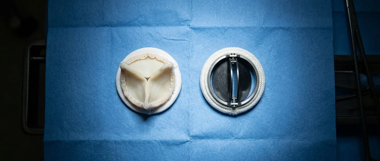
The two families of replacement valves. A replacement valve is either mechanical — made of durable artificial materials such as pyrolytic carbon — or biological (tissue), made from animal or human tissue. A Stented Valve has its tissue leaflets mounted on a supporting frame, while a Stentless Valve dispenses with the frame and relies on the patient's own aortic root for support, which can allow better blood flow in some cases. Tissue valves may be a Porcine Valve (from pig tissue) or a Bovine Pericardial Valve (from the protective sac around a cow's heart); both are treated to work safely in the human body.
The trade-off in one paragraph. A mechanical valve lasts a lifetime but requires warfarin blood-thinning medication every day for the rest of your life, with regular blood tests to keep the dose in a safe range. A tissue valve avoids lifelong blood thinners but wears out over time and eventually needs replacement — sooner in younger patients, whose metabolism ages the tissue faster. For rheumatic patients the decision has an extra layer: many are young, and some also have reasons to prefer or avoid blood thinners, such as plans for pregnancy (tissue valves are generally favored for women who may become pregnant) or a history of bleeding problems.
Special options worth knowing. Among mechanical valves, some designs allow somewhat lower blood-thinning targets in certain patients after the aortic valve — a genuine quality-of-life advantage for someone facing decades of anticoagulation. Transcatheter valves belong to a different category entirely: they are delivered through a catheter, which matters mainly in specific situations such as a failing tissue valve, where a Valve-in-valve procedure can place a new valve inside the old one without open surgery. Where a rheumatic patient fits among these options depends on age, lifestyle, other conditions and the anatomy of their own valve — which is exactly why the choice should be made case by case with a team that handles all of them.
What Are the Minimally Invasive Options for Rheumatic Valves?
Smaller incisions, same surgery. Conventional valve surgery uses an incision down the middle of the chest through the breastbone. Minimally invasive surgery achieves the same operation through a small incision between the ribs or at the edge of the breastbone, using long instruments and a camera — or, in robotic surgery, instruments controlled with robotic arms that translate the surgeon's movements with great precision. For suitable patients, this means less pain, faster return to work and a smaller scar. Not every valve problem can be treated this way, and rheumatic disease with multiple valves or heavy calcification sometimes requires the conventional approach — the honest answer is always anatomy-specific.
Transcatheter options and their place. Transcatheter Aortic Valve Implantation — known as TAVR — delivers a Balloon-expandable Valve or a self-expanding one through a catheter, usually from an artery in the groin, with no chest incision at all. TAVR was developed mainly for older or higher-risk patients with aortic stenosis, and in rheumatic disease its role is more selective: heavily calcified valves, previously operated patients and certain Valve-in-valve situations are where it most often fits. Mitral stenosis from rheumatic disease has its own catheter option — balloon mitral valvuloplasty — which in selected patients with suitable valve anatomy can delay or avoid surgery entirely.
Our team's experience with these techniques. Dr Molina trained in cardiothoracic surgery at the Universidad de Guadalajara, in cardiac, minimally invasive, robotic and structural surgery at the University of Montreal, and in structural cardiology and TAVR at CHU Bordeaux, and the team also trained at Jena, Bad Berka and Buffalo. Dr Molina performed the first robotic mitral valve repair in the Ángeles Health System and the first robotic heart surgery in Guadalajara. That background matters for one specific reason: it means the team can genuinely weigh conventional, minimally invasive, robotic and transcatheter approaches against each other for your particular valve, rather than defaulting to whichever technique happens to be available.

Can You Live a Long Life With Rheumatic Heart Disease?
Yes — with discipline and follow-up. People with rheumatic heart disease routinely live long, full lives when three things stay in place: continued preventive antibiotics, regular echocardiographic surveillance, and timely intervention when the evidence says the valve needs it. Many patients who had valve surgery in their twenties or thirties are walking, working and traveling decades later. The disease becomes a managed condition, like diabetes or high blood pressure, rather than a sentence.
What lifestyle changes are recommended for people with rheumatic heart disease? The pillars are the same as for most heart conditions, adapted to valve disease:
- Dental and infection care: keep teeth and gums healthy and tell your dentist about your valve — some patients need antibiotics before certain dental procedures, and untreated infections can threaten a repaired or replaced valve.
- Heart-friendly eating: plenty of vegetables, fruits, whole grains and fish, with salt limited if there is fluid retention — the recommended diet for people with rheumatic fever or established valve disease is essentially a heart-healthy one, and a nutritionist can tailor it.
- Consistent, moderate activity: walking, swimming or cycling most days, with intensity agreed with your cardiologist; severe valve disease may require adjusting intensity, not abandoning exercise.
- No smoking: smoking accelerates nearly every form of cardiovascular disease and directly worsens the outlook after valve surgery.
- Medication reliability: take preventive penicillin and any prescribed heart medications exactly as scheduled, and keep every follow-up echo appointment even when you feel perfectly well.
You do not have to manage this alone. Families facing this diagnosis do better when they understand it, and connecting with others helps: Are there any online support groups for people with heart failure? Yes — patient organizations focused on rheumatic heart disease and heart conditions offer communities where patients and parents share practical experience of injections, surgery, recovery and daily life. And for the medical side, the first step is a real conversation with a qualified team. The first contact with our practice is a free video consultation: we get to know the patient, talk through the condition, and say which studies are still needed so no unnecessary ones are ordered; a face-to-face visit at our consulting rooms in Guadalajara, Querétaro or Hermosillo follows when the patient decides to go ahead. Rheumatic heart disease treatment works best when it starts early and when the patient is a true partner in the decisions — if a murmur, a past episode of rheumatic fever or any of the symptoms above describes you or your child, we invite you to book a consultation and talk it through before deciding anything on your own.