

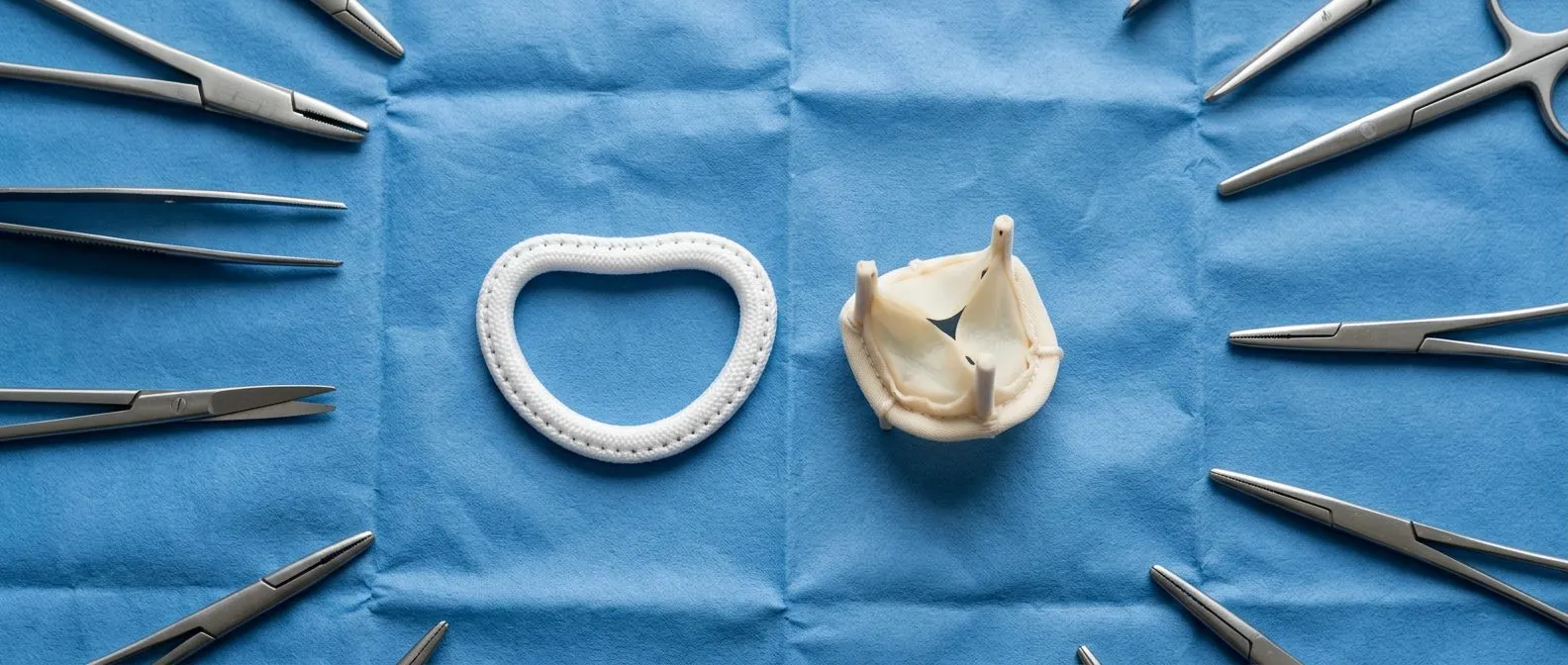
Rheumatic Mitral Disease: Repair, Balloon or Replacement
Rheumatic mitral disease treatment: compare valve repair, balloon valvuloplasty and replacement.
When a childhood sore throat leaves scars on the heart decades later, patients often arrive confused and frightened. Rheumatic mitral disease treatment today offers three well-defined paths: repairing the valve, stretching it open with a balloon, or replacing it. The right choice depends on the valve's anatomy, not on fear or marketing. This guide walks you through how rheumatic heart disease damages the mitral valve, what each option involves, and how a qualified team decides which one fits you.
What Is Rheumatic Mitral Disease?
Rheumatic mitral disease is long-term damage to the mitral valve caused by Rheumatic Fever, an inflammatory illness that can follow an untreated strep throat infection. The body's immune system attacks the bacteria, but in some people the antibodies also target heart tissue. Over years, the valve leaflets stiffen, thicken and fuse at the commissures, the points where they meet.
The result is Valvular Heart Disease that appears 10 to 20 years after the original infection, most often in adults who had rheumatic fever as children. Oxford Academic journals describe rheumatic mitral disease as the most common form of valve disease among young adults in much of the world. It remains a leading reason for heart surgery in patients under 40.
This pattern is common: adults in their thirties and forties who never knew a childhood sore throat changed their heart. Knowing your heart: the mitral valve is a one-way door between the left atrium and the left ventricle. When scar tissue narrows or leaks that door, the whole heart has to work harder.
What Is Mitral Valve Disease?
The mitral valve has two leaflets that must open wide for blood to fill the heart and then close tightly when the heart pumps. Mitral valve disease means one of two problems, or both at once. Stenosis means the opening is too narrow. Regurgitation means the valve leaks blood backward.
How the two problems differ. In stenosis, the heart upstream of the valve becomes congested, causing breathlessness and fluid in the lungs. In Mitral Regurgitation, part of every heartbeat is wasted, so the heart enlarges and weakens over time. Rheumatic damage often causes a mix of both.
Why the cause matters. Mitral Valve Prolapse, a common degenerative condition, behaves differently from rheumatic disease. Rheumatic valves are stiff, thickened and often involve the chords and muscle beneath them. That anatomy shapes whether rheumatic mitral valve repair is even possible, or whether replacement is the safer plan.
What Is Heart Valve Stenosis?
Heart valve stenosis means a valve opening has become narrowed, so blood struggles to pass through. Imagine a door that will not swing fully open: everything behind it backs up. In mitral stenosis, pressure builds in the left atrium and then in the lung circulation.
Doctors measure severity with an echocardiogram, an ultrasound of the heart. The key number is the valve area. A normal mitral valve opens to about 4 to 5 square centimeters. Severe stenosis is typically a valve area of 1.5 square centimeters or less, and critical stenosis below 1.0.
What patients feel. Early on, nothing. Stenosis progresses slowly, so the heart adapts. Symptoms usually start with breathlessness on stairs, a racing or irregular pulse, or coughing at night when lying flat. These symptoms, not just the numbers, drive the decision to treat.
What Are the Symptoms of Rheumatic Mitral Valve Disease?
Rheumatic mitral valve disease symptoms usually appear gradually. Many patients dismiss them for years as aging or poor fitness. Recognizing the pattern matters because early treatment preserves the heart muscle.

Breathlessness and fatigue. The most common complaints are shortness of breath with exertion and unusual tiredness. As pressure backs up into the lungs, patients may wake at night gasping or need extra pillows to sleep.
Irregular heartbeat. The stretched left atrium often develops atrial fibrillation, a chaotic rhythm. Patients feel a fluttering or pounding pulse. This complication raises stroke risk and often appears before other symptoms.
Other warning signs. Coughing with pink-tinged sputum, chest discomfort, swelling of the ankles, and hoarseness from an enlarged atrium pressing on a nerve all occur. If you notice several of these together, an echocardiogram is the sensible first step. In our first free video consultation, we listen to your story and tell you exactly which studies are needed — so you never pay for tests you do not require.
How Do Doctors Choose Between Repair, Balloon and Replacement?
The decision in rheumatic mitral stenosis treatment follows the valve's anatomy. Three factors dominate: how mobile the leaflets are, how much they are thickened, and whether the chords beneath them are fused or the surrounding structures are diseased.
The Wilkins score. The Wilkins score is used in mitral stenosis to grade valve anatomy on echocardiography. It scores four features — leaflet mobility, thickening, calcification, and the subvalvular chords — from 1 to 4 each, for a total of 4 to 16. A low score means the valve is pliable and a good candidate for a balloon procedure. A high score means the valve is stiff and deformed, favoring surgery.
Other decisive factors. Doctors also check for clot in the left atrium, significant regurgitation, and heavy calcium. Any of these rules out the balloon route. The patient's age, surgical risk, and rhythm history matter too.
- Pliable valve, low Wilkins score, no clot: mitral balloon valvuloplasty, a catheter procedure, is usually first choice.
- Deformed valve with leak or heavy calcium: surgical repair or rheumatic mitral valve replacement.
- Mixed disease: the multidisciplinary team weighs both procedures against each patient's goals.
Every case at our practice is reviewed by the whole multidisciplinary team against international guidelines before we propose anything, and you receive the alternatives in writing — the final decision is always yours.
What Are the Surgical Options for Treating Mitral Stenosis?
When a balloon is not suitable, surgery offers two main options, each with real trade-offs.
Open commissurotomy and repair. The surgeon opens the chest — or works through a small right-chest incision in minimally invasive mitral repair — and carefully divides the fused commissures and frees the chords. This can restore good valve function for years and avoids a prosthesis. Its success depends on the valve's condition, and scar tissue can eventually narrow the valve again.
Replacement. When the valve is too destroyed to save, rheumatic heart disease surgery means Mitral Valve Replacement with either a mechanical or tissue prosthesis. Mechanical valves last decades but require lifelong blood thinners. Tissue valves avoid thinners but wear out sooner, especially in younger patients.
Regurgitation is different. For rheumatic mitral regurgitation repair, surgical Mitral Valve Repair techniques are used where the anatomy allows, because a repaired native valve usually outlasts any prosthesis. But rheumatic tissue is less favorable for repair than degenerative tissue, and the team must judge this honestly on each echocardiogram.
What Is Mitral Balloon Valvuloplasty and What Should You Expect?
Mitral balloon valvuloplasty — sometimes called percutaneous mitral commissurotomy — stretches the narrowed valve open from inside, without open surgery. A thin catheter is threaded from a vein in the groin, punctured through the wall between the atria, and a balloon is inflated across the valve to split the fused commissures.
It is a procedure, not an operation. Most patients go home within a day or two and return to normal activity within a week. There is no chest incision and no heart-lung machine. For a carefully selected patient with a pliable valve, results rival surgery.
Its limits are real. The balloon cannot remove calcium or repair a leak. If the valve is badly deformed, or regurgitation worsens during the procedure, surgery becomes necessary — sometimes urgently. Long-term restenosis is also more common in younger patients with active rheumatic disease. We perform transcatheter procedures ourselves when the evidence supports that route, and we are equally candid when surgery would serve you better.
Rheumatic Mitral Valve Repair vs Replacement: Which Is Better?
When repair is possible, most guidelines favor it: a repaired native valve preserves the natural anatomy and avoids prosthetic complications. But rheumatic valves test this principle, because scar tissue makes durable repair harder than in degenerative disease.
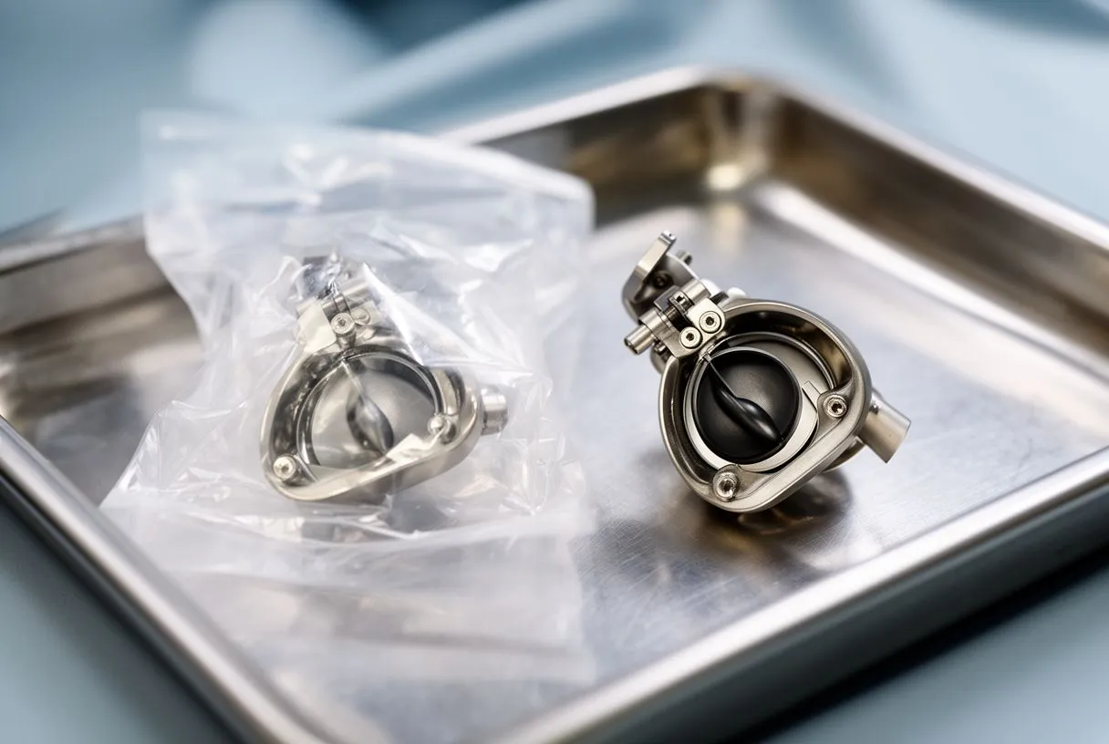
Where repair works well. Techniques developed for degenerative valves have evolved considerably. Endoscopic and minimally invasive approaches achieve excellent outcomes in selected degenerative disease, as documented in a study of endoscopic mitral repair for degenerative mitral regurgitation (ScienceDirect), though rheumatic anatomy remains more challenging and demands careful patient selection.
Where replacement wins. A heavily calcified, destroyed valve cannot be reliably repaired. Forcing repair in a poor candidate invites early failure and a second operation. In these patients, rheumatic mitral valve replacement with a durable prosthesis is the lower-risk choice for their situation.
Shared decision-making. The honest answer is that neither option is universally better. Dr Molina trained in cardiac surgery at the University of Montreal and in structural cardiology at CHU Bordeaux, and every case is reviewed by the team against international guidelines before recommending repair, replacement or a transcatheter route. We also encourage second opinions — seeking one is a normal, healthy part of deciding about heart surgery.
What Does Recovery After Mitral Valve Surgery Look Like?
Mitral valve repair recovery follows a predictable arc, and knowing the milestones reduces anxiety.
The hospital phase. Open mitral valve surgery typically means three to seven days in hospital: a day or two in intensive care, then progressive mobilization. Breathing exercises and early walking are central. Minimally invasive patients usually move faster and go home sooner.
The first six weeks. Sternum healing restricts driving, lifting and pushing for four to six weeks after conventional surgery. Cardiac rehabilitation — supervised, progressive exercise — is strongly recommended and helps most patients return to work and daily life. Heart valve replacement recovery time is broadly similar, though patients on mechanical valves add the routine of blood-thinner monitoring.
Long-term follow-up. Rheumatic patients need ongoing care: periodic echocardiograms, dental hygiene to prevent infection of the valve, and continued rheumatic fever prophylaxis with antibiotics in younger patients. Structured follow-up after every procedure matters, because patients who stay in surveillance catch problems while they are still small. In the consultation we explain what the studies show in plain words and answer every question before any decision.
How Do You Choose a Rheumatic Valve Disease Cardiologist?
Choosing where to have your valve treated matters as much as the procedure itself. A rheumatic valve disease cardiologist and surgical team should offer three things: honest anatomy assessment, a full menu of options, and transparent communication.
Ask about the team, not just the surgeon. Mitral valve surgery options should be presented by a group that can genuinely perform repair, replacement, minimally invasive and transcatheter approaches — not a practice that offers only one and therefore recommends only one. Our multidisciplinary team, led by Dr Alejandro Molina with consulting rooms in Guadalajara, Querétaro and Hermosillo, performs conventional, minimally invasive, robotic and transcatheter procedures, and recommends the less invasive route only where the evidence supports it for that patient. Dr Molina performed the first robotic mitral valve repair in the Ángeles Health System and the first robotic heart surgery in Guadalajara.
Ask about costs and logistics. For international patients considering mitral valve surgery in Mexico, request a written estimate covering surgeon, hospital, prosthesis and follow-up, and confirm what your insurance covers. Reputable teams provide this without pressure. 
Start with a conversation. If you have been told you have rheumatic mitral disease, do not self-diagnose the severity or panic. Book an echocardiogram review with a qualified cardiac team, bring your questions, and make sure you understand each option before you decide. If you would like a free video consultation to talk through your situation, we are here to help you take that first calm step.






