


Serology Tests for Heart Infections and Endocarditis
Serology tests endocarditis diagnosis explained: learn how blood cultures and cardiac infection markers detect heart valve infections.
What Is Endocarditis and Why Is It Dangerous?
Endocarditis is an infection of the endocardium, the smooth inner lining of your heart chambers and valves. When bacteria or, less commonly, fungi enter the bloodstream and settle on a heart valve — especially one that is already damaged or prosthetic — they can form vegetations, which are clusters of microorganisms mixed with blood clots. These growths damage valve tissue, obstruct blood flow, and can break off and travel to the brain, lungs, or other organs. This condition, formally called infective endocarditis, is a medical emergency that requires prompt diagnosis and sustained antibiotic treatment, often in a hospital setting for four to six weeks.
Why heart valves are especially vulnerable: The normal heart has four valves that open and close to keep blood flowing in one direction. The atrium (each of the two upper chambers) pushes blood through a valve into the ventricle below it, and the ventricle then pumps blood out through another valve. Valve surfaces have a relatively poor blood supply compared to other tissues, which means the immune system's cells and injected antibiotics have a harder time reaching infections there. Bacteria that settle on a valve can multiply in a protected environment, which is precisely why blood tests alone are rarely sufficient and imaging is always needed.
Some patients face higher risk than others. People with prosthetic heart valves, congenital heart defects, a history of endocarditis, or certain conditions like atrial flutter that require frequent vascular access are more susceptible. The infection does not announce itself with a single dramatic symptom — instead, it builds slowly with fevers, fatigue, and vague aches that many people dismiss. That slow, deceptive onset is why understanding the role of serology and blood work is so important for patients and families navigating cardiac care.
How Serology Tests Help Diagnose Endocarditis
Serology tests examine the liquid portion of your blood — the serum — for evidence that your immune system is fighting an infection. When bacteria or fungi invade the heart, your body produces specific proteins called antibodies to identify and neutralize the organism. Serology detects these antibodies, and in some cases detects fragments of the microorganism itself, giving clinicians indirect but powerful evidence that an infection is active. Unlike a blood culture, which tries to grow the organism in a laboratory dish, serology looks for the immune response the organism has already triggered inside you.
What serology can detect that blood cultures miss: Approximately 2 to 10 percent of endocarditis cases are culture-negative, meaning the standard blood culture does not grow any organism even though the infection is real. This can happen if the organism is particularly slow-growing, if it requires special nutrients the lab does not supply, or if the patient has already taken antibiotics before blood was drawn. In these scenarios, serology for organisms like Coxiella burnetii (the cause of Q fever), Bartonella species, and Brucella becomes the primary diagnostic tool. These atypical organisms are not rare in certain parts of the world, and missing them delays treatment.
It is worth noting that serology is not a single test but a panel of targeted assays. Each test is ordered based on clinical suspicion — your cardiac team decides which organisms to look for based on your history, exposure to animals, travel, and surgical background. For a patient who has had recent dental work and developed a fever, the search may focus on oral streptococci. For someone who works with livestock, the panel might include Q fever serology. This tailored approach is why an experienced cardiac team is essential, and why no single blood test can rule endocarditis in or out on its own. For broader patient-facing information on blood infection testing, Medical News Today publishes regularly updated overviews.
What Are the Blood Tests for Endocarditis?
When your cardiologist suspects endocarditis, the first round of laboratory work typically includes a complete blood count (CBC), inflammatory markers, blood cultures, and sometimes specific serology panels. The complete blood count looks for an elevated white blood cell count, which signals infection, and may also show anemia, which is common in chronic endocarditis because persistent inflammation suppresses red blood cell production. Inflammatory markers — C-reactive protein (CRP) and erythrocyte sedimentation rate (ESR) — rise when tissue is inflamed, and while they are not specific to the heart, they give your team a way to track whether treatment is working over days and weeks.
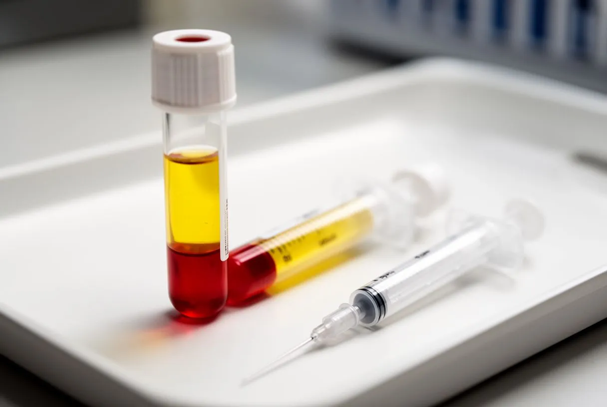
The core blood panel typically ordered includes:
- Blood cultures: Three sets drawn from different venipuncture sites, spaced at least one hour apart, before antibiotics are started. These are the single most important test because they identify the exact organism and its antibiotic sensitivities.
- CRP and ESR: Inflammatory markers tracked over time. A rising trend suggests the infection is not controlled; a falling trend suggests antibiotics are working.
- Serology for atypical organisms: Antibody tests for Coxiella, Bartonella, Brucella, Chlamydia, and Legionella — ordered when blood cultures are negative but clinical suspicion remains high.
- Complete blood count and basic metabolic panel: Detects anemia, kidney dysfunction from septic emboli, and electrolyte imbalances that complicate treatment.
These tests do not stand alone. A patient with endocarditis may have normal white blood cell counts, particularly if the infection is indolent or the immune system is suppressed. CRP can be elevated by many conditions unrelated to the heart. Blood cultures can be negative if antibiotics were given even briefly before sampling. This is why the modified Duke criteria — a diagnostic scoring system that combines blood findings with imaging and clinical signs — forms the backbone of how cardiologists confirm or reject the diagnosis. The blood panel is one pillar of that system, not the entire structure.
How Blood Cultures Work in Endocarditis Diagnosis
A blood culture is the process of taking a sample of your blood and placing it in a nutrient-rich broth that encourages bacteria or fungi to multiply. The laboratory monitors the broth for several days, watching for cloudiness or gas production that indicates growth. Once an organism grows, the lab identifies it and tests it against a panel of antibiotics to determine which drugs will kill it — a process called susceptibility testing. This result directly guides your cardiologist's choice of antibiotic, the dose, and the duration of therapy.
Why three sets are drawn, not one: Endocarditis produces bacteria in the bloodstream in a continuous but low-grade way. A single sample might miss the organism by chance, or might be contaminated by skin bacteria at the needle site. By drawing three separate sets from three different locations, the laboratory can distinguish true bloodstream infection from contamination. If the same organism grows in two or more bottles, the likelihood of true bacteremia — bacteria circulating in the blood — is very high. This is one of the most important quality measures in the diagnostic process, and a patient should always ask how many sets were drawn and whether they were taken before antibiotics were administered.
The timing matters enormously. If a patient has already received even a single dose of antibiotics, the chance of a positive culture drops sharply. In some cases, the lab may need to use special media or extend the incubation period beyond the standard five days to recover slow-growing organisms. Research on managing acute cardiac conditions, including the safety of using the intravenous beta-1-adrenoblocker esmolol in patients with ST-segment elevation myocardial infarction before primary percutaneous coronary intervention, highlights how time-sensitive interventions in cardiology depend on accurate, early laboratory data — and blood cultures are no exception.
Endocarditis Symptoms and When to Seek Testing
Endocarditis rarely begins with chest pain. More often, the first signs are a low-grade fever that comes and goes, night sweats, unexplained weight loss, fatigue, and muscle or joint aches. Some patients notice small, tender spots on their fingers or toes called Osler nodes, or painless red spots on the palms and soles called Janeway lesions. Auscultation — the act of listening to the heart with a stethoscope — may reveal a new or changed heart murmur, which is often the first clue a clinician picks up during a physical exam. Because these symptoms overlap with influenza, rheumatoid arthritis, and many other conditions, endocarditis is frequently diagnosed late, sometimes weeks after the infection began.

When symptoms warrant urgent blood work: Any patient with a known heart valve condition, prosthetic valve, or history of endocarditis who develops a persistent fever above 38°C (100.4°F) lasting more than three days should have blood cultures drawn and be evaluated by a cardiologist. Do not take antibiotics before blood cultures are collected unless a physician explicitly instructs you to do so, because even a single dose can sterilize the bloodstream and make identifying the organism far more difficult. If you have recently had dental extraction, surgery, or a procedure involving the urinary tract and you develop fever and a new murmur, the index of suspicion should be even higher.
Patients who have undergone valve replacement should be particularly vigilant. Prosthetic valves are more susceptible to infection because the foreign material provides a surface that bacteria can adhere to without being swept away by normal blood flow. If you have a prosthetic valve and develop fever, chills, or any sign of systemic illness, you should contact your cardiac team immediately rather than waiting for symptoms to resolve. Early blood testing — ideally within hours of the fever — significantly improves the chance of identifying the organism and tailoring treatment before the infection causes irreversible valve damage.
Cardiac Infection Markers: What Your Blood Work Shows
Cardiac infection markers are blood test results that specifically point toward infection or inflammation involving the heart. Unlike a simple cold, where a white blood cell count might be mildly elevated, endocarditis often produces a distinctive pattern: a moderately elevated white count, a high CRP (frequently above 50 mg/L and sometimes above 100), an elevated ESR, and evidence of anemia of chronic disease. When blood cultures are positive, the specific organism and its antibiotic sensitivity profile become the most important markers of all, because they tell your team exactly which drug will work and which will not.
Tracking markers during treatment: Once antibiotic therapy begins, your team will repeat inflammatory markers every few days. CRP should begin to fall within one to two weeks of effective treatment. If it does not, the cardiologist may question whether the antibiotic is working, whether the dose is correct, or whether the infection has spread or formed an abscess. In some cases, a persistently elevated inflammatory marker despite appropriate antibiotics is the first sign that surgery will be needed to remove infected tissue or replace a damaged valve. Research on how metabolic disturbances affect cardiac function — such as the influence of electrolyte imbalance on regional wall motion abnormalities in STEMI patients of North Indian origin — underscores how closely the body's chemistry and the heart's mechanical function are linked during acute illness.
Other markers your team may monitor include renal function (kidney tests), because septic emboli can damage the kidneys; liver enzymes, which may rise if right-sided endocarditis causes venous congestion; and blood counts to watch for antibiotic-induced bone marrow suppression. If you are on aspirin or other blood thinners, your team will also track clotting studies, because some antibiotics interact with these medications and alter their effect. The takeaway is that blood work in endocarditis is not a one-time event — it is a continuous monitoring process that guides every clinical decision from the first day of treatment to the last.
How to Diagnose Endocarditis: The Full Diagnostic Pathway
The diagnosis of endocarditis is never based on a single test. Cardiologists use the modified Duke criteria, a scoring system that weighs clinical findings, blood culture results, imaging evidence, and serology. A patient is classified as having definite endocarditis, possible endocarditis, or rejected endocarditis based on how many major and minor criteria are met. Two major criteria — for example, two positive blood cultures for a typical organism plus an echocardiogram showing a vegetation — confirm the diagnosis. One major and three minor criteria, or five minor criteria, also confirm it.

Major and minor criteria in plain terms: The major criteria are straightforward — typical organisms in blood cultures and evidence of endocardial involvement on echocardiography. Minor criteria include a pre-existing heart condition that predisposes to infection (such as a prosthetic valve or atheromatous plaque in the aortic root), a fever above 38°C, evidence of blood vessel involvement (like septic emboli visible on imaging), and immunological signs such as positive serology or certain skin findings. Your cardiologist assembles these pieces like a puzzle, and the picture becomes clearer as more data accumulates over the first few days of hospital evaluation.
Echocardiography — ultrasound imaging of the heart — is central to this process. A transthoracic echocardiogram (performed through the chest wall) is done first, but it may miss small vegetations, particularly on prosthetic valves. If suspicion remains high, a transesophageal echocardiogram (performed via a probe passed down the esophagus) provides much clearer images because the esophagus sits directly behind the heart. This test can detect vegetations as small as two to three millimetres and can identify abscesses forming in the valve ring. Procedures such as balloon angioplasty or catheter-based interventions, while primarily used for coronary artery disease, illustrate the same principle: direct visualization of cardiac structures drives accurate diagnosis and treatment decisions in modern cardiology.
Serology Tests for Heart Infections: What Results Mean
When serology results come back, your cardiac team interprets them in context — not in isolation. A single elevated antibody level can mean a past infection, a current infection, or cross-reactivity with another organism. For this reason, serology is often repeated two to four weeks later to look for a rising titre, which indicates an active immune response rather than a lingering antibody from an old encounter. A fourfold rise in antibody levels between two samples taken weeks apart is strong evidence of active infection, even when blood cultures remain negative.
Understanding positive and negative results: A negative serology result does not exclude endocarditis if the clinical picture is convincing, because the organism may not be one for which serology was ordered, or the immune response may not yet have produced detectable antibody levels. Conversely, a positive result must be interpreted alongside blood cultures, echocardiography, and the physical exam. In culture-negative endocarditis, a high antibody titre against Coxiella burnetii (phase I IgG greater than 1:800) is one of the major Duke criteria and can confirm the diagnosis on its own. The same applies to high titres for Bartonella in patients with appropriate exposure history.
For patients and families, the key message is patience. Serology results may take several days to a week because some assays require specialized laboratories that are not available in every hospital. During that time, your team may begin empirical antibiotic treatment — a broad-spectrum regimen designed to cover the most likely organisms — while waiting for definitive results. This is standard practice and does not mean your doctors are uncertain; it means they are acting responsibly to prevent valve damage while the laboratory finishes its work. Once the organism is identified and its sensitivities are known, the antibiotic regimen is narrowed to target that specific organism precisely.
Endocarditis Prevention and Screening for At-Risk Patients
Prevention of endocarditis starts with identifying which patients are at highest risk and ensuring that antibiotic prophylaxis is used appropriately before procedures that can introduce bacteria into the bloodstream. Current guidelines recommend prophylactic antibiotics before dental procedures involving manipulation of the gingival tissue or the periapical region of teeth for patients with prosthetic heart valves, a history of endocarditis, congenital heart disease managed with prosthetic material within the past six months, or cardiac transplant recipients with valvular disease. For most other patients, prophylaxis is not recommended because the risk of adverse reactions to antibiotics may exceed the benefit.

Practical prevention strategies for high-risk patients:
- Dental hygiene: Maintain regular dental check-ups every six months. Poor dental health and gingivitis are the most common sources of bacteria that cause endocarditis. Brushing twice daily and flossing once daily reduce bacterial load in the mouth.
- Skin and wound care: Clean cuts, abrasions, and puncture wounds promptly. Avoid tattoos and body piercings if you have a prosthetic valve, because these procedures introduce bacteria into the bloodstream.
- Antibiotic prophylaxis: Take the prescribed antibiotic exactly as directed before dental procedures — typically a single dose of amoxicillin taken one hour before the procedure. If you are allergic to penicillin, your cardiologist will prescribe an alternative.
- Avoid unnecessary procedures: Cosmetic injections, unsterile manicures, and self-administered intravenous lines can all introduce bacteria. Discuss any planned procedure with your cardiac team first.
Screening for endocarditis is not the same as screening for coronary disease. There is no routine blood test that catches endocarditis before symptoms appear, because the condition is relatively rare and the tests are not specific enough for population-wide screening. Instead, screening means vigilance: knowing your risk category, recognizing early symptoms, and seeking evaluation promptly when fever or new symptoms develop. For patients in Guadalajara, Querétaro, and Hermosillo, having a relationship with a cardiac team who knows your valve history means that if symptoms arise, you can move quickly from suspicion to diagnosis to treatment without unnecessary delays.
Endocarditis Testing: Next Steps for Patients
If you or a family member has a heart valve condition and is experiencing persistent fever, fatigue, or any of the symptoms described in this guide, the next step is a clinical evaluation with a cardiac specialist. Diagnostic evaluation for suspected endocarditis usually includes blood cultures, inflammatory markers, serology panels, and both transthoracic and transesophageal echocardiography. The goal is not merely to run tests but to assemble the full diagnostic picture so that treatment — whether antibiotics alone or surgery combined with antibiotics — is tailored to your specific situation.
What to expect at your consultation: Bring a complete medical history, including any prior valve surgery, prosthetic valve details (manufacturer and model if known), recent dental or surgical procedures, current medications, and any recent antibiotic use. If you have had blood work done at another facility, bring those results with you. Your cardiologist will perform a physical examination, listen for heart murmurs, and order the initial blood panel and imaging studies. Depending on the results, you may be admitted to hospital for intravenous antibiotics, or you may be managed as an outpatient with close monitoring while results are pending. Every decision is guided by the modified Duke criteria and current international guidelines for infective endocarditis management.
Endocarditis is a serious condition, but with timely diagnosis and appropriate treatment, outcomes can be good. The most important factor is time — the sooner the infection is identified and targeted therapy begins, the lower the risk of permanent valve damage and the better the long-term prognosis. If you are experiencing symptoms and you have a known heart valve condition, do not wait. Contact a qualified cardiac team, ask about blood cultures and echocardiography, and let the laboratory data guide the next step. Your heart deserves a deliberate, evidence-based approach — and that begins with a conversation with a specialist who understands your full cardiac history.






