


Stroke Volume and Heart Function: Measuring Each Beat
Stroke volume heart function explained: learn how much blood your heart pumps per beat, measurement methods, normal ranges.
What Is Stroke Volume Heart Function?
When your cardiologist talks about stroke volume heart function, they are referring to the specific amount of blood your heart pumps out of its main pumping chamber—the left ventricle—with every single beat. Think of it as the engine's displacement per cycle. It is a crucial measurement because it tells us exactly how much oxygen-rich blood is reaching your body each time your heart contracts. A healthy heart maintains a steady stroke volume to keep your organs functioning optimally, adapting seamlessly whether you are resting or walking up a flight of stairs. Understanding this baseline measurement is often the first step in evaluating overall cardiac health.
Why ventricular filling matters: Before your heart can pump blood out, the left ventricle must fill with blood returning from the left Atrium. This resting phase, known as diastole, sets the stage for the forceful contraction that follows. If the ventricle does not fill adequately due to restrictions or stiffness, the stroke volume naturally drops, leaving you feeling fatigued. Evaluating these filling pressures is a standard part of cardiac function assessment, often monitored closely in ongoing clinical research available at clinicaltrials.gov.
How the left ventricle contracts: During systole, the heart muscle squeezes tightly to push blood out through the aorta. The strength of this squeeze, or contractility, determines how much blood leaves the heart. If the muscle is weakened by disease or an Atypical Infarction (a silent or unusual heart attack), the squeezing force diminishes, directly lowering your stroke volume and impairing heart function.
How Does Stroke Volume Relate to Cardiac Output?
Stroke volume and cardiac output are deeply connected, but they are not the same thing. Cardiac output is the total volume of blood your heart pumps in one minute, while stroke volume is the amount pumped in a single beat. To find the cardiac output, doctors multiply the stroke volume by the heart rate (beats per minute). If your stroke volume decreases because the heart muscle is weakened, your heart must beat faster to maintain the same overall cardiac output, which can lead to a racing pulse and exhaustion.
The role of heart rate compensation: When the heart senses a drop in stroke volume, it often tries to compensate by increasing the heart rate. This is why you might experience palpitations or a rapid heartbeat when your heart pumping efficiency is compromised. However, beating faster is only a temporary fix; a chronically fast heart rate can eventually wear out the heart muscle, making the underlying stroke volume problem worse over time.
Impact on the ascending aorta: The blood ejected with each stroke volume forcefully enters the Ascending Aorta, the first major artery carrying blood away from the heart. A healthy, strong stroke volume ensures robust blood flow through this vessel. If the stroke volume is weak, blood can stagnate, increasing the risk of complications like Atheromatous Plaque buildup in the arterial walls, which complicates overall heart function.

What Is the Stroke Volume Formula?
Calculating stroke volume involves a straightforward clinical formula: End-Diastolic Volume (EDV) minus End-Systolic Volume (ESV). The end-diastolic volume is the amount of blood in the left ventricle just before it contracts, while the end-systolic volume is the blood remaining after the contraction. The difference between these two numbers is the exact amount of blood ejected with each beat. For example, if your ventricle holds 120 milliliters before contracting and 50 milliliters after, your stroke volume is 70 milliliters.
Understanding end-diastolic volume: This is the maximum capacity of your ventricle during the resting phase. Factors like hydration levels, blood volume, and the elasticity of the heart muscle all influence EDV. If you are dehydrated, there is less blood available to fill the ventricle, which decreases the EDV and subsequently lowers your stroke volume, a common reason for feeling lightheaded during cardiac assessments.
Understanding end-systolic volume: This represents the blood left behind after the heart has done its best to squeeze. A strong, healthy heart will leave very little blood behind, resulting in a low ESV and a high stroke volume. Conversely, a heart struggling with Atrial Flutter or weak muscle contractions will have a higher ESV, meaning less blood makes it out to the body with each beat.
How Do We Measure Heart Beat Volume?
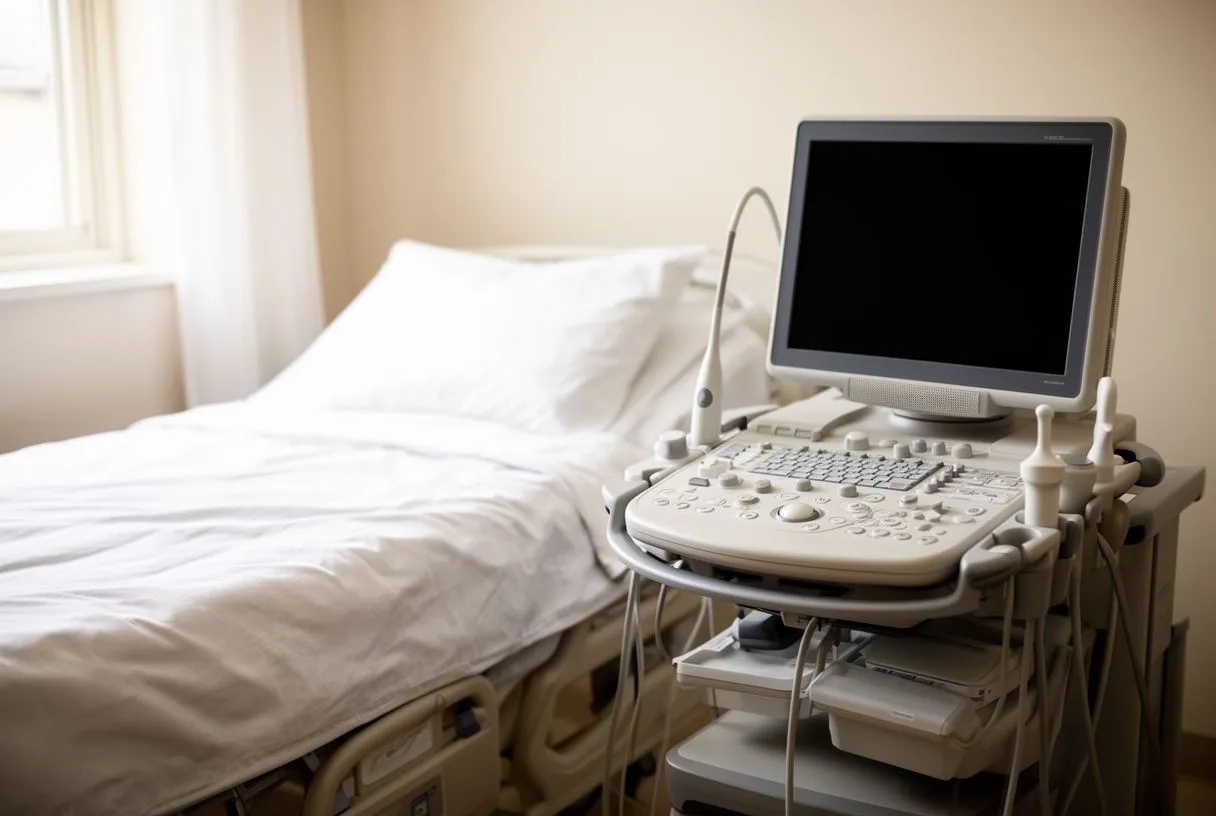
Measuring heart beat volume is most commonly done using an echocardiogram, a safe and painless ultrasound of the heart. This imaging technique allows cardiologists to visually measure the exact size of the left ventricle before and after contraction, applying the stroke volume formula to get an accurate number. Auscultation, or listening to the heart with a stethoscope, can provide clues about valve function and blood flow, but precise volume measurements require detailed imaging.
Using advanced imaging: In complex cases, doctors might use cardiac MRI or CT scans to get a highly detailed view of the ventricular chambers. These tools are particularly useful when planning interventions for severe heart function decline. Evaluating these parameters is vital, especially when researching outcomes for conditions like unstable coronary syndromes, where a Safety and efficacy of angioplasty with intracoronary stenting in patients with unstable coronary syndromes. Comparison with stable coronary syndromes is considered.
Direct arterial monitoring: In intensive care settings, doctors might use an Arterial Line or an Arterial Cannula to continuously monitor blood pressure and estimate stroke volume variations in real-time. This is crucial for critically ill patients where minute-to-minute changes in heart pumping efficiency can dictate immediate medical decisions, allowing for rapid adjustments in medications or fluids.
What Is a Normal Stroke Volume?
A normal stroke volume for a healthy adult at rest typically ranges between 60 and 100 milliliters per beat. However, what is considered normal can vary slightly based on body size, age, and fitness level. Athletes, for instance, often have a higher normal stroke volume because their heart muscle has adapted to pump more blood with less effort, which is why their resting heart rate is often much lower than average.
- Resting Range: 60 to 100 mL per beat for average adults.
- Athletic Hearts: Often exceeds 100 mL per beat due to enhanced ventricular filling.
- Stroke Volume Index: Adjusts the volume based on body surface area for precise comparisons.
Body size and stroke volume index: Because larger bodies require more blood, doctors often use the Stroke Volume Index, which divides the stroke volume by the body's surface area. This gives a more personalized view of heart function metrics, ensuring that a petite individual and a tall individual are evaluated against appropriate, individualized benchmarks rather than a single rigid standard.
How Do Ejection Fraction and Stroke Volume Compare?
Ejection fraction and stroke volume are two sides of the same coin. While stroke volume measures the absolute volume of blood pumped (in milliliters), ejection fraction measures the percentage of blood pumped out relative to the total volume in the ventricle. A normal ejection fraction is typically between 50% and 70%. You can have a normal ejection fraction but a low stroke volume if the ventricle itself is unusually small, which is why both measurements are vital for a complete cardiac function assessment.
When percentages are misleading: Relying solely on ejection fraction can sometimes mask underlying issues. For instance, in patients with severe valve disease who undergo a Balloon-expandable Valve procedure (often part of transcatheter aortic valve replacement, or TAVR), the ejection fraction might look acceptable, but the actual stroke volume could be critically low due to restricted blood flow prior to the intervention.
Silent threats to heart function: Sometimes, heart function declines without obvious symptoms. For example, research on Cardiac Autonomic Dysfunction and Risk of Silent Myocardial Infarction Among Adults With Type 2 Diabetes highlights how silent heart attacks can gradually damage the heart muscle, reducing stroke volume over time without the dramatic chest pain typically associated with a heart attack. Recognizing these silent shifts is crucial for long-term cardiac care.
What Factors Influence Stroke Volume Regulation?
Three primary factors dictate stroke volume regulation: preload, afterload, and contractility. Preload is the stretch of the ventricle at the end of filling—more stretch generally means a stronger contraction, a principle known as the Frank-Starling mechanism. Afterload is the resistance the heart must overcome to eject blood; high blood pressure increases afterload, making it harder for the heart to pump and thereby reducing stroke volume.
The impact of afterload: When blood pressure is high, the heart has to push against a tighter, narrower vascular system. Over time, this increased resistance tires the heart muscle, lowering the amount of blood ejected per beat. Managing blood pressure with medications or lifestyle changes is often the most effective way to improve stroke volume without surgical intervention, helping to lower the resistance the heart faces with every contraction.
The role of contractility: This is the inherent strength of the heart muscle's squeeze, independent of preload and afterload. Conditions like Arrhythmias, valve defects, or prior heart attacks weaken contractility. If an Arterial Graft is needed to bypass blockages caused by high Apolipoprotein B levels, restoring blood flow through procedures like Balloon Angioplasty can help recover some of this inherent squeezing power and stabilize stroke volume.

How Does Heart Pumping Efficiency Affect Daily Life?
When heart pumping efficiency drops, the effects ripple through your entire body. A low stroke volume means your tissues and muscles are not receiving enough oxygenated blood to function normally. This leads to persistent fatigue, shortness of breath, and a decreased tolerance for physical activity. Simple tasks like carrying groceries or walking up an incline can suddenly feel like monumental efforts, signaling that your heart function is compromised.
Recognizing physical symptoms: Beyond fatigue, poor heart pumping efficiency can manifest as swelling in the legs and ankles, known as edema. This happens because blood backs up in the venous system when the heart cannot move it forward efficiently. Some patients also experience unusual discomfort, such as Arm Pain during physical exertion, which can be an atypical sign of cardiac distress rather than a simple muscle strain.
Mental and emotional toll: Living with reduced heart function can be incredibly stressful. The constant fatigue and breathlessness can lead to anxiety and a reduced quality of life. It is important to recognize that these emotional responses are a normal reaction to a physical limitation, and addressing the underlying cardiac issue often improves both physical stamina and mental well-being.

When Should You Assess Cardiac Function Parameters?
You should consider assessing your cardiac function parameters if you experience any persistent symptoms of poor heart pumping efficiency, such as unexplained shortness of breath, chronic fatigue, or swelling in your lower extremities. Even without symptoms, individuals with a history of heart disease, high blood pressure, or diabetes should have regular assessments. Early detection of declining stroke volume allows for proactive management before severe damage occurs.
Monitoring chronic conditions: Patients managing chronic cardiac issues often require routine monitoring. For instance, those taking Aspirin (evaluated in major clinical trials like ARISTOTLE for antiplatelet therapy) or those recovering from procedures like Balloon Aortic Valvuloplasty need their heart function metrics tracked to ensure the treatments are working. Regular echocardiograms provide a clear picture of whether the stroke volume is maintaining, improving, or declining over time.
Preparing for surgical interventions: Before any major cardiac surgery, a thorough assessment of cardiac function parameters is mandatory. Surgeons need to know the exact stroke volume and ejection fraction to plan the safest approach. Whether considering an Autograft for valve replacement or preparing for complex bypass surgery, these measurements guide the surgical team in determining the lowest risk strategy for your specific situation.
How Is Stroke Volume Optimized Before Surgery?
Optimizing stroke volume before surgery involves a combination of medication management, fluid balance, and sometimes mechanical support. Cardiologists may adjust diuretics to ensure the heart is not overloaded with fluid, or prescribe medications to reduce afterload, making it easier for the heart to pump. The goal is to create the most stable, efficient baseline heart function possible before the stress of an operation.
- Medication Adjustments: Tuning blood pressure and fluid medications to reduce cardiac strain.
- Autologous Blood Management: Planning for blood conservation to maintain optimal volume during procedures.
- Advanced Support: In severe cases, utilizing Awake ECMO or an Automated External Defibrillator standby to support circulation.
Surgical planning and recovery reality: During surgery, tools like an Autologous Blood recovery system might be used to maintain your own blood volume, keeping your preload stable. After surgery, recovery timelines depend heavily on how well the stroke volume improves. Patients must follow strict recovery milestones, gradually increasing physical activity as the heart muscle heals and its pumping efficiency returns to a stable, healthy baseline.
In conclusion, understanding stroke volume and heart function is essential for navigating cardiac health. From recognizing the subtle signs of a dropping stroke volume to understanding how it is measured and optimized before surgery, this knowledge empowers you to take an active role in your care. If you or a loved one are experiencing symptoms of poor heart pumping efficiency or are facing a cardiac intervention, do not self-diagnose. We encourage you to discuss your specific situation with a qualified cardiac team. Please book a consultation with Cirugía de Corazón, the practice of Dr Alejandro Molina in Guadalajara, Querétaro, and Hermosillo, Mexico, to receive a thorough evaluation and a personalized plan.






