

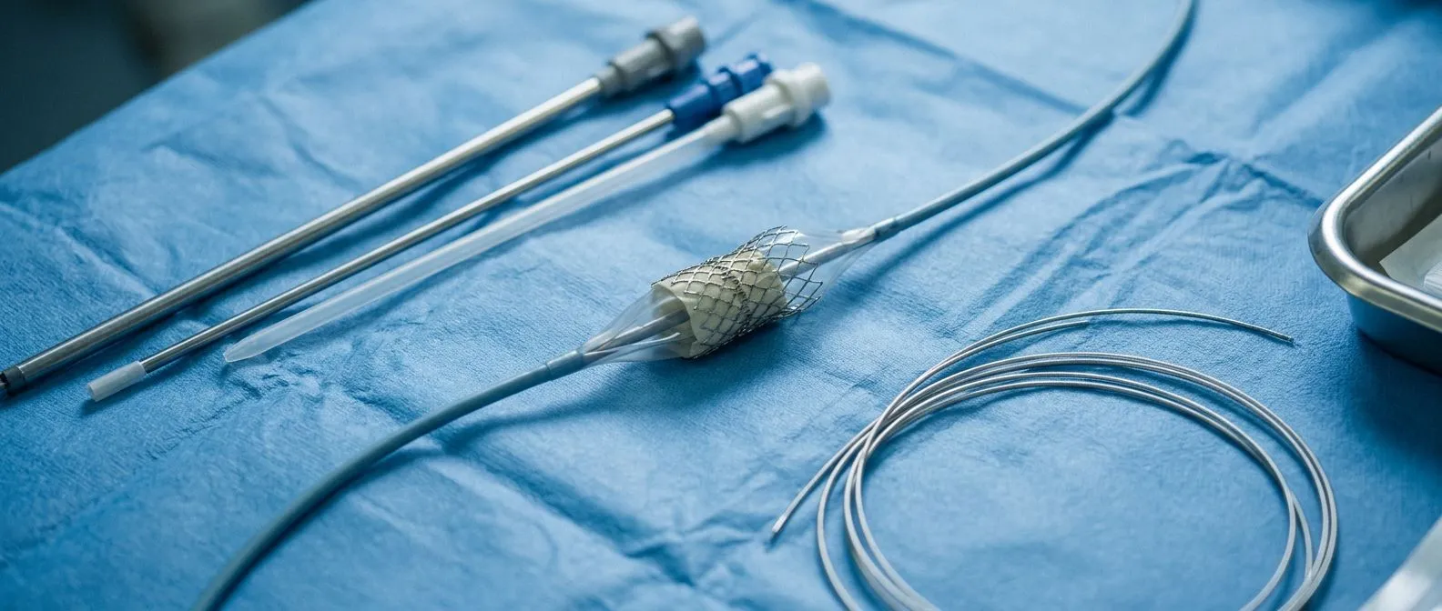
Structural Cardiology: Fixing the Heart Through a Catheter
Structural cardiology procedures repair heart valves and defects through a catheter—no open surgery needed.
Many people are told they have a heart valve problem and assume that open-heart surgery is the only answer. In the last two decades, that has changed. Structural cardiology procedures repair or replace parts of the heart — valves, holes, narrowed openings — using thin tubes called catheters, threaded through an artery in the wrist or groin, often without opening the chest at all. This guide walks through what these procedures are, which conditions they treat, what recovery actually looks like, and how to decide whether one is right for you or a family member.
What Is Structural Cardiology?
Structural cardiology is the branch of heart medicine that deals with the physical "hardware" of the heart: the valves that control blood flow, the walls that separate its chambers, and the large vessels attached to it. Rather than treating the heart's rhythm or its arteries with medication alone, structural cardiology procedures physically repair or replace the parts that have worn out, narrowed, or never formed correctly.
How it differs from other branches of cardiology: General cardiology manages heart disease with medicines and monitoring. Interventional cardiology opens blocked coronary arteries with stents. Structural cardiology goes one step further, using similar catheter tools to fix valves, close holes, and rebuild structural defects — work that once required a sternum cracked open and a heart-lung machine.
Why it matters for older or higher-risk patients: Many people with severe valve disease are older, frail, or have other conditions that make open surgery risky. Catheter-based treatments offer an option with a smaller incision, no heart-lung machine in most cases, and a much shorter hospital stay. For these patients, the choice between surgery and a catheter procedure can genuinely change what treatment is possible.
Who Is a Structural Cardiologist?
A structural cardiologist is a heart specialist trained specifically in catheter-based repair of the heart's structure. These doctors sit at the intersection of cardiology and cardiac surgery — they understand the anatomy a surgeon knows and command the catheter tools an interventional cardiologist uses. In many cases they work side by side with a cardiac surgeon during the procedure, a model called the "heart team."
What their training looks like: The path is long. It typically includes a general cardiology fellowship followed by additional dedicated training in structural interventions, often in high-volume centers where these procedures are performed regularly. Dr Molina trained in cardiothoracic surgery at the Universidad de Guadalajara, in cardiac, minimally invasive, robotic and structural surgery at the University of Montreal, and in structural cardiology and TAVR at CHU Bordeaux; the team also trained at Jena, Bad Berka and Buffalo. This depth of training matters, because structural heart procedures reward experience — the more of these procedures a team has done, the better the outcomes tend to be.
What the most competitive cardiology fellowships involve: Structural cardiology and electrophysiology are among the most competitive cardiology fellowships because the technology is advancing quickly and demand is growing. That competition reflects a real shift: patients increasingly want effective treatment with less physical trauma, and structural cardiology delivers it.
What Are the Structural Components of the Heart?
To understand what these procedures fix, it helps to picture the heart's four chambers — two upper atria and two lower ventricles — and the parts that keep blood moving in one direction. When any of them fails, symptoms follow, and a structural heart procedure may be the answer.
- Four valves: the aortic, mitral, tricuspid and pulmonary valves act as one-way doors between chambers and vessels, opening and closing with every heartbeat.
- Walls and septum: the muscular wall (septum) separates left from right. Some people are born with a small hole in it — a defect that can often be closed with a catheter.
- The aorta and great vessels: the aorta is the body's largest artery, carrying all the blood the heart pumps out. Its narrowing or widening can be treated with catheter-delivered devices.
- The heart muscle itself: thickening or stiffening of the heart muscle affects how the valves and chambers work and often shapes the treatment plan.
What Is the Most Common Structural Heart Disease?
Among adults, the most common structural heart disease is aortic valve stenosis — a narrowing of the aortic valve caused by calcium buildup, wear, or a valve that was abnormal from birth. In younger people, mitral valve disease and congenital holes in the heart are frequent. The three types of cardiology — general, interventional, and structural (alongside surgical) — all come into play here, because valve disease often needs both monitoring and, eventually, intervention.
What patients feel first: Aortic stenosis often announces itself with a heart murmur a doctor hears on a stethoscope, then with shortness of breath, chest tightness, dizziness or fainting spells. The valve narrows slowly over years, so many people adapt without realizing — until climbing stairs becomes a struggle. In our consultations in Guadalajara, Querétaro and Hermosillo, we often see patients who delayed a year or more before being told their valve was severe — because the symptoms had quietly become "normal" for them.
When treatment becomes necessary: The decision to treat depends on the severity of the narrowing and the presence of symptoms. Guidelines recommend intervention when the valve is severely narrowed and symptoms are present, or when certain measurements show the heart is already being strained. Our team reviews every case against international practice guidelines before recommending anything, and the patient always receives a clear proposal with the alternatives — the final decision is theirs.
Types of Structural Heart Procedures Done by Catheter
Structural heart procedures catheter-based span a wide range. The common thread is that all of them use thin tubes inserted, usually, through the femoral artery at the groin or the radial artery at the wrist, guided by X-ray imaging and ultrasound inside the heart.
- Valve replacement without surgery: a new valve mounted on a spring-like frame (stent) is carried through the circulation and deployed inside the old one — TAVR for the aortic valve is the best known example.
- Valve repair by catheter: the mitral valve can be repaired with a small clip that brings its leaking edges together, a non-surgical heart valve repair used when the valve leaks rather than narrows.
- Closure of holes: atrial septal defects and patent foramen ovale — openings between the upper chambers — can be closed with a small umbrella-like device through a catheter in the vein.
- Widening narrowed valves: balloon catheters can stretch narrowed valves, sometimes as a bridge to later valve replacement.
- Left atrial appendage closure: in patients with atrial fibrillation who cannot take blood thinners, a small plug can be implanted to close the pouch where clots form.
TAVR Procedure Explained: Aortic Valve Replacement by Catheter
TAVR — transcatheter aortic valve replacement — is the flagship structural heart procedure. It replaces a severely narrowed aortic valve with a new tissue valve, delivered through a catheter, usually without opening the chest. The heart catheterization repair approach means the new valve is compressed onto the catheter, advanced to the heart, positioned inside the old valve, and expanded.
How the procedure actually goes: The patient is usually under general anesthesia or heavy sedation. The team accesses the femoral artery in the groin (most commonly), or another access point if the arteries are not suitable. A temporary pacemaker wire is placed because the heart rate is briefly slowed during valve deployment. The new valve is expanded, and the team immediately checks that it sits well and that the valve is not leaking. Most patients go home within a few days.
Who it suits best: TAVR was first approved for patients too frail for open surgery. Evidence has since expanded its use, and in many guidelines it is now an accepted option for most patients with severe aortic stenosis, with age and anatomy deciding the route. Our team performs both conventional surgery and transcatheter procedures, and we recommend the less invasive route only where the evidence supports it for that specific patient — that is why every case is reviewed by the multidisciplinary team before a recommendation is made.
What we explain in the consultation: We take time to show patients their own imaging and explain, in plain words, what the studies actually show and what the alternatives are. I encourage patients to ask every question they have before deciding — a confident decision comes from understanding, not from pressure.
Mitral Valve Catheter Repair: What to Expect
The mitral valve sits between the left atrium and the left ventricle. When it doesn't close fully, blood leaks backward — a condition called mitral regurgitation. Severe leakage can lead to breathlessness, fatigue, and eventual heart failure. Not every leak is suitable for catheter repair, but for the right patients, it avoids open-heart surgery entirely.
How the catheter repair works: The most established technique uses a clip delivered through a vein, puncturing through the wall between the atria to reach the valve. The clip fastens the middle of the two leaking leaflets together, creating a double opening instead of one wide leaking one. The result is a significantly reduced leak. Dr Molina performed the first robotic mitral valve repair in the Ángeles Health System and the first robotic heart surgery in Guadalajara, and our team performs conventional, minimally invasive, robotic and transcatheter procedures.
Who qualifies: Suitability depends on the specific anatomy of the leak — which part of the valve is leaking, how much, and why. Echocardiography (an ultrasound of the heart) determines whether the clip approach is feasible, and which patients should instead be considered for surgery or, in some cases, a new transcatheter valve in the mitral position.
Are Structural Heart Procedures Safe?
Every heart procedure carries risk, and structural cardiology is no exception. But for many patients, the risk of a catheter procedure is substantially lower than the risk of leaving severe valve disease untreated — or of open surgery in someone frail. The honest answer is: safety depends on the patient, the procedure, and the experience of the team.
Common risks and how they're managed: Bleeding or bruising at the access site, temporary heart rhythm problems (some patients need a pacemaker afterwards, especially after TAVR), stroke, and rare but serious complications such as vascular injury. Structured heart teams monitor for these closely and have protocols for each.
What reduces risk: Choosing a high-volume center with an experienced multidisciplinary team, full pre-procedure imaging (CT scan, echocardiogram), and careful selection of the right procedure for the right patient all matter. Our team reviews each patient's imaging and risk profile together before recommending a procedure — and we actively encourage second opinions, treating them as a normal part of deciding about heart surgery.
Structural valve deterioration: One specific consideration is that tissue valves, whether implanted surgically or by catheter, can gradually degenerate over years — a process called structural valve deterioration. This is part of every long-term conversation with patients, because the durability of the new valve matters for younger patients especially.
Catheter Heart Procedure Recovery: Timelines and Milestones
Recovery after a catheter heart procedure is typically much faster than after open-heart surgery. But "faster" is not "instant," and knowing what to expect helps families plan.
The first days: Most patients stay in the hospital one to three days, sometimes longer depending on the procedure and their overall health. The groin or wrist access site needs to be watched for bleeding. Walking usually begins the same day or the next day. Soreness at the access site is normal for a few days.
The first weeks: Most patients are back to everyday activities within one to two weeks. Driving typically resumes once cleared by the doctor. Gradual return to exercise follows, guided by how the patient feels and any post-procedure instructions.
Long-term follow-up: The new valve or repair needs periodic check-ups, usually with echocardiography, to confirm it's working well over time.
How to Choose a Structural Heart Team in Mexico
Choosing where and with whom to have a structural heart procedure is one of the most important decisions in the process. Here's a practical framework for patients in Mexico or traveling there for care.
- Training and credentials: ask where the team trained in structural interventions and how many procedures they perform.
- Multidisciplinary review: the best outcomes come from a heart team — surgeon, cardiologist, imaging specialist — reviewing your case together, not one person deciding alone.
- Clear explanation of alternatives: a good team will lay out the surgical option and the catheter option, with honest trade-offs, and support your choice.
- Second opinions: seek one. It is normal, expected, and protects you.
- Transparent costs and logistics: for international patients, ask about travel planning, the expected length of stay in Mexico, and what the hospital stay includes.
We offer a free first video consultation: we get to know the patient, talk through the condition, and say which studies are still needed so no unnecessary ones are ordered. A face-to-face visit at our consulting rooms in Guadalajara, Querétaro or Hermosillo follows only when the patient decides to move forward.
What Is Considered a Structural Heart Procedure?
In short, any catheter-based treatment that repairs or replaces a physical part of the heart — valve replacement, valve repair, closure of holes, or widening of narrowed structures — is a structural heart procedure. The aortic valve is the most common target, but the mitral valve, septal defects, and the left atrial appendage are also treated this way.
Structural cardiology procedures have changed the conversation for patients who once had no safe option. The procedures are real tools, with real evidence behind them, and they deserve a serious look — not as an automatic replacement for surgery, but as one of the routes a well-informed heart team can offer.
If you or a family member has been told about a valve problem, a heart murmur, or a structural defect, the most valuable next step is a conversation with a qualified cardiac team that can review your specific case. Don't self-diagnose from a symptom list — and don't accept "there's nothing we can do" without a second opinion. A discussion with a team that performs both conventional surgery and catheter-based procedures will give you the full picture of what your options are.
What Are the Three Foods Cardiologists Say Not to Eat?
This is a popular question, and the honest answer is that it's a simplification — but it points at a real principle. Cardiologists consistently emphasize limiting processed foods high in sodium, foods high in saturated and trans fats, and sugary drinks. For a patient with valve disease or after a structural heart procedure, the goal is the same as for any heart patient: support the heart's workload, not add to it.
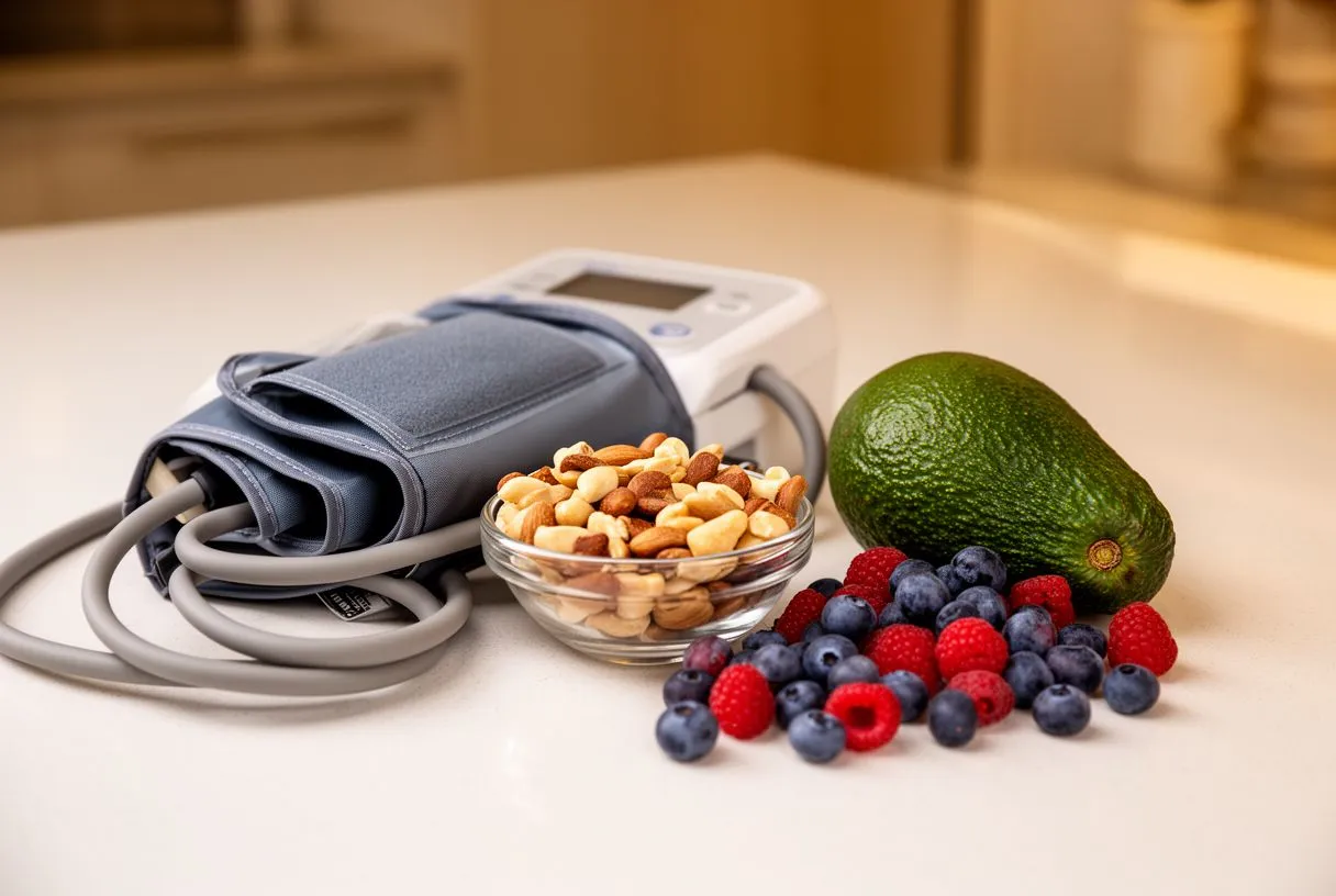
What actually matters for valve patients: For someone with a valve condition, diet is not the treatment — but it matters. Controlling blood pressure, avoiding excess salt, and maintaining a healthy weight all reduce the strain on the heart. Blood pressure control is especially important after a valve procedure, because high blood pressure can affect how the new or repaired valve performs over time.
Practical guidance: Rather than an absolute list of forbidden foods, focus on patterns — mostly vegetables, whole grains, lean protein, and limited processed foods. A cardiologist can help tailor this to a patient's individual condition, kidney function, and medications.


Structural cardiology procedures — from TAVR to mitral valve clip repair to closure of congenital holes — have given patients options that were unthinkable a generation ago. They are not a cure-all, and they are not right for every patient in every situation. But for those with valve disease or structural defects, especially those facing high surgical risk, they offer an alternative with less physical trauma and faster recovery.
The best decision is made when the patient understands the full picture: the anatomy of the problem, the surgical and catheter options, the risks, and the recovery. A multidisciplinary team that reviews your case against international guidelines and explains everything in plain words is the right partner for that decision. We encourage you to book a consultation with a qualified cardiac team — including ours — and to get a second opinion if any doubt remains. Your heart deserves a decision made with clarity, not fear.






