Superior Vena Cava in Heart Surgery and Catheters
Superior vena cava heart surgery treats blockages, tumors, and catheter problems.
If you or a family member has been told that a problem with the superior vena cava is part of a heart condition — or that a catheter or tumor is pressing on it — the terminology can feel overwhelming. The superior vena cava is a large vein in the chest, and it matters enormously in heart surgery and catheter procedures. This guide explains what the superior vena cava does, how superior vena cava heart surgery works, what treatments are available when this vein becomes blocked, and what recovery honestly looks like. Our goal is that you finish this article able to ask your cardiac team sharper questions and understand the answers.
What Is the Superior Vena Cava in the Heart?
The body's main return highway. The superior vena cava is one of the two largest veins in your body. It collects oxygen-poor blood from your head, neck, arms, and upper chest, and delivers it into the right atrium — the upper right chamber of the heart. From there, the blood is pumped to the lungs to pick up fresh oxygen.
Think of it as a two-lane highway. The inferior vena cava handles the lower half of the body, while the superior vena cava handles everything above the heart. Both are essential, but the superior vena cava is particularly vulnerable because it sits in a tight space in the middle of the chest, surrounded by lymph nodes, the windpipe, the aorta, and the right lung.
Because it has thin, soft walls and carries blood at low pressure, the superior vena cava can be squeezed shut by anything growing next to it. That is why it appears so often in discussions of chest tumors, central venous catheter placement, and heart surgery planning. It is also why imaging of the chest almost always includes a close look at this vein.
How Is the Superior Vena Cava Formed?
A meeting of two big branches. The superior vena cava is formed when two large veins — the right and left brachiocephalic veins, each draining one side of the head, neck, and arm — join together behind the right first rib. This confluence happens just above the heart, and the resulting single vessel then travels a short distance downward to empty into the right atrium.
In simple terms: many smaller veins from the upper body merge into two brachiocephalic veins, and those two merge into the superior vena cava. It is only about 5 to 7 centimeters long, yet it carries a remarkable volume of blood every minute of your life.
Some people are born with anatomic variants — for example, a persistent left superior vena cava alongside the normal right one. These variants are usually harmless and are often discovered by chance on imaging done for another reason. Knowing about them matters before any heart surgery or device placement, because a variant changes how a surgeon or cardiologist plans access to the heart.
What Is Superior Vena Cava Syndrome?
When the highway gets blocked. Superior vena cava syndrome is what doctors call the collection of symptoms that appear when this vein becomes narrowed or completely blocked. Blood leaving the head, neck, and arms backs up, and pressure builds in the vessels of the upper body. It is a serious condition that deserves prompt medical attention, though it usually develops gradually rather than as a sudden emergency.
The classic signs are easy to recognize once you know them. Patients often notice swelling of the face, neck, and arms — especially noticeable in the morning or after lying flat. Many describe a feeling of fullness or tightness in the head, prominent veins on the chest wall, a persistent cough, hoarseness, or shortness of breath when bending over or lying down.
In the United States and worldwide, the great majority of cases are caused by cancer — most often lung cancer or lymphomas growing in the middle of the chest. Benign causes such as blood clots, fibrosis from radiation, or scar tissue around long-term catheters account for a meaningful share as well, and these benign causes have become more common with the widespread use of implanted devices. An overview of the condition published by Medical News Today notes that recognizing the pattern of symptoms early is key to getting the right treatment started.
What Causes SVC Obstruction and When Is Surgery Needed?
Cancer-related compression. Tumors in the mediastinum — the central compartment of the chest between the lungs — are the leading cause of SVC obstruction. Lung cancers and lymphomas can grow until they press directly on the vein, narrowing it from the outside. When the tumor itself is treatable and resectable, SVC tumor removal together with superior vena cava reconstruction may be considered as part of the cancer operation.
Benign and device-related causes. Not every blockage comes from cancer. Long-term central venous catheters, pacemaker leads, and previous chest radiation can all scar or narrow the vein over years. A blood clot forming inside the vein — SVC thrombosis — is another important cause, particularly in patients with catheters or clotting disorders.
When surgery enters the conversation. The decision to operate depends on the cause, the severity of symptoms, and the patient's overall condition. A practical way to understand the options:
- Tumor-related obstruction: treatment starts with the cancer itself — chemotherapy, radiation, or resection — with stenting often used for fast symptom relief.
- Clot-related obstruction: blood thinners and, in selected cases, catheter-directed clot removal come first; surgery is reserved for cases that do not respond.
- Structural narrowing: a stent placed through a vein in the groin or arm can reopen the vessel without any open operation at all.
Every case with suspected SVC involvement is reviewed by the multidisciplinary team against international guidelines before a recommendation is made, because the right answer differs so much from patient to patient. In our practice, every case with suspected SVC involvement is reviewed by the multidisciplinary team against international practice guidelines before we recommend anything, because the right answer differs so much from patient to patient.
How Does a Superior Vena Cava Stent Procedure Work?
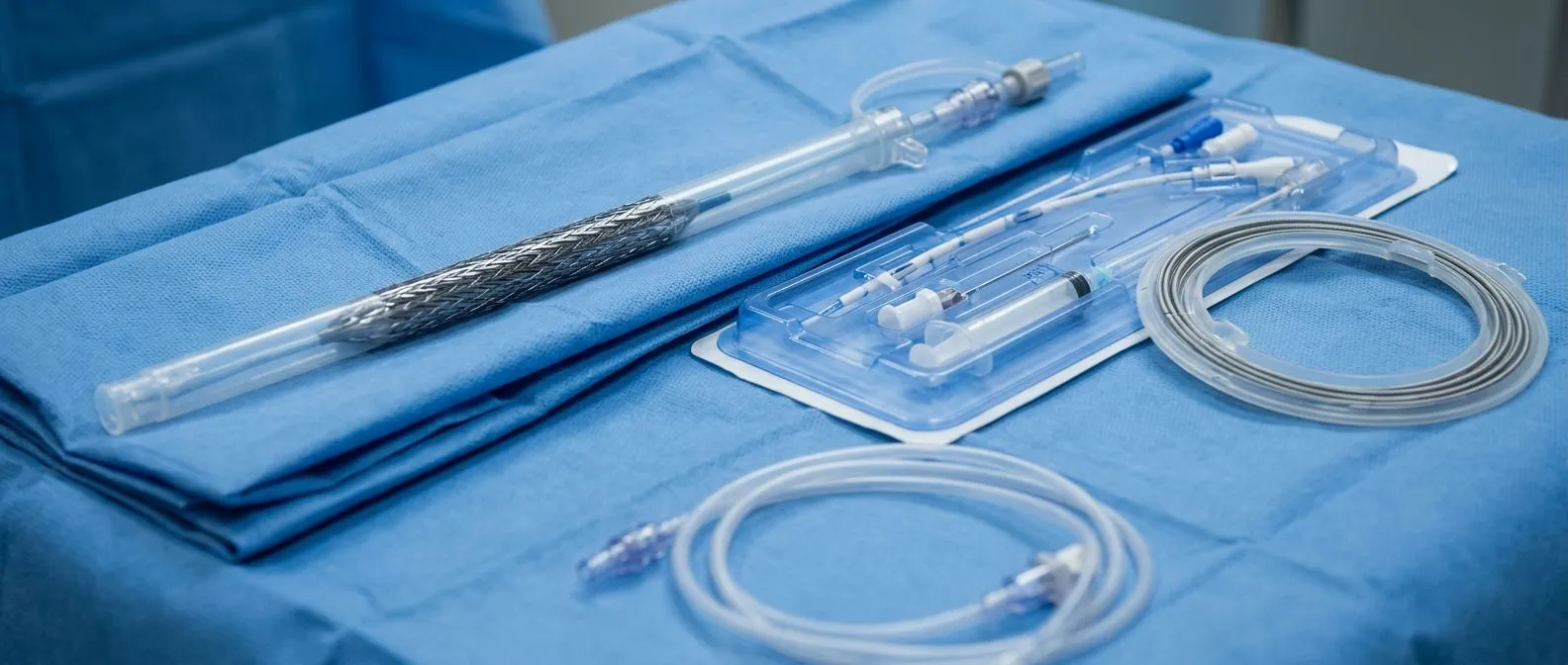
A stent without open surgery. The superior vena cava stent procedure is one of the most common and effective treatments for SVC obstruction today. A specialist — usually an interventional cardiologist or radiologist — threads a thin tube through a vein in the groin or arm, crosses the narrowed segment, inflates a small balloon to open it, and leaves behind a metal mesh tube called a stent that holds the vein open permanently.
Why patients often prefer it. Endovascular SVC repair requires no chest incision, no rib spreading, and usually only a day or two in hospital. Relief of head and neck swelling is often felt within hours to days, which is why stenting is frequently the first choice when symptoms are severe and fast relief matters — for example, before cancer treatment begins.
What to know beforehand. A stent relieves symptoms but does not treat the underlying cause. If a tumor is compressing the vein, cancer treatment must still address it, because a growing tumor can squeeze a stent shut from outside. Patients also need follow-up imaging to confirm the stent stays open, and blood thinners or antiplatelet medication may be recommended for a period after the procedure. I always tell patients that a stent is an excellent tool for the vein, but the plan for the disease behind it is just as important.
What Are SVC Bypass Surgery and Superior Vena Cava Reconstruction?
When the vein must be rebuilt. SVC obstruction surgery — a true open operation — is reserved for situations where a stent will not solve the problem: a tumor that is being surgically removed along with a segment of the vein, or a vein damaged beyond what a stent can hold open. Superior vena cava reconstruction means the surgeon replaces or patches the affected segment, often using a section of the patient's own tissue or a synthetic graft.
Bypassing the blockage. In SVC bypass surgery, the surgeon creates a new route for blood to travel around the obstruction, connecting an upper-body vein to a lower one — most often the left or right atrium or a large lower vein — so the backed-up blood can return to the heart. These are major operations performed with cardiopulmonary bypass or careful circulatory management, and they belong in the hands of experienced thoracic or cardiac surgical teams.
Combined and complex cases. The most demanding scenario is a mediastinal mass SVC surgery situation, where a chest tumor is removed together with reconstruction of the vein it has invaded. Published case reports — including an account of simultaneous pectus repair and aortic root surgery in Heart Vessels and Transplantation, and a report of aortic root replacement in a patient with severe pectus excavatum in the Journal of Cardiovascular and Thoracic Research — show how chest anatomy itself can complicate access to the great vessels, and how careful preoperative planning makes even unusual combinations of repairs possible. These operations carry real risks, and no honest surgeon will describe them as routine.
Why Does Heart Catheter Placement Involve the Superior Vena Cava?
The standard route to the heart. Nearly every heart catheterization and catheter-based procedure in modern practice travels through the veins of the upper body. During heart catheter SVC placement, a thin tube is advanced from an arm or neck vein down the superior vena cava and into the right side of the heart. From there, a Swan-Ganz catheter — a special tube used to measure pressures in the heart and lungs — can be floated into position, or contrast dye can be injected to study the coronary arteries and chambers.
Everyday procedures that use this route. If you have ever had a right heart catheterization, a pacemaker implanted, or an intravenous line placed for chemotherapy or long-term medication, the superior vena cava was part of your procedure whether you knew it or not. Pacemaker and defibrillator leads, dialysis catheters, and ports all pass through this vein to reach the heart.
Why it matters to patients. Because so many devices travel this route, the vein can develop scar tissue or clots over the years — especially in patients with repeated catheter placements. Before a device implantation or catheter procedure, the patient's previous imaging and catheter history are usually reviewed first, because a narrowed or occluded superior vena cava changes the approach. Before a device implantation or catheter-based procedure, the patient's previous imaging and catheter history are usually reviewed first, because a narrowed or occluded superior vena cava changes which approach is chosen.
What Central Venous Catheter Complications Affect the SVC?
The vein remembers every line. A Central Venous Catheter is a long, thin tube placed into a large vein so medications, nutrition, or dialysis can be given over weeks or months. Because these catheters sit in — or their tips rest near — the superior vena cava, they are a well-known cause of central venous catheter complications involving this vessel.
The problems fall into a few familiar categories. Mechanical irritation can cause vein wall inflammation and pain. Clots can form around the catheter and grow until they block the vein. Over long periods, the catheter can scar the vein wall until it narrows permanently. And rarely, the catheter tip can erode through the thin vein wall, which is an emergency recognized by chest discomfort and sudden swelling.
Practical warning signs families should know:
- New swelling of the face, neck, or arm on the side where the catheter was placed.
- Fever or chills together with neck discomfort — possible catheter infection that needs same-day attention.
- Sudden breathlessness or chest fullness — could signal a serious catheter complication and requires emergency care.
If a catheter-related blockage is found, the treatment usually starts with anticoagulation — blood-thinning medication — and removal of the catheter when it is no longer needed. In more severe narrowing, the same stenting techniques used for tumor-related obstruction can reopen the vein.
What Does SVC Syndrome Treatment and Recovery Look Like?
Relief usually comes quickly. For most patients, superior vena cava syndrome treatment begins with symptom relief and runs in parallel with treatment of the underlying cause. Steroid medication and head elevation can reduce swelling in the short term. A stent, when appropriate, typically relieves the characteristic head and neck fullness within a day or two. When cancer is the cause, chemotherapy or radiation shrinks the tumor over the following weeks and the pressure eases further.
Recovery after stenting. Most patients who undergo a stent go home the next day. You will be asked to avoid heavy lifting for a few days where the catheter was inserted, keep the site clean, and attend follow-up imaging to confirm the stent remains open. Many people return to normal daily activities within a week.
Recovery after open surgery. When a bypass or reconstruction is needed, recovery is that of major chest surgery: several days in hospital, six to eight weeks of gradual return to activity, and cardiac rehabilitation to rebuild strength. In the first consultations we take the time to explain, in plain words, what each study shows and what the recovery will realistically involve, so patients can plan work, travel, and family life around it. In our first consultations we take the time to explain in plain words what each study actually shows and what the recovery will realistically involve, so our patients can plan work, travel, and family life around it.

What Is the Prognosis After Superior Vena Cava Heart Surgery?
Prognosis follows the cause, not the vein. The honest answer about superior vena cava syndrome prognosis after surgery is that outcomes depend far more on the underlying disease than on the vein procedure itself. When a stent or bypass successfully relieves the obstruction, the symptoms of SVC syndrome almost always improve and quality of life rises significantly. When the cause is cancer, long-term outlook is driven by how that cancer responds to treatment.
What follow-up involves. Patients treated for SVC obstruction need ongoing surveillance. Imaging confirms the stent or graft stays open. Anticoagulation may be continued long term in clot-related cases. Any return of swelling or breathlessness warrants prompt imaging, because recurrence of narrowing is treatable — but only if it is caught.
The value of a second opinion. Because treatment ranges from a day-procedure stent to a major open reconstruction, and because the right choice depends on so many individual factors, seeking more than one expert view is a genuinely reasonable step, not a sign of distrust. We encourage second opinions and treat them as a normal part of deciding about heart surgery. We encourage second opinions and treat them as a normal part of deciding about heart surgery.
When Should You Talk to a Cardiac Team About the Superior Vena Cava?
Listen to your body's earliest signals. Persistent puffiness of the face in the morning, new visible veins across your chest, or breathlessness when lying flat are symptoms that deserve a proper evaluation — not necessarily because they are emergencies, but because they can be explained and often treated. Bring a list of your previous hospitalizations, any catheters or pacemakers you have had, and any cancer treatments, because all of these shape how your cardiac team evaluates the superior vena cava.
Getting started is easier than you think. The first contact with our team is a free video consultation: we get to know you, talk through your condition, and identify exactly which studies are still needed so no unnecessary tests are ordered. A face-to-face visit at our consulting rooms in Guadalajara, Querétaro, or Hermosillo follows when you decide to move forward. Every recommendation comes with the alternatives clearly laid out, and the final decision is always yours.
The superior vena cava heart surgery questions in this guide — what the vein does, why it gets blocked, and how it is treated — are ones patients commonly ask. If any part of this article describes your situation, the most important next step is a conversation with a qualified cardiac team rather than self-diagnosis. Book a consultation, bring your questions, and let the multidisciplinary team help you choose the lowest-risk path for your specific situation.