

The Sterile Field in Heart Surgery: Preventing Infection
Sterile field heart surgery protocols protect patients from life-threatening infections.
What Is the Sterile Field in Heart Surgery?
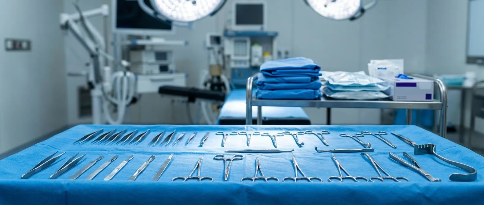
When preparing for a major cardiac procedure, understanding the sterile field heart surgery protocols can ease much of the anxiety patients feel. The sterile field refers to the highly controlled, meticulously cleaned physical area surrounding the surgical site and the sterile instruments used during the operation. It includes the patient's skin, the surgical drapes, the operating table, and the surgical team's gowns and gloves. By creating an invisible but strict barrier against microorganisms, the surgical team ensures that no external bacteria or pathogens enter the open chest cavity, which is highly vulnerable during an operation. Establishing and maintaining this environment is the foundation of modern cardiac care and patient safety.
Cardiac surgery involves direct contact with the body's most vital organs and blood vessels, making any breach in cleanliness a life-threatening risk. The underlying complexity of these operations is well-documented in medical literature, including discussions surrounding the Diagnosis and Treatment of Fetal Cardiac Disease. When a surgeon operates on the heart, whether to clear a blocked artery or repair a damaged valve, they are creating an entry point directly into the circulatory system. If bacteria were to enter this space, they could rapidly multiply in the bloodstream, leading to severe complications like sepsis or endocarditis, an infection of the heart's inner lining.
How does the sterile field interact with existing cardiac conditions? Patients often arrive in the operating room with pre-existing cardiovascular challenges, such as the buildup of an atheromatous plaque in their arteries. This fatty, calcified deposit narrows the blood vessels and requires precise surgical intervention, such as a coronary artery bypass. Because the surgical team is working around compromised tissue, introducing any foreign bacteria could worsen the patient's prognosis significantly. The sterile environment acts as a protective shield, allowing the surgeon to focus entirely on the intricate repair of the heart and vessels without the added danger of microbial interference. Every detail, from the air filtration in the room to the way the team moves, is calibrated to protect you.
How Does Heart Surgery Sterile Technique Work?
Heart surgery sterile technique relies on a strict set of physical barriers and behavioral protocols that separate the clean surgical zone from the potentially contaminated outside world. At its core, the technique dictates that only sterile objects can touch other sterile objects. If a sterile object comes into contact with a non-sterile surface, it is instantly considered contaminated and must be removed or replaced. This principle applies to everything in the operating room, from the instruments used to cut through the sternum to the delicate tools required to suture delicate heart tissue. The operating team is highly trained to recognize these boundaries in real-time, constantly monitoring themselves and each other to prevent accidental breaches.
What happens when operating near major vessels? When the surgical team accesses the chest, they often work near the ascending aorta, the large artery that carries oxygen-rich blood from the heart to the rest of the body. This vessel is under high pressure, and any manipulation requires absolute precision. If a non-sterile instrument were to accidentally touch this area, the risk of introducing a severe systemic infection is immense. To prevent this, surgeons use specialized clamps and cannulas that have been sterilized using high-pressure steam or chemical processes. The technique is not just about cleanliness; it is about choreographing the entire surgical team's movements to maintain a safe perimeter around these critical structures.
The behavioral aspect of sterile technique is just as important as the physical barriers. The surgical team moves in deliberate, predictable ways to avoid creating air currents that might carry dust or microbes into the sterile zone. They keep their hands above waist level and within their direct line of sight at all times. Conversations are kept to a minimum, and doors to the operating room are locked during the procedure to limit traffic. Furthermore, routine auscultation, or listening to the internal sounds of the body using a stethoscope, is performed with sterilized equipment before the chest is opened to ensure no external pathogens are introduced. This rigorous discipline ensures that the environment remains controlled from the moment the first incision is made until the final suture is placed.
Why Is Operating Room Sterile Field Critical for Cardiac Procedures?
The operating room sterile field is particularly critical for cardiac procedures because the consequences of contamination are immediately life-threatening. Unlike superficial surgeries where an infection might cause localized redness or delayed healing, an infection inside the chest cavity can rapidly overwhelm the body's immune system. When the chest is open, the heart and lungs are exposed to the environment, and any bacteria introduced can directly enter the bloodstream. This can lead to mediastinitis, a severe infection of the chest cavity that requires aggressive treatment and prolonged hospitalization. Maintaining an immaculate environment is the primary defense against these catastrophic outcomes.
How do underlying conditions affect infection risk? Patients with complex anatomies or connective tissue disorders, such as those identified in the The revised Ghent nosology for the Marfan syndrome: Table 1, often require specialized cardiac interventions. These patients may have weakened tissues that are more susceptible to infection and slower to heal. For these individuals, the sterile field is not just a standard precaution; it is a crucial element of their survival and recovery. The surgical team must account for the unique fragility of these tissues when preparing the sterile environment, ensuring that even the gentlest manipulations do not compromise the protective barrier or introduce pathogens.
Another critical reason for maintaining this environment is the use of foreign materials during cardiac surgery. Surgeons frequently implant synthetic grafts, mechanical heart valves, or pacing wires. These artificial materials do not have a blood supply, meaning the body's immune system cannot easily fight off bacteria that settle on them. If a sterile field is breached and bacteria colonize a newly implanted valve, the infection is incredibly difficult to clear and often requires a second surgery to remove the contaminated device. To protect these implants, the team utilizes autologous blood, the patient's own blood, whenever possible, and handles all synthetic materials with the highest level of sterile precaution to prevent any microbial attachment.
What Are the Steps for Maintaining Sterile Field Surgery?
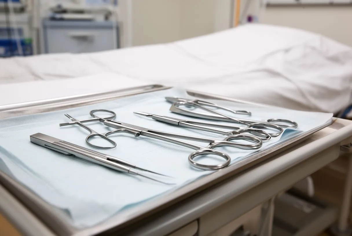
Maintaining sterile field surgery involves a continuous, step-by-step process that begins long before the patient enters the operating room and continues until they are safely transferred to the intensive care unit. The first step is the physical preparation of the room itself, which includes terminal cleaning of all surfaces and ensuring the air handling systems are functioning correctly to filter out airborne particles. Once the room is prepared, the surgical instruments, which have been sterilized in specialized autoclaves, are carefully unwrapped and arranged on a sterile back table. The scrub nurse meticulously organizes these tools so that the surgeon can access them without breaking the sterile perimeter.
What are the key phases of sterile maintenance? The process can be broken down into several distinct phases that the surgical team monitors closely. The team follows a strict checklist to ensure no step is missed.
- Pre-operative preparation: The patient's skin is cleaned with a specialized antiseptic solution, and the team performs a comprehensive surgical scrub.
- Intra-operative vigilance: Continuous monitoring of the sterile field is maintained, with immediate replacement of any contaminated instrument.
- Environmental control: Room doors remain closed, and foot traffic is minimized to prevent airborne contamination from entering the surgical zone.
During the procedure, the team must remain acutely aware of their surroundings. If a surgeon is performing a balloon angioplasty or a bypass, they may need to reposition their hands or the operating table. Every adjustment is coordinated so that no part of the sterile team crosses into the unsterile zone. For example, if a surgeon experiences arm pain or fatigue during a long procedure, they must step back carefully, keeping their hands in view, while the assistant takes over. The scrub nurse acts as the gatekeeper, constantly observing for any accidental touches and passing instruments in a way that keeps them within the sterile boundary. This unwavering attention to detail is what makes maintaining the field a dynamic and demanding responsibility.
How Is Sterile Draping Heart Surgery Performed?
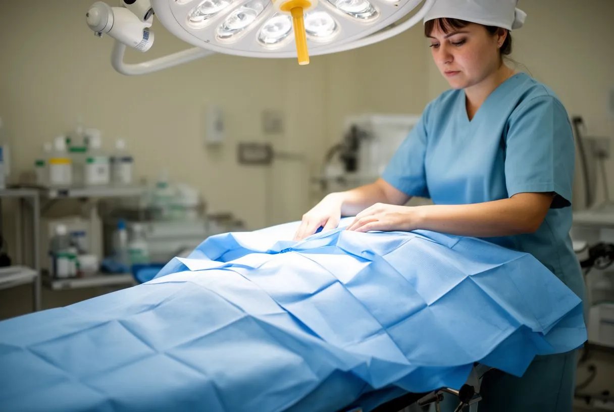
Sterile draping heart surgery is the physical process of covering the patient and the operating table with specialized sterile barriers to create a continuous, microbe-free zone. After the patient's skin is prepped with an antiseptic solution, the surgical team begins draping. They start by placing a sterile towel around the edges of the planned incision site, typically down the center of the chest. Then, large, adhesive-backed plastic drapes are applied directly to the skin. These drapes serve a dual purpose: they secure the sterile barrier to the patient and prevent any moisture or bacteria from migrating from the unprepared skin into the surgical wound.
How do drapes accommodate complex cardiac anatomy? Modern surgical drapes are highly engineered to meet the specific needs of cardiac operations. They often feature clear, plastic windows that allow the anesthesiologist to monitor the patient while keeping the surrounding areas covered. Research published by Frontiers highlights how advanced material science has improved these barriers, making them more resistant to fluid penetration and tearing. For instance, if a patient is being treated for atrial flutter, an irregular heart rhythm, the drapes must allow access to the chest for defibrillation pads while maintaining a continuous sterile field around the primary incision. The drapes are layered systematically, ensuring that any instrument dropped outside the draped area is immediately recognized as contaminated.
The application of the drapes is a highly choreographed routine. The team members handle the drapes only by their folded edges, carefully unfolding them outward without allowing the material to drag across non-sterile surfaces. Once the drapes are in place, they form a definitive boundary. The area beneath the drapes, including the patient's body and the operating table, becomes part of the unsterile zone, while the top surface of the drapes is entirely sterile. This clear demarcation allows the surgeon to work with confidence, knowing that as long as their hands and instruments remain on the drape's upper surface, the risk of contamination is minimized. Any tear or gap in the draping is addressed immediately by applying additional sterile patches.
What Are the Surgical Site Infection Prevention Cardiac Protocols?
Surgical site infection prevention cardiac protocols encompass a comprehensive set of guidelines that extend beyond the operating room. These protocols begin in the preoperative clinic, where patients are screened for existing infections, such as a urinary tract infection or a skin condition, that could compromise their surgery. If an infection is found, the operation is typically postponed until it is resolved. Patients are also advised to wash with a special antibacterial soap in the days leading up to the procedure to reduce the natural bacterial load on their skin. These proactive steps are essential because the patient's own flora is the most common source of surgical site infections.
What happens during the critical intra-operative window? One of the most important protocols is the timely administration of prophylactic antibiotics. The anesthesiologist delivers these antibiotics intravenously within one hour before the surgical incision is made. This ensures that the patient's tissues are saturated with the medication, providing a strong defense against any bacteria that might be introduced during the operation. Even with the most stringent sterile techniques, microscopic breaches can occur, and the antibiotics act as a fail-safe. If the surgery extends beyond the antibiotic's effective duration, additional doses are administered to maintain the protective blood levels throughout the procedure.
Another crucial aspect of these protocols is temperature and glucose control. Patients are actively warmed using specialized blankets to prevent hypothermia, which can impair the immune system and increase the risk of infection. Blood sugar levels are carefully managed, as elevated glucose can promote bacterial growth and impair wound healing. In the event of a cardiac arrest during the procedure, the team uses a sterile automated external defibrillator system to restore a normal heart rhythm without compromising the sterile field. Every element of the protocol, from the air temperature in the room to the patient's metabolic state, is optimized to support the body's natural defenses and the physical sterile barriers.
How Does Aseptic Technique Cardiovascular Surgery Protect Patients?
Aseptic technique cardiovascular surgery protects patients by providing a systematic framework that minimizes the risk of microbial contamination at every stage of the procedure. Unlike sterilization, which is the complete destruction of all microorganisms, aseptic technique is the method used to prevent the transmission of those organisms to the patient. It is a comprehensive philosophy that governs how the operating room is set up, how equipment is handled, and how the surgical team interacts. By adhering strictly to these principles, the team creates a protective bubble around the patient's open chest, ensuring that the complex repair of the heart proceeds without the complication of infection.
How does this technique manage invasive lines? During cardiac surgery, the body is supported by various invasive lines and tubes that monitor vital signs and deliver medications. An arterial line is often placed in the wrist to provide real-time blood pressure readings, while large intravenous catheters are inserted in the neck to deliver fluids and anesthetics. The aseptic technique dictates that these lines are inserted using maximal sterile barriers, including full draping and sterile gloves, much like the main surgical procedure. The insertion sites are continuously monitored throughout the operation to ensure they remain secure and free from contamination, as these lines provide a direct pathway to the bloodstream if not managed correctly.
The protection offered by aseptic technique also extends to the handling of surgical specimens. If a piece of tissue or a valve is removed for analysis, it is immediately passed off the sterile field in a controlled manner to prevent any back-contamination. Sometimes, patients present with an atypical infarction, a heart attack that does not show classic symptoms, requiring urgent exploration. Even in these high-pressure, time-sensitive situations, the surgical team does not abandon aseptic principles. They may move faster, but the fundamental rules of maintaining the sterile boundary remain intact. This unwavering commitment ensures that patients receive the safest possible care, regardless of the clinical urgency.
What Is the Surgical Scrub Protocol Cardiac Teams Follow?
The surgical scrub protocol cardiac teams follow is a rigorous, timed process designed to remove as many microorganisms as possible from the hands and forearms before donning sterile gowns and gloves. This protocol has evolved over decades, moving from traditional brush scrubbing to modern waterless antiseptic rubs. The goal is to reduce the resident bacterial flora on the skin to an absolute minimum. The team enters a designated scrub area outside the operating room, where they clean under their fingernails and then apply a specialized antiseptic solution, working it thoroughly over their hands and arms up to the elbows for a specific duration, ensuring no area is missed.
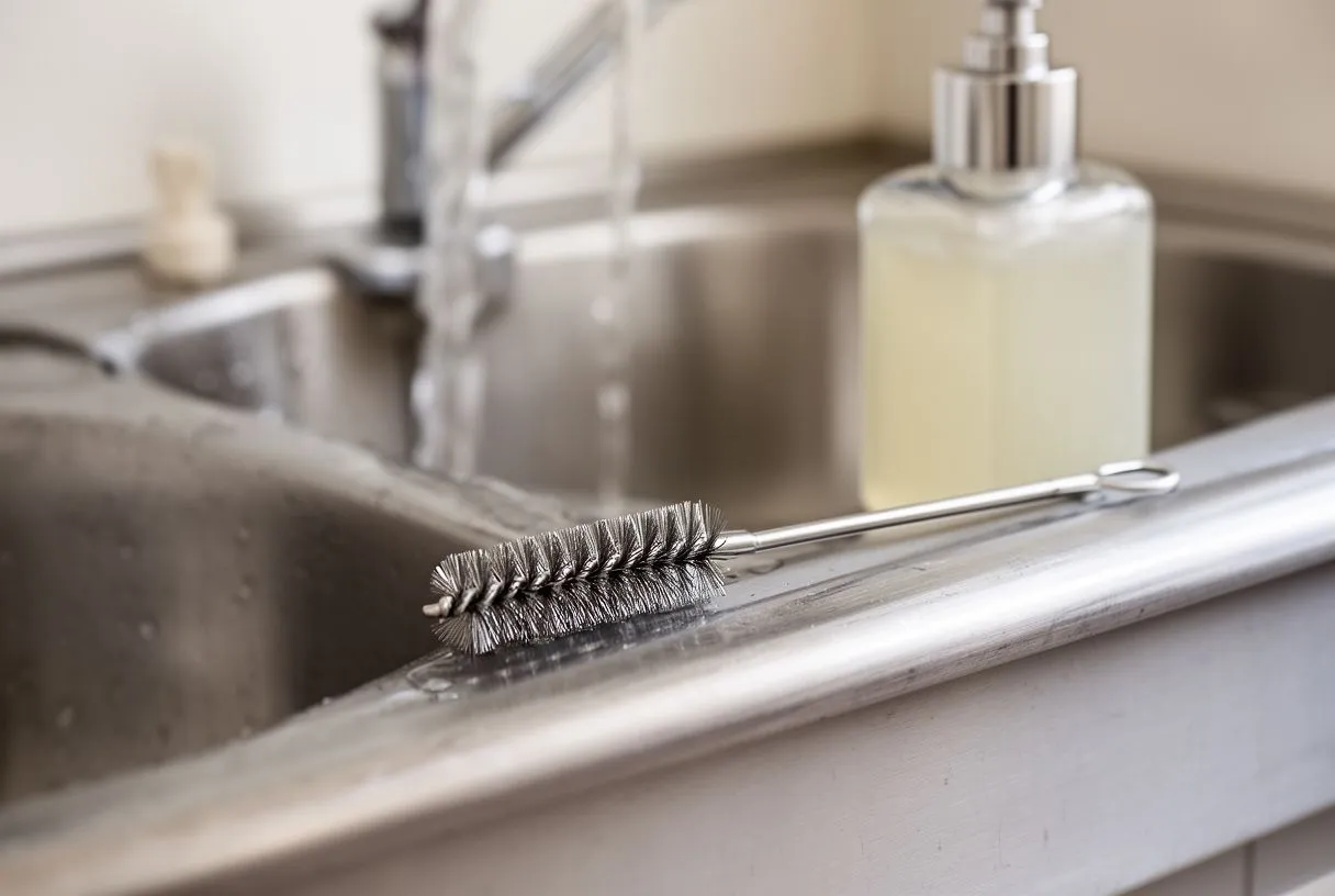
What are the critical elements of the scrub routine? The effectiveness of the scrub relies on both the chemical agent used and the mechanical action of the rubbing. Traditional protocols used a sponge with an iodine-based solution, scrubbing for five to ten minutes. Modern protocols often use alcohol-based chlorhexidine rubs, which are just as effective and gentler on the skin, reducing the risk of dermatitis that can harbor bacteria.
- Chlorhexidine gluconate: Provides long-lasting antimicrobial activity, continuing to kill bacteria for hours after the scrub is complete.
- Povidone-iodine: A broad-spectrum antiseptic that remains a reliable option, particularly for surgeons with specific skin sensitivities.
- Contact time: The solution must remain wet on the skin for the specified duration to ensure maximum bacterial reduction before drying.
After the scrub, the team enters the operating room with their hands held above their elbows, allowing water to drip away from the clean hands. They use a sterile towel to dry their hands, wiping from the fingertips down to the elbows without retracing their steps. Then, they are assisted into a sterile gown and gloves. The process of gloving is done carefully so that the outside of the gloves, which will touch the patient and the sterile field, never comes into contact with the bare skin. Even with patients who have high levels of apolipoprotein B, a protein associated with cardiovascular risk, the fundamental scrub protocol remains unchanged, as it is the cornerstone of preventing surgical site infections for every patient, regardless of their underlying metabolic profile.
What Are the Operating Room Contamination Risks During Cardiac Bypass?
Operating room contamination risks during cardiac bypass are heightened due to the complexity and duration of the procedure, as well as the specialized equipment required. Cardiopulmonary bypass, often called a heart-lung machine, takes over the function of the heart and lungs, allowing the surgeon to operate on a still, bloodless field. This machine requires a network of large tubes, pumps, and oxygenators. The setup and management of this massive circuit present multiple opportunities for contamination if not handled with extreme care. The perfusionist, who operates the heart-lung machine, must work in close coordination with the surgical team to ensure the circuit remains completely sterile.
How is the bypass circuit kept sterile? The components of the heart-lung machine that come into contact with the patient's blood, such as the arterial cannula and the tubing, are single-use, pre-sterilized, and sealed in packaging. The perfusionist assembles these components using strict aseptic technique on a separate sterile table. When connecting the cannula to the patient's blood vessels, the surgeon works meticulously to ensure the open ends of the tubes do not touch any non-sterile surfaces. Even in advanced setups like awake ECMO, where a patient may remain conscious during extracorporeal membrane oxygenation, the connection points are treated as critical sterile zones. Any breach in this circuit can introduce bacteria directly into the bloodstream, leading to immediate and severe systemic infection.
Another risk during bypass is the potential for contamination from the prolonged open wound. Cardiac bypass surgeries can last several hours, increasing the time the internal tissues are exposed to the operating room environment. To mitigate this, the surgical team frequently irrigates the wound with sterile saline to wash away any settling particles. The air handling system in a cardiac operating room is also designed to provide positive pressure and laminar flow, directing filtered air upward and away from the surgical site. These engineering controls are vital, as even a small amount of airborne contamination settling into the chest cavity can lead to devastating consequences, a risk profile sometimes categorized under clinical trial codes like ARISTOTLE to standardize reporting and management of complex surgical complications.
How Is Heart Surgery Wound Infection Prevention Managed After Discharge?
Heart surgery wound infection prevention does not end when the patient leaves the operating room; it is a critical focus throughout the hospital stay and after discharge. Before a patient goes home, the nursing team provides comprehensive education on how to care for the incision site. Patients are taught to inspect their chest wound daily for signs of redness, swelling, or unusual drainage. They are instructed to wash their hands thoroughly before touching the area and to keep the incision clean and dry. These practical steps empower patients to take an active role in preventing an infection that could compromise their surgical recovery.

What lifestyle factors influence wound healing? Postoperative infection prevention is heavily influenced by the patient's overall health and lifestyle. Proper nutrition, particularly adequate protein intake, is essential for tissue repair. Managing underlying conditions like diabetes is crucial, as high blood sugar impairs healing. Patients are often advised to continue taking medications like aspirin, which prevents blood clots and supports overall cardiovascular health, but they must follow their surgeon's specific instructions regarding all their prescriptions. They are also warned to avoid activities that could strain the chest muscles or expose the healing wound to dirt, such as gardening or heavy lifting, until the surgeon grants clearance. Understanding the anatomy, such as the location of the atrium or the graft sites, helps patients appreciate why protecting the chest wall is so important.
The final phase of wound infection prevention involves knowing when to seek medical help. Patients are given clear guidelines on warning signs that require immediate attention, such as a fever above a certain threshold, increased pain at the incision site, or a wound that begins to separate. Early intervention is key to treating a superficial infection before it progresses into a deeper, more serious complication. We encourage you to discuss your specific risks and recovery plan with a qualified cardiac team. Book a consultation with our practice to receive personalized guidance on navigating your heart surgery journey safely and effectively, ensuring your long-term health and well-being.






