

Thoracic Aorta Dilation: When It Needs Surgery
Thoracic aorta dilation can be silent until it becomes dangerous.
What Is Aortic Dilation and Why Does It Happen?
Aortic dilation is a gradual widening of the aorta, the largest artery in your body, which carries oxygen-rich blood from the heart to every organ. When the aortic wall stretches beyond its normal diameter, the vessel becomes thinner and weaker over time. This widening is not a disease by itself — it is a sign that something is changing the structural integrity of the aortic wall, whether from inherited connective tissue conditions, long-standing high blood pressure, or age-related degeneration. Understanding what drives this process is the first step in deciding whether watchful monitoring or surgery is the right path for you.
Where in the aorta does dilation typically occur? The most common site is the ascending aorta, the segment that rises directly from the left ventricle and curves toward the head. Dilation can also develop at the aortic root, where the valve meets the vessel, or in the aortic arch and descending thoracic aorta. Each location carries different implications for symptoms, monitoring, and surgical approach. When dilation involves the root, it often stretches the valve opening as well, which can cause leakage — a condition your surgeon will evaluate carefully before recommending a repair strategy.
How fast does the aorta dilate? Growth rates vary from person to person, but most thoracic aortas expand by less than one millimetre per year. A rapid increase — for example, more than five millimetres in twelve months — is a red flag that prompts closer imaging or earlier surgical consideration. Published cardiovascular research catalogued through DOAJ shows that growth rate and absolute diameter are the two variables cardiologists weigh most heavily when timing intervention. The key message is that dilation is usually slow and predictable, which is why regular imaging gives your team the ability to act before the vessel becomes dangerous.
What Are the Symptoms of Aortic Dilation?
Most people with aortic dilation feel completely well and discover the condition incidentally during a chest X-ray, echocardiogram, or CT scan ordered for another reason. This is one of the challenges of managing the condition: the vessel can grow silently for years without producing any warning signs. When symptoms do appear, they tend to reflect either compression of nearby structures or, more urgently, a tear in the aortic wall. Knowing the difference between vague discomfort and a true emergency helps you and your family respond appropriately.
What does aortic dilation feel like when symptoms begin? Patients may notice a deep, pressing chest pain that can radiate to the back, between the shoulder blades, or into the neck. Unlike the crushing central chest pressure of a classic heart attack, this pain is often described as tearing or ripping, and it may shift position as it progresses. Some people also experience hoarseness from pressure on the recurrent laryngeal nerve, a persistent cough, or difficulty swallowing if the dilated segment presses against the oesophagus or airway.
When should you go to the emergency department? Sudden, severe chest or back pain that reaches maximum intensity within moments is a medical emergency — it may indicate an acute aortic dissection, a tear in the inner layer of the vessel that allows blood to split the wall. Related warning events such as atypical infarction patterns on an electrocardiogram, or arm pain that appears alongside chest symptoms, should never be dismissed. If you have a known dilated aorta and develop any of these signs, call emergency services immediately rather than waiting for your next scheduled appointment.
What Causes the Aorta to Dilate?
The aorta dilates when the smooth muscle and elastic fibres in its wall weaken and can no longer resist the force of each heartbeat. Several factors can drive this weakening, and in many patients more than one factor is at play. Long-standing hypertension is the single most common contributor: every systolic pulse pounds against the aortic wall, and over decades the vessel gradually stretches. Understanding your personal risk profile helps your cardiac team tailor both monitoring intervals and blood-pressure targets.

Can inherited conditions cause aortic dilation? Yes — connective tissue disorders such as Marfan syndrome, Loeys-Dietz syndrome, and vascular Ehlers-Danlos syndrome alter the structural proteins that give the aorta its elasticity. People with a bicuspid aortic valve — a congenital condition where the valve has two leaflets instead of three — also have a higher lifetime risk of dilation, even if the valve itself functions normally. In these families, screening first-degree relatives with an echocardiogram is standard practice because dilation can appear at a younger age and grow more quickly.
Do lifestyle and age-related changes contribute? Absolutely. Atherosclerosis — the buildup of atheromatous plaque within the arterial wall — reduces the flexibility of the aorta and is linked to higher dilation risk. Elevated levels of apolipoprotein B, a cholesterol-carrying particle, are associated with more aggressive plaque formation. Smoking accelerates wall degeneration, and poorly controlled blood pressure compounds every other risk factor. The following list summarises the most significant contributors your team will evaluate:
- Long-standing hypertension: chronic high pressure stretches the wall with every heartbeat.
- Genetic connective tissue disorders: Marfan, Loeys-Dietz, and Ehlers-Danlos weaken structural fibres.
- Bicuspid aortic valve: congenital valve abnormality raises dilation risk independent of valve function.
- Smoking and atherosclerosis: plaque buildup stiffens the wall and accelerates enlargement.
- Advancing age: natural loss of elastic fibres over decades slowly widens the vessel.
How Is Aortic Dilation Diagnosed?
Diagnosis begins with imaging. Because the aorta sits deep in the chest, it cannot be assessed by physical examination alone, though your doctor may detect a murmur during auscultation — listening to the chest with a stethoscope — if the dilation has affected the aortic valve. The standard first test is a transthoracic echocardiogram, a painless ultrasound that visualises the aortic root and ascending aorta and measures the diameter to the nearest millimetre. If the echo suggests dilation beyond approximately 4.0 centimetres, your cardiologist will arrange more detailed cross-sectional imaging.
Which imaging tests give the clearest picture? A computed tomography angiogram (CTA) or magnetic resonance angiogram (MRA) provides precise three-dimensional measurements of the entire thoracic aorta. These scans show the exact diameter at each segment, the presence of any thrombus or plaque, and whether the vessel is tortuous or straight. Most guidelines recommend a baseline CTA or MRA once dilation is confirmed, followed by serial imaging at intervals of six to twenty-four months depending on size, growth rate, and underlying cause. The diameter thresholds that guide surgical timing are based on these measurements, so consistency in imaging technique matters.
What else will your team check during evaluation? Beyond imaging, your cardiologist will review your blood pressure control, order lipid studies including apolipoprotein B, and assess valve function. If a genetic syndrome is suspected, genetic counselling and testing may be recommended, and first-degree relatives may be screened with their own echocardiograms. The goal is to build a complete picture — not just the size of the aorta today, but the trajectory it is on and the factors you can modify to slow that trajectory.
When Does Aortic Dilation Need Surgery?
The decision to operate is driven primarily by the maximum diameter of the aorta, its rate of growth, and the presence of symptoms. For most adults without a genetic connective tissue disorder, the widely accepted surgical threshold is 5.5 centimetres for the ascending aorta or aortic root. For patients with Marfan syndrome, Loeys-Dietz syndrome, or a bicuspid aortic valve, the threshold is lower — typically 5.0 centimetres — because these vessels carry a higher risk of dissection at smaller sizes. Your surgical team will also factor in EuroSCORE II, a validated risk calculator that estimates operative mortality based on age, comorbidities, and cardiac function.
What if your aorta is growing rapidly but has not reached the threshold? A documented growth rate exceeding five millimetres per year is an independent indication for surgery even if the absolute diameter is below the standard cut-off. Likewise, any dilation accompanied by chest or back pain attributed to the aorta warrants urgent surgical evaluation regardless of size. A case report published in Revista Brasileira de Cirurgia Cardiovascular illustrates how progressive dilation and complications from prior coronary stenting can converge to produce end-stage ischaemic cardiomyopathy, underscoring why timely aortic intervention matters before secondary damage accumulates.
Does valve leakage change the timing? Yes. When aortic root dilation pulls the valve leaflets apart and produces moderate to severe aortic regurgitation — backward flow of blood into the left atrium and ventricle — surgery is recommended even at smaller diameters. The rationale is that repairing or replacing both the aorta and the valve in a single operation prevents progressive heart chamber enlargement and protects long-term cardiac function. Your surgeon will discuss whether a valve-sparing root repair is feasible or whether valve replacement is the safer option for your anatomy.
What Are the Surgical Options for Aortic Dilation?
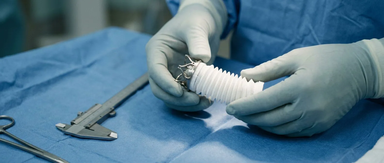
Surgery for aortic dilation is not a single procedure — it is a family of operations tailored to the segment of the aorta involved and the condition of the valve. The most common operation for ascending aorta and root dilation is the open repair, performed through a median sternotomy — an incision along the front of the chest — with the patient on cardiopulmonary bypass, which temporarily takes over the work of the heart and lungs so the surgeon can work on a still, bloodless field. Your team will explain which specific reconstruction fits your anatomy and why.
What is a valve-sparing aortic root replacement? In carefully selected patients whose valve leaflets are healthy despite the dilated root, the surgeon can replace the diseased wall of the aortic root with a synthetic graft while reattaching your own valve inside it — a technique often called the David or Yacoub procedure. This avoids lifelong anticoagulation, the blood-thinning medication required when a mechanical valve is used. When the valve itself is diseased or congenitally abnormal, a combined aortic root and valve replacement using a biological or mechanical valved conduit may be the better choice, and the surgeon will discuss the trade-offs of each.
What about the descending thoracic aorta? When dilation involves the segment beyond the arch, surgeons may offer an open surgical repair or a less invasive endovascular approach, in which a fabric-covered stent graft is delivered through a small incision in the groin and deployed inside the aorta to reinforce the weakened wall. During open repairs, an arterial cannula and arterial line provide precise pressure monitoring throughout the procedure, and autologous blood — your own blood, collected and reinfused during surgery — reduces the need for donor transfusion. The right approach depends on the location, length, and geometry of the dilation, as well as your overall fitness for open surgery.
What Is the Recovery After Aorta Surgery?
Recovery from elective aortic surgery follows a predictable but demanding path. Most patients spend one to two days in the intensive care unit, where monitors track every heartbeat, and then move to a standard cardiac ward for another four to seven days. During the first week, the priorities are pain control, gentle mobilisation, and lung exercises to prevent pneumonia. By the time you go home — typically seven to ten days after surgery — you should be able to walk short distances, climb a few steps with assistance, and manage your own medications.
What is the timeline for returning to normal activities? Full recovery generally takes six to twelve weeks. Most surgeons advise no driving for four to six weeks, avoidance of heavy lifting (anything over five kilograms) for eight to twelve weeks, and a gradual return to exercise guided by a cardiac rehabilitation programme. Your sternum needs about three months to heal completely, so contact sports and vigorous upper-body training must wait. Many patients are surprised by how fatigued they feel in the first month — this is normal, and stamina rebuilds steadily with daily walking and structured rehabilitation.
What medications will you need afterwards? Nearly all patients leave hospital on a beta-blocker or similar blood-pressure medication to protect the repaired aorta and any remaining native aortic tissue from high pulse pressure. If you received a mechanical valve, you will also take warfarin, and your team will explain how this interacts with other common cardiac drugs — for example, the anticoagulation trial known as ARISTOTLE helped establish modern dosing and monitoring standards. Some patients also continue aspirin for coronary protection if they have coexisting artery disease managed with balloon angioplasty or stents.
Are There Alternatives to Surgery for Aortic Dilation?
When the aorta has not reached surgical size and is growing slowly, the alternative to surgery is careful medical management combined with serial imaging. This is not a passive approach — it is an active strategy designed to reduce wall stress and slow progression. Blood pressure control is the cornerstone: guidelines recommend keeping systolic pressure below 130 mmHg in most patients, and below 120 mmHg in those with genetic syndromes. Beta-blockers are the first-choice medication because they lower both pressure and the force of each heartbeat, directly reducing the mechanical load on the aortic wall.
Can lifestyle changes actually slow dilation? Yes, within realistic limits. Stopping smoking is the single most impactful modifiable step — nicotine and combustion by-products damage the endothelial lining and accelerate wall stiffness. Maintaining a healthy weight, avoiding heavy isometric weightlifting, and managing stress all contribute to lower blood pressure. Your cardiologist may also address other cardiovascular risk factors: patients with atrial flutter or other rhythm disturbances need rate control to prevent irregular, forceful contractions, and those with extensive atheromatous plaque may benefit from statin therapy to stabilise existing lesions.
Is there a role for catheter-based treatments before surgery? Not for the aorta itself — dilation of the ascending aorta cannot be treated with stents or balloons in the way coronary arteries can. However, patients who have coexisting coronary disease may undergo balloon angioplasty or balloon aortic valvuloplasty as bridge procedures while awaiting definitive aortic surgery. A study comparing procedural results and long-term outcomes of percutaneous coronary intervention for in-stent restenosis versus de novo chronic total occlusion, available through Dove Medical Press, highlights how managing coronary blockages in the setting of aortic disease requires careful coordination between cardiologist and surgeon to avoid delays in definitive repair.
What Are the Risks of Aortic Dilation Surgery?
Elective aortic surgery at a high-volume centre carries a perioperative mortality risk generally between one and three percent for straightforward ascending aorta replacement, rising to four to six percent for more complex root or arch repairs. These figures come from large registries and should be interpreted alongside your individual EuroSCORE II calculation, which accounts for age, renal function, lung disease, and previous cardiac operations. The most serious complications include stroke, bleeding requiring reoperation, and infection of the surgical site or the implanted graft material.
How does your team minimise these risks? Meticulous intraoperative technique is paramount. Surgeons use hypothermic circulatory arrest — cooling the body to a low temperature and briefly stopping circulation — to protect the brain during arch repairs. Advanced neuromonitoring tracks brain function in real time. In select centres, awake ECMO — extracorporeal membrane oxygenation managed with minimal sedation — can support patients with marginal cardiac function before or after surgery. Your anaesthesiologist will discuss the plan for anticoagulation reversal, blood conservation with autologous blood recovery, and the small but real risk of needing an arterial graft if coronary bypass is combined with aortic repair.

What are the long-term considerations? Most patients who undergo elective repair return to a full and active life, but the reconstructed aorta and any implanted valve require lifelong follow-up. Annual imaging — usually a CT scan or MRI — confirms that the graft remains intact and that other segments of the aorta are not dilating. If a biological valve was used, it will eventually degenerate and may need replacement; if a mechanical valve was chosen, lifelong anticoagulation monitoring is essential. The following list summarises the key risks your surgeon will discuss during informed consent:
- Stroke: related to air or plaque debris during circulatory arrest; risk roughly 1–3%.
- Bleeding requiring reoperation: more common in complex arch repairs; managed with blood-conservation protocols.
- Cardiac complications: arrhythmias such as atrial flutter, low output syndrome, or need for postoperative circulatory support.
- Graft infection: rare but serious; requires long-term antibiotics and sometimes reoperation.
- Valve-related issues: biological valve degeneration over time or anticoagulation management for mechanical valves.
How to Live Well With Aortic Dilation
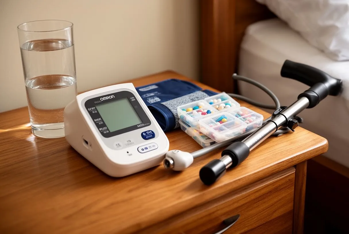
Living well with aortic dilation — whether before surgery, after repair, or during long-term monitoring — means building habits that protect your aorta and staying alert to changes that warrant a call to your doctor. The foundation is blood pressure control: measure it regularly, take your medications consistently, and keep a log to share at every appointment. If you have been prescribed a beta-blocker, an angiotensin receptor blocker, or another agent, stopping it abruptly can cause dangerous pressure spikes, so always discuss changes with your cardiologist first.
What activities should you modify or avoid? Heavy isometric exercise — such as maximal bench presses, deadlifts, or overhead presses — generates sharp spikes in blood pressure that stress the aortic wall and are best avoided. Endurance exercise at moderate intensity, such as brisk walking, cycling, or swimming, is generally encouraged and benefits overall cardiovascular health. If you have an implanted device such as a pacemaker or have been trained to use an automated external defibrillator in an emergency, discuss specific exercise guidelines with your team. In some cases, a balloon-expandable valve or other implant may have unique imaging or activity restrictions your surgeon will outline.
Ultimately, aortic dilation is a condition you can live with confidently when you have the right information and the right team. The decision to monitor, to operate, or to adjust your medications is never made in isolation — it is a conversation between you, your cardiologist, and your cardiac surgeon. If you or a family member have been told your aorta is enlarged, do not wait for symptoms to appear. Contact Cirugía de Corazón to schedule a consultation with Dr Alejandro Molina and his team in Guadalajara, Querétaro, or Hermosillo, and take the next step with expert guidance tailored to your situation.






