Transthoracic Echocardiogram: What to Expect
Learn what a transthoracic echocardiogram procedure involves, from preparation to results.
If your cardiologist has recommended a transthoracic echocardiogram procedure, it is entirely normal to feel a mix of curiosity and apprehension. This non-invasive test is one of the most common and valuable tools in modern cardiology, allowing your medical team to see your heart in motion without making a single incision. For patients and families navigating complex cardiac concerns, understanding exactly what this test involves can transform anxiety into empowered decision-making.
What is a transthoracic echocardiogram procedure
A transthoracic echocardiogram procedure is a sophisticated yet completely non-invasive imaging test that uses high-frequency sound waves to create real-time moving pictures of your heart. According to the European Society of Cardiology, ultrasound remains the cornerstone of cardiovascular diagnostics. By placing a small device called a transducer on your chest, the technician can bounce sound waves off your heart structures, capturing detailed images of the chambers, valves, and blood flow without any radiation or surgical intervention.
If you have reported chest discomfort, unusual shortness of breath, or Arm Pain that radiates from your chest, this test helps determine if the heart is the root cause. Sometimes, an irregular heartbeat detected during an Auscultation, which is the medical term for listening to the heart with a stethoscope, will prompt an echo to rule out structural problems. It is a critical diagnostic tool for catching issues early, before they progress to severe complications.
Understanding this test begins with understanding your own anatomy. The heart is a complex muscular pump divided into four chambers, and a transthoracic echocardiogram procedure allows your cardiologist to watch these chambers contract and relax in real time. It provides a clear view of the pericardium, the sac surrounding the heart, and helps identify any fluid buildup that might be causing symptoms. By giving us a window into the heart's mechanical function, this ultrasound test bridges the gap between a patient's physical symptoms and the underlying anatomical reality driving them.
How does a transthoracic echocardiogram work
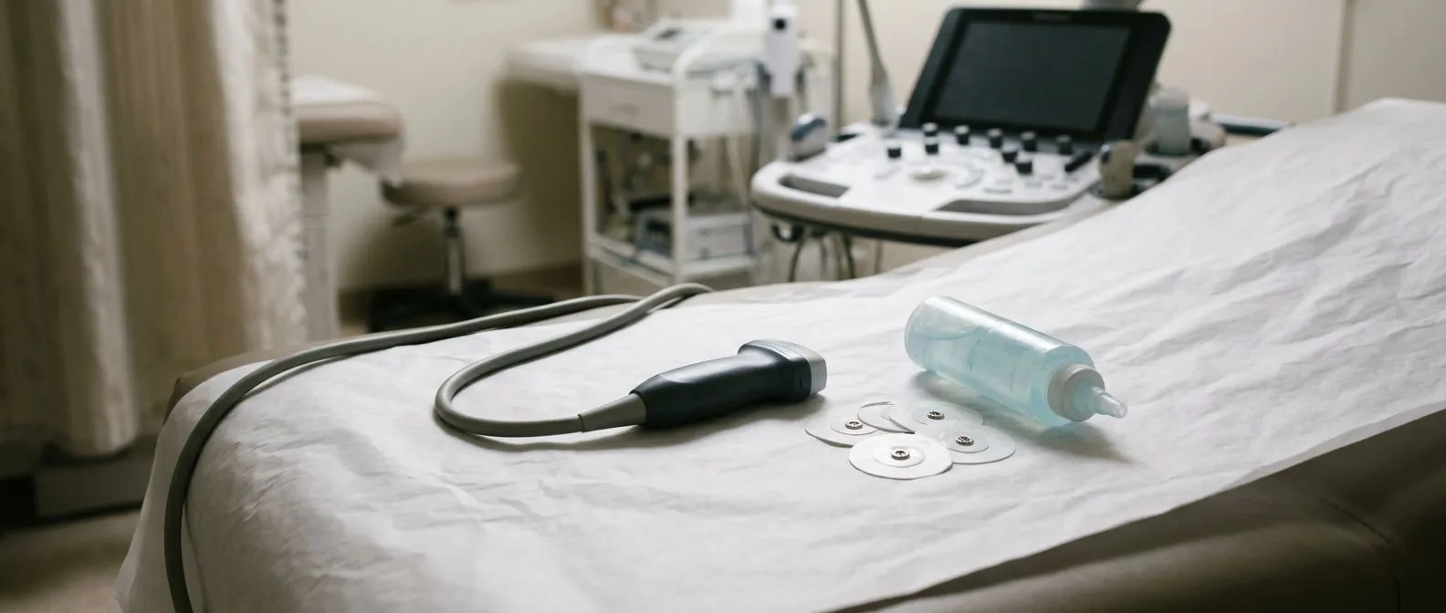
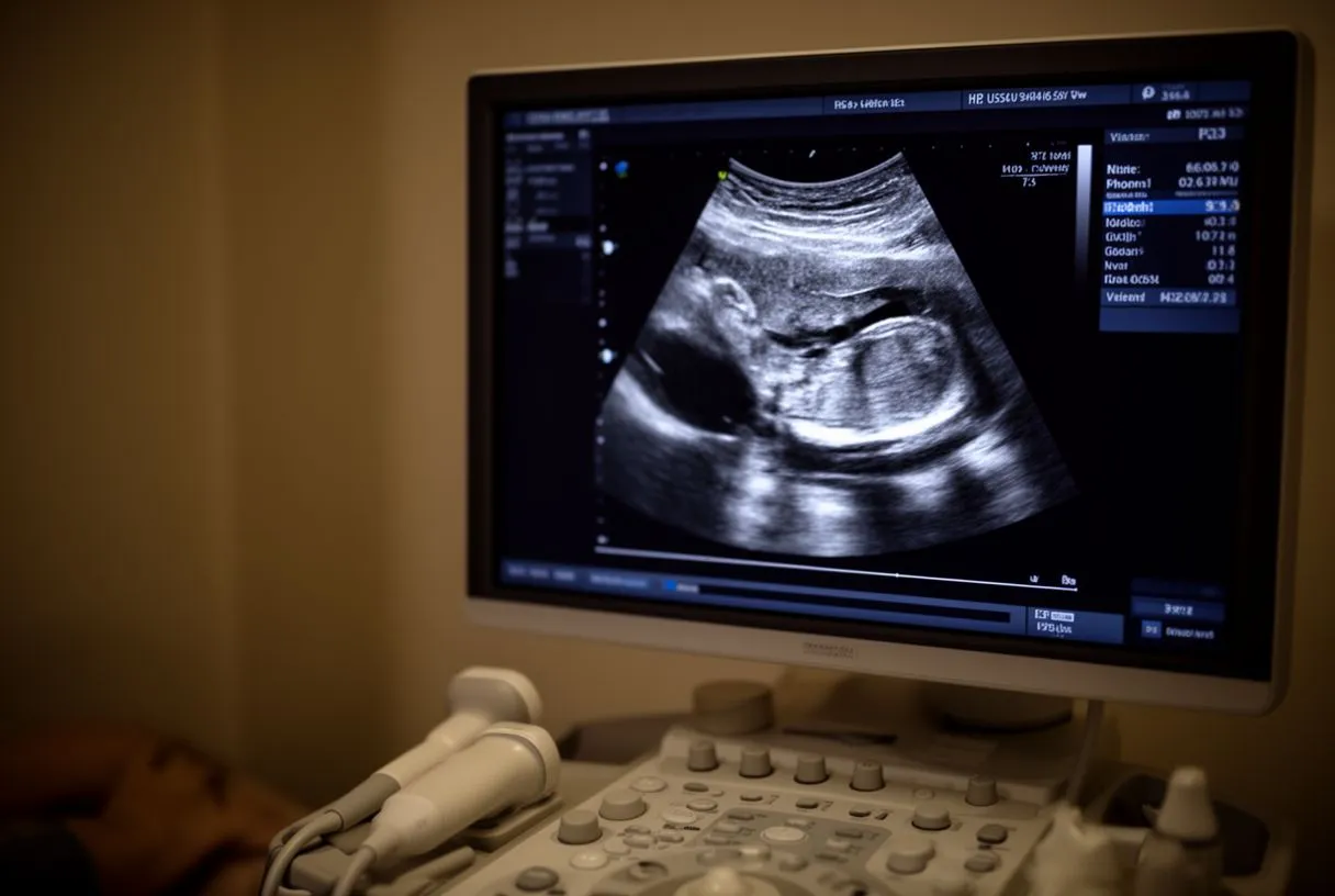
The fundamental technology behind a transthoracic echocardiogram procedure relies on piezoelectric crystals inside the transducer. When electricity is applied to these crystals, they vibrate at incredibly high frequencies, emitting sound waves that travel through the chest wall and into the heart. As these sound waves encounter different densities of tissue—such as muscle, blood, or bone—they bounce back to the transducer, which then translates these returning echoes into detailed digital images displayed on a monitor. A conductive gel is applied to the skin to eliminate air pockets, ensuring the sound waves travel smoothly into the body.
Doppler ultrasound capabilities: Beyond simply showing the physical structure of the heart, the transthoracic echocardiogram procedure utilizes the Doppler effect to measure the speed and direction of blood flow. By analyzing how the frequency of the sound waves changes as they bounce off moving red blood cells in the Ascending Aorta, the machine can determine if blood is flowing normally or if there is turbulence. This is crucial for identifying leaky valves or narrowed passages, as turbulent flow produces distinct color patterns on the screen.
The entire system is designed to capture the heart in motion rather than static images. Because the heart beats roughly 60 to 100 times per minute, a transthoracic echocardiogram procedure records continuous loops of video. This allows the interpreting physician to evaluate how the heart muscle relaxes and fills with blood, a phase known as diastole, and how it forcefully pumps blood out, known as systole. If you experienced an Atypical Infarction, an unusual presentation of a heart attack, the echo will reveal how much muscle damage occurred.
Why you might need a transthoracic echocardiogram procedure
There are many reasons a physician might order a transthoracic echocardiogram procedure, most commonly stemming from new or unexplained symptoms. If your doctor suspects an Atheromatous Plaque is affecting your coronary arteries, this test evaluates how the heart muscle is compensating. By assessing the heart's overall pumping function, your medical team can determine if reduced blood flow has damaged the heart muscle, which is a crucial step in diagnosing ischemic heart disease.
Monitoring existing conditions: If you have already been diagnosed with a heart condition, regular echocardiograms are essential for tracking the disease's progression. For example, if you have an enlarged Atrium due to an arrhythmia like Atrial Flutter, your cardiologist needs to monitor the chamber's size and pumping efficiency over time. Clinical trials such as ARISTOTLE have shaped how these arrhythmias are managed, and ongoing echocardiographic monitoring helps confirm that those management strategies are working.
Finally, a transthoracic echocardiogram procedure is frequently used as a preoperative assessment. Before undergoing any major surgery, your medical team must ensure your heart is strong enough to handle the stress of anesthesia and the operation itself. By evaluating your cardiac function beforehand, surgeons can mitigate risks and plan the safest surgical approach. This proactive evaluation is standard protocol and provides both the patient and the surgical team with necessary peace of mind.
How to prepare for your transthoracic echocardiogram
One of the greatest advantages of a transthoracic echocardiogram procedure is that it requires almost no specialized preparation. Unlike procedures that require fasting or bowel preparation, you can generally eat, drink, and take your usual medications before your appointment. To ensure the smoothest experience, keep these practical points in mind:
- Wear loose clothing: You will need to undress from the waist up, so a two-piece outfit is highly recommended.
- Take your usual medications: Unless specifically instructed otherwise, continue your normal schedule, including Aspirin or other blood thinners.
- Eat normally: There is no fasting required for a standard echo, so you can have your regular meals.
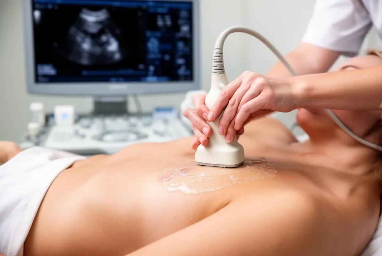
What to expect regarding the gel: The ultrasound gel used during the transthoracic echocardiogram procedure is water-based and will not stain your clothing, but it can feel cool and slightly wet. Because the technician will need to place the transducer in several different positions across your chest, the gel may spread to your neck or upper abdomen. It is easily wiped off at the end of the test, but knowing about it beforehand helps prevent any surprise or discomfort during the imaging process.
Before you arrive, take a moment to write down any questions you have for your technician or physician, though the technician performing the test typically cannot discuss the results with you in real-time. You should also bring a list of your current medications and any previous cardiac imaging reports if you have them. Arriving a few minutes early will allow you to complete the necessary registration paperwork and take a few deep breaths to relax before the test begins, ensuring your heart rate is as calm and natural as possible.
What happens during the transthoracic echocardiogram procedure
Once you are in the exam room, you will be asked to lie on your back and then roll slightly onto your left side. This specific positioning is crucial for a transthoracic echocardiogram procedure because it brings your heart closer to the chest wall. When you lie on your left side, the heart naturally falls forward, resting against the sternum and providing the ultrasound transducer with the clearest, least obstructed pathway. The technician will dim the lights in the room to better see the images on the monitor.
The technician will apply the conductive gel and place the transducer firmly against your chest in various locations. Unlike an Arterial Line, which continuously monitors blood pressure from inside an artery, the TTE uses external gel and sound waves to gather its data. During the transthoracic echocardiogram procedure, you may be asked to hold your breath for a few seconds at a time, take deep breaths, or change positions slightly. These maneuvers help move the lungs out of the way and provide sharper images of the heart's upper chambers and major vessels.
The entire imaging process usually takes between 30 and 45 minutes. The transthoracic echocardiogram procedure is entirely painless, though the pressure from the transducer might feel slightly uncomfortable if the technician has to press hard to see past your ribs. If a contrast agent is needed to improve image quality—usually a saline solution injected into an IV line—it may cause a brief, metallic taste or a warm flushing sensation, but this fades quickly and is completely normal.
What the echocardiogram shows about your heart valves
Your heart contains four valves that ensure blood flows in one direction: the mitral, tricuspid, aortic, and pulmonary valves. A transthoracic echocardiogram procedure provides exquisite detail of these structures, showing whether they open fully and close tightly. If you have a history of an Arterial Graft from a previous bypass surgery or an Autograft used in a valve repair, the echo is the primary method used to ensure the surgical sites are healing and functioning correctly. It is also vital for monitoring artificial implants like a Balloon-expandable Valve used in TAVR.
Assessing stenosis and regurgitation: The Doppler component of the transthoracic echocardiogram procedure is especially useful for evaluating valve diseases. If a valve is narrowed, a condition known as stenosis, the ultrasound detects high-velocity, turbulent blood flow pushing through the tight opening. If a valve is leaking, called regurgitation, the color Doppler will display blood flowing backward into the chamber it just left. These precise measurements help your cardiologist determine if a valve needs surgical repair or replacement.
In addition to the valves themselves, the echo evaluates the surrounding structures and chamber sizes. When a valve is diseased, the heart chambers behind it often have to work harder, causing them to enlarge or thicken over time. By measuring the precise dimensions of your heart's chambers during a transthoracic echocardiogram procedure, your doctor can gauge the severity and chronicity of a valve problem. This detailed anatomical mapping dictates whether intervention is required immediately or if watchful waiting is safe for your specific situation.
Understanding the results of your heart ultrasound
When your physician reviews the results of your transthoracic echocardiogram procedure, one of the most critical measurements they look at is the ejection fraction. This is the percentage of blood that leaves the heart's main pumping chamber, the left ventricle, with each beat. A normal ejection fraction is typically between 50% and 70%. A number lower than this range may indicate that the heart muscle is weakened, a condition known as heart failure, which requires specific medical management and sometimes correlates with abnormal blood tests for Apolipoprotein B.
Identifying structural anomalies: The results also detail the physical structure of your heart. The transthoracic echocardiogram procedure can reveal hypertrophy, which is an abnormal thickening of the heart muscle often caused by long-standing high blood pressure. It can also identify the presence of blood clots, particularly in the upper chambers, which is a major concern for patients with irregular heartbeats. If severe arrhythmias are detected, the echo data helps determine if a patient might require an Automated External Defibrillator to prevent sudden cardiac arrest.
Understanding these results requires clinical correlation, meaning your doctor must view the echo findings alongside your symptoms and medical history. For instance, research such as the study on Screening of Multiple Biomarkers Associated With Ischemic Stroke in Atrial Fibrillation highlights how cardiac imaging helps stratify risk in complex patients. The raw data from a transthoracic echocardiogram procedure is most valuable when integrated into the broader picture of your overall cardiovascular health.
Are transthoracic echocardiogram procedures safe
The safety profile of a transthoracic echocardiogram procedure is exceptional. Because it relies entirely on sound waves rather than ionizing radiation, it does not carry the risks associated with X-rays or CT scans. There is no established risk of cellular damage from diagnostic ultrasound, making it safe for pregnant women, infants, and patients who require repeated imaging over many years. The procedure is classified as non-invasive, meaning it does not break the skin or enter the body's cavities.
Absence of radiation exposure: The lack of radiation is a primary reason the transthoracic echocardiogram procedure is the preferred monitoring tool for chronic conditions. Patients who need annual checkups for valve disease or heart failure can undergo dozens of these tests over a lifetime without accumulating radiation exposure. The only discomfort typically reported is the pressure of the transducer against the ribs, which resolves the moment the test is completed. There are no needles, no incisions, and no recovery time required.
The only minor risk involves the use of contrast agents, which are occasionally needed if the standard images are unclear, often due to a thick chest wall or lung disease. The contrast used is usually microbubbles of saline, which are extremely safe and are filtered out of the bloodstream naturally. Rarely, patients might have a mild allergic reaction, but this is highly unusual. Your medical team will thoroughly review your allergy history before the transthoracic echocardiogram procedure begins to ensure absolute safety.
Recovering after a transthoracic echocardiogram procedure
Recovery from a standard transthoracic echocardiogram procedure is immediate. There is no anesthesia, sedation, or physical alteration to the body, meaning you can return to your normal activities the moment you walk out of the exam room. You will simply need to wipe the remaining ultrasound gel off your skin, which can be done with a towel provided by the clinic. You can drive yourself home, return to work, eat a full meal, and engage in exercise without any restrictions.
If you received an IV for contrast enhancement during your transthoracic echocardiogram procedure, the recovery is still minimal. Unlike complex procedures requiring an Arterial Cannula or Awake ECMO for life support, or surgeries that utilize Autologous Blood to minimize donor transfusions, the TTE recovery is instant. The IV will be removed, and a small bandage applied to the puncture site. You might be advised to keep the site dry for a few hours, but otherwise, your day continues uninterrupted.
While physical recovery is instantaneous, the emotional wait for results can be a different matter. The images from your transthoracic echocardiogram procedure are not usually read instantly by a cardiologist; they are often sent to a specialized interpreting physician who generates a formal report. This process can take anywhere from 24 hours to a few days. It is important to remember that the technicians performing the test are not permitted to give you a preliminary diagnosis, so try to remain patient and avoid letting anxiety build while waiting for your official follow-up.

When to discuss your echo results with a cardiac specialist
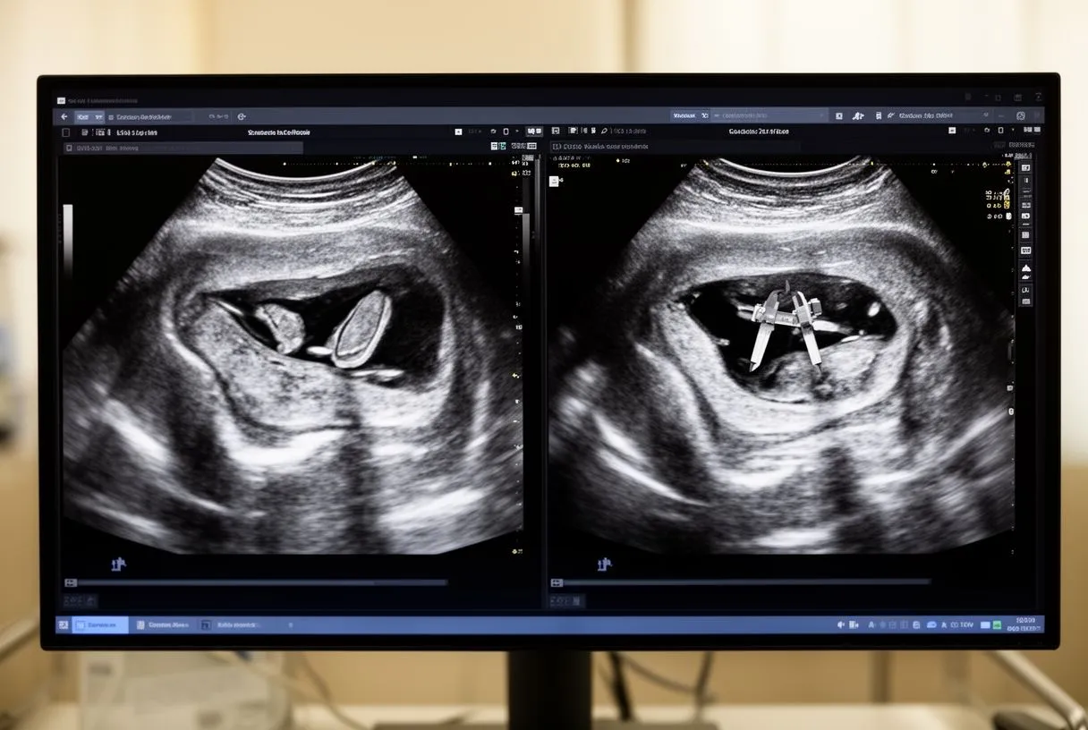
Once the report from your transthoracic echocardiogram procedure is available, the next crucial step is scheduling a consultation with your cardiologist. This appointment is not just to hear a diagnosis; it is an opportunity to understand the "why" behind your symptoms and the "how" of your treatment plan. Your specialist will explain the findings in plain language, showing you the actual images of your heart and pointing out any areas of concern. They will correlate the structural findings with your physical symptoms to build a complete clinical picture.
- Clarify your diagnosis: Ask exactly what the images mean for your specific heart condition.
- Understand your trajectory: Discuss whether your condition is stable, improving, or requires intervention.
- Review medications: Ensure your current prescriptions are optimized based on the new echo data.
Planning the next clinical steps: If the transthoracic echocardiogram procedure reveals an issue, your cardiologist will outline the safest and most effective path forward. Depending on the findings, this could range from simple lifestyle adjustments to treatments like Balloon Angioplasty or a Balloon Aortic Valvuloplasty. For patients managing complex arrhythmias, literature such as the article Rivaroxaban in stroke prevention in patients with non-valvular atrial fibrillation: what facts are important for the clinician? demonstrates how imaging influences blood thinner decisions. Understanding these options requires a guided conversation.
Ultimately, an echo is just data until a skilled physician interprets it for your specific situation. At Cirugía de Corazón, Dr Alejandro Molina and his team are dedicated to translating these complex results into clear, calm, and actionable plans for patients across Mexico. Do not let an abnormal or inconclusive echo result send you into a spiral of internet searches. Instead, book a consultation with a qualified cardiac team to discuss your results and take an informed, confident step toward protecting your heart health.