

Tricuspid Valve Leak: Symptoms and Treatment
Tricuspid valve leak treatment ranges from monitoring to minimally invasive surgery.
A tricuspid valve leak treatment plan usually starts with something simple: an ultrasound of the heart. If you or a family member has just been told the tricuspid valve is leaking, you may be feeling confused and a little anxious. The tricuspid valve sits between the two chambers on the right side of the heart, and it opens with every heartbeat to let blood flow forward. When it leaks, blood flows backward instead, and over time that can tire the heart out. This guide walks you through what a leak means, what symptoms to expect, and which options exist, from monitoring to medication to tricuspid valve leak surgery.
What Is a Tricuspid Valve Leak?
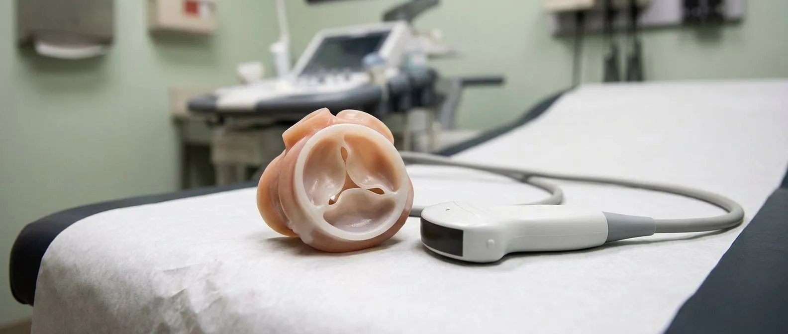
The heart has four valves, and the Tricuspid Valve is one of them. It has three thin leaflets, so it is often described as Tricuspid (three-leaflet) in anatomy texts. Its job is to keep blood moving in one direction, from the right upper chamber into the right lower chamber, which then pumps blood to the lungs. A leak, which doctors call tricuspid regurgitation, means some blood slips backward with each beat.
How common is it? Very common. Small leaks show up on heart ultrasounds in many healthy people and usually mean nothing. The concern begins when the leak becomes moderate or severe, because the right side of the heart then has to work harder to do the same job. According to WebMD, tricuspid regurgitation is one of the more frequent valve findings on routine echocardiograms.
Why the tricuspid valve behaves differently. Unlike valves on the left side of the heart, the tricuspid valve lives in a low-pressure zone. It also anchors to a ring of muscle, the annulus, that stretches easily. When the right heart chamber enlarges for any reason, the ring stretches too, and the leaflets can no longer meet in the middle. That gap is the leak. Understanding this anatomy helps explain why so much of tricuspid valve leak treatment focuses on timing, because early disease often needs watching, not operating.
Tricuspid Valve Leak Symptoms to Watch For
Tricuspid valve leak symptoms are often subtle at first. Because the leak affects the right side of the heart, which handles blood going to the lungs, the signs tend to appear in the body rather than in breathing alone. Many people with mild leakage feel perfectly well and only learn of the problem on a scan done for another reason.
- Swelling: puffiness in the ankles, feet, or legs, caused by fluid building up when blood backs up in the veins.
- Fullness and fatigue: a sense of heaviness, low energy, or discomfort under the ribs, especially on the right side.
- Advanced signs: abdominal swelling, reduced appetite, and noticeable neck vein pulsing, which suggest the leak is affecting circulation.
When symptoms matter. Doctors pay close attention when symptoms begin to limit daily life, because that point often marks the moment the leak is becoming severe. A patient who can no longer walk a few blocks without their legs swelling is telling the heart team something important. We take the time in consultation to explain what the studies actually show, in plain words, so patients know which of these signs apply to them and which do not.
Rhythm changes. A leaky tricuspid valve also stretches the upper right chamber, and that chamber is where atrial fibrillation, a common irregular rhythm, tends to start. Some people first notice a fluttering or racing heartbeat. If you feel a new irregular pulse alongside swelling or fatigue, that combination deserves a prompt cardiology visit rather than waiting for the next annual check.
What Causes Tricuspid Valve Leaks?
Tricuspid valve leak causes fall into two broad groups. The first is primary disease, where the valve leaflets themselves are damaged. The second, and by far the more common, is secondary or "functional" leakage, where the valve structure is fine but the heart around it has changed shape.
Primary causes. These include infection of the valve, called infective endocarditis, which is more common in people who use intravenous drugs or have certain devices. Rheumatic fever, still seen in parts of the world, can scar the valve and cause mixed problems; management of mixed rheumatic tricuspid disease is described in detail in Kardiochirurgia i Torakochirurgia Polska. Rare congenital conditions also exist, such as tricuspid atresia, in which the valve never formed at all and babies need surgery in the first days of life.
Secondary causes. Most leaks in adults are secondary. The right pumping chamber enlarges when the lungs raise blood pressure over years, when the left side of the heart is diseased, or after long-standing rhythm problems. As the chamber grows, it pulls the valve ring outward, and the leaflets drift apart. This is why a leak that appears after another cardiac problem often reflects the bigger picture, not just the valve itself.
Leak after other valve surgery. One frequent scenario is a new leak appearing on the right side after an operation on the left. A leak that develops following mitral surgery is common enough that surgeons plan for it, which we discuss below in its own section.
How an Echocardiogram Measures a Tricuspid Leak
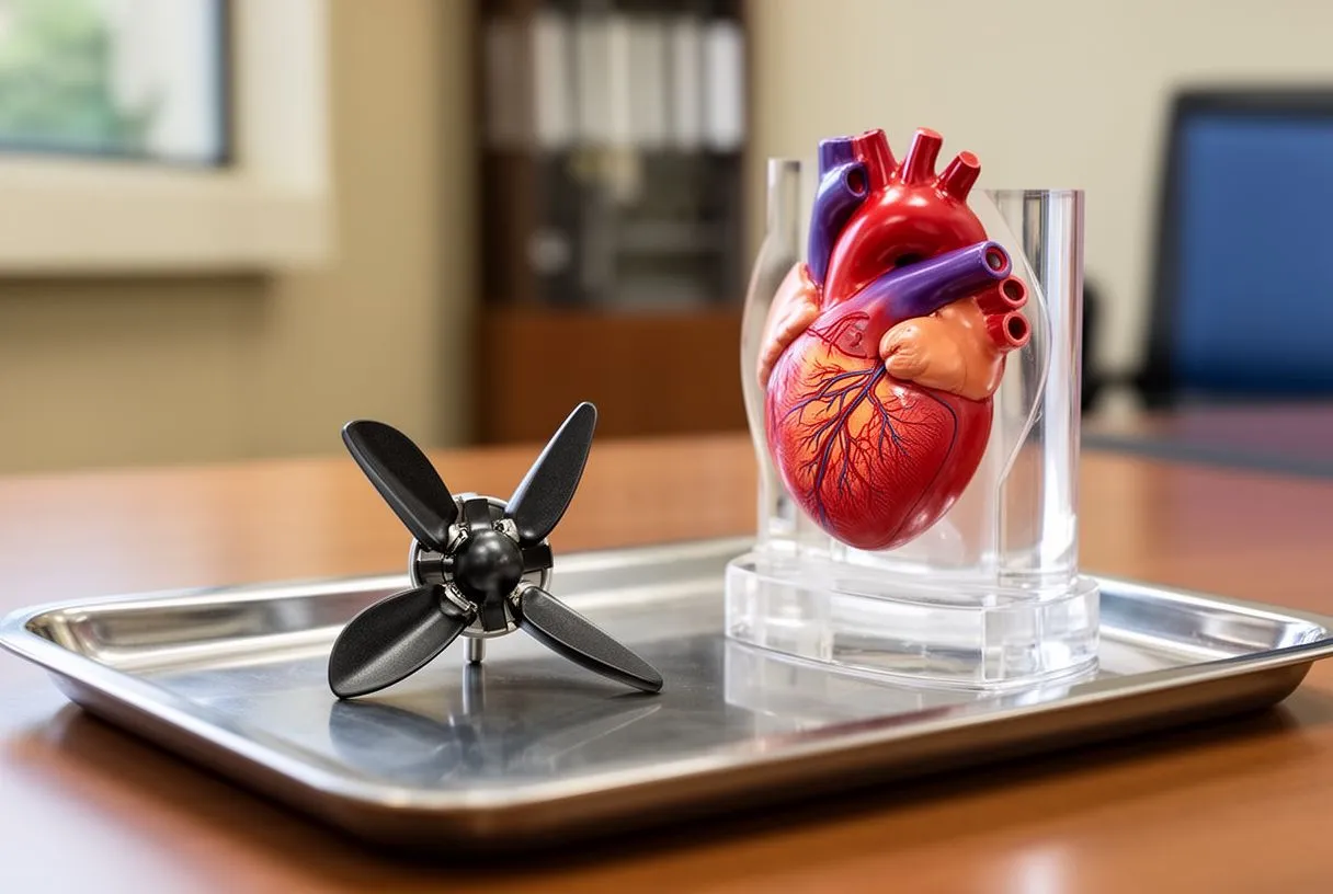
A tricuspid valve leak echocardiogram is the cornerstone of diagnosis. This painless ultrasound scan shows the valve moving in real time, measures the size of the right chambers, and estimates the pressure in the lungs. There is no radiation and no preparation; gel on the chest, a probe, and a picture of the heart.
What the numbers mean. The report will grade the leak as trivial, mild, moderate, or severe. It will also report the size and squeeze of the right ventricle, often using a measurement called TAPSE, which tracks how far the right ventricle wall moves with each beat. Research has shown that this simple measure after mitral valve surgery predicts long-term survival, as reported in Indonesian Journal of Cardiology. In plain terms, the scan does more than grade the leak; it tells the team how well the right heart is coping.
Additional studies. When the picture is unclear, or when intervention is being considered, the team may add a transesophageal echocardiogram, where a slim probe is passed down the throat for a close-up view, or a cardiac CT and MRI. A stress echocardiogram can reveal a leak that only becomes significant under exertion. Every case is reviewed by our multidisciplinary team against international practice guidelines before a recommendation is given, and the patient receives a clear proposal with the alternatives, and the final decision is always theirs.
Monitoring schedule. Mild leaks are usually rechecked every few years. Moderate leaks need closer follow-up, often yearly, with attention to chamber size and symptoms. Severe tricuspid valve leak calls for evaluation by a valve team, because symptoms can appear late even when the heart is already straining.
Tricuspid Regurgitation Treatment Options
Tricuspid regurgitation treatment options range from watchful waiting to medication to catheter-based repair to open surgery. The right choice depends on the grade of the leak, the shape of the heart, your symptoms, and whether the leak is primary or secondary. What follows is a map of the territory.
- Observation: mild and many moderate leaks need no treatment beyond regular scans and attention to blood pressure and rhythm.
- Medication: diuretics, or "water pills," ease swelling and breathlessness by helping the body shed excess fluid.
- Catheter procedures: clips or stents delivered through a vein can reduce the leak without open surgery in selected patients.
- Surgery: repair or replacement through the chest, either with a sternum-splitting approach or through smaller incisions.
Treating the underlying driver. Because most leaks are secondary, treatment often starts elsewhere. If the leak stems from pressure in the lungs, from atrial fibrillation, or from disease on the left side of the heart, fixing or controlling that problem may shrink the leak without touching the valve. This is why the first step in tricuspid valve leak treatment is usually a full picture of the whole heart, not the valve alone.
Medication limits. Tricuspid valve leak medication relieves symptoms but does not close the leak. Diuretics can dramatically improve comfort in someone with swollen legs, and treating rhythm disturbance matters for long-term heart health. But once a leak is severe and symptoms persist despite good medical therapy, interventional options enter the conversation.
Tricuspid Valve Repair vs Replacement
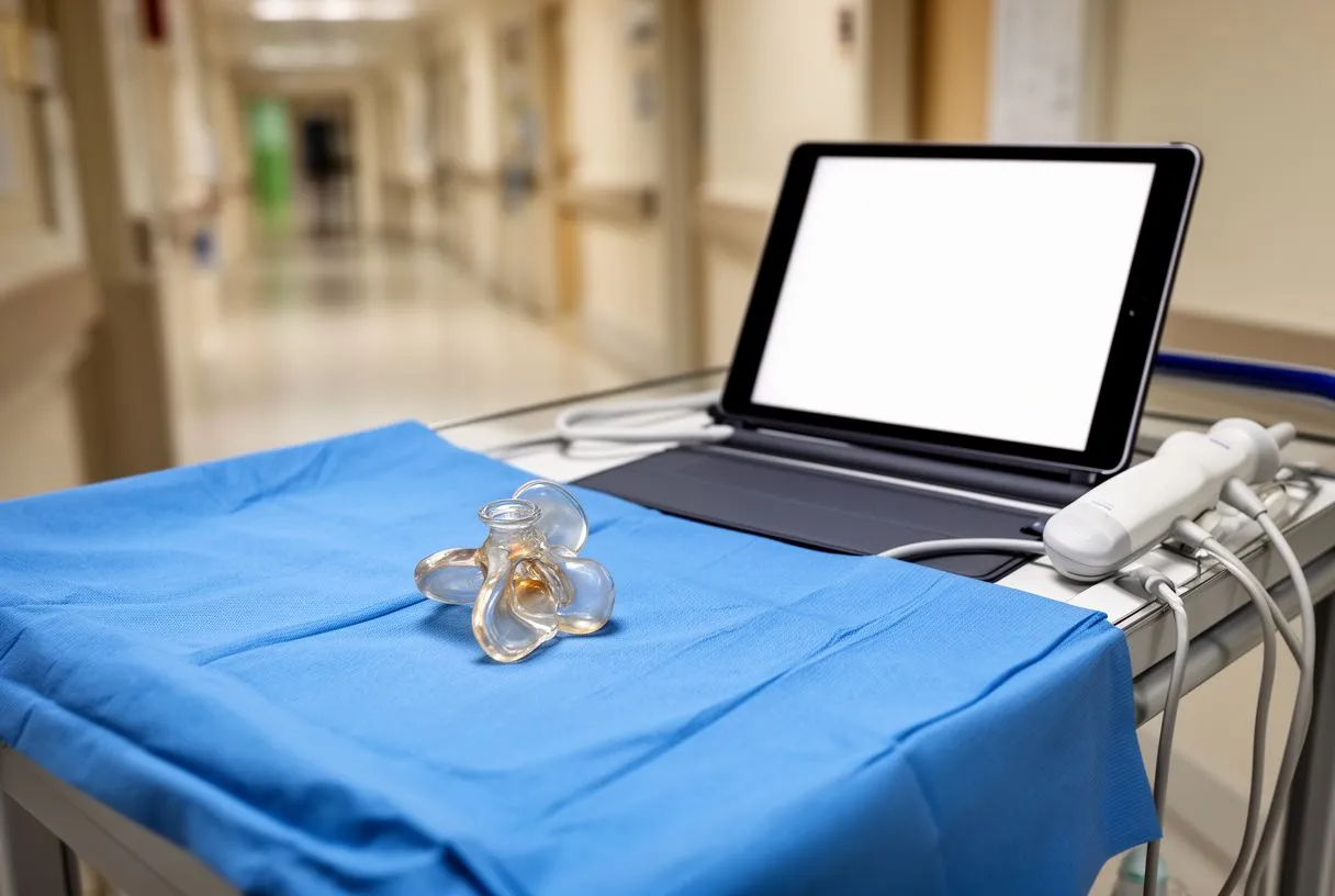
The question of tricuspid valve repair vs replacement is one surgeons weigh carefully. Repair, whenever it is possible, is generally preferred. A repaired valve keeps your own tissue, avoids the lifelong considerations of an artificial valve, and tends to carry fewer long-term complications.
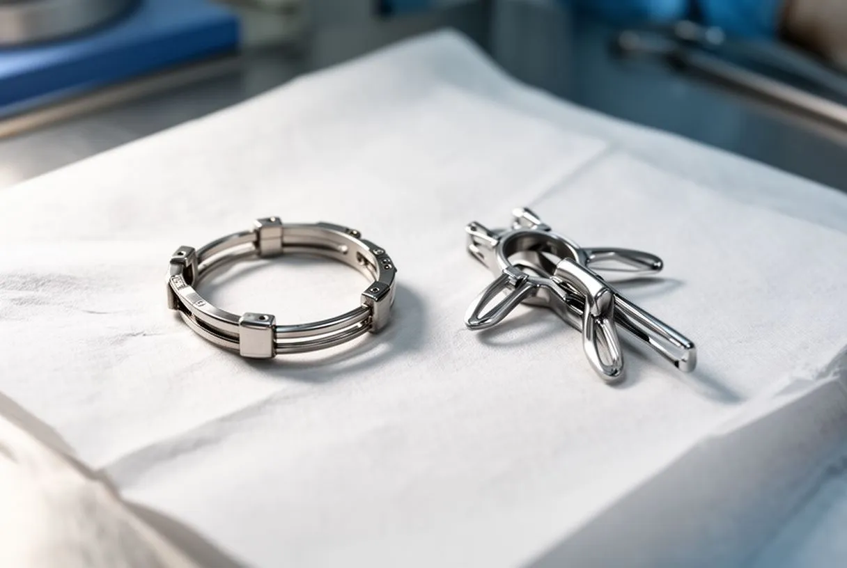
Repair techniques. The most common repair is an annuloplasty, where a fabric ring is sewn around the valve ring to restore its natural shape and bring the leaflets back together. Tricuspid valve annuloplasty recovery follows the usual cardiac surgery pattern: a few days in hospital, weeks of gradual activity, and steady improvement over two to three months. When leaflets are structurally damaged, surgeons may add techniques such as suturing the leaflets' free edges or, in selected cases, using leaflet tissue from a processed animal source.
When replacement is needed. If the valve is destroyed by infection, too deformed to repair, or has failed after a previous repair, replacement becomes the option. Artificial valves are either mechanical valves or tissue valves such as a Porcine Valve or a Bovine Pericardial Valve. Mechanical valves last decades but require blood-thinning medication for life; tissue valves avoid that but may wear out sooner. A Stented Valve includes a supportive frame, while a Stentless Valve is sewn in without one, and a Supra-annular Valve or Intra-annular Valve describes where the artificial valve sits relative to the natural ring. Notably, the tricuspid valve is often cited as among the hardest to replace well, because its ring is broad and flexible and sits in a low-pressure system where artificial valves are more prone to clotting.
Catheter alternatives. For patients at high surgical risk, Transcatheter Tricuspid Intervention offers options without open surgery. These include edge-to-edge clipping systems and transcatheter valve replacements, as well as Valve-in-valve procedures to fix a failing artificial valve from the inside. Paravalvular Leak Closure addresses leaks that develop beside an implanted valve. We perform conventional, minimally invasive, robotic and transcatheter procedures, and we recommend the less invasive route only where the evidence supports it for that patient.
Minimally Invasive Tricuspid Surgery
Minimally invasive tricuspid surgery replaces the traditional midline chest incision with a small cut, often on the right side of the chest, through which the surgeon works with long instruments and a camera. In robotic approaches, the surgeon controls articulated instruments from a console nearby.
What changes for the patient. The valve work is the same inside; the difference is the access. Smaller incisions usually mean less pain, a lower chance of wound complications, and a faster return to driving and daily activity. Not every patient is a candidate, however. Chest shape, prior surgery, lung function, and the urgency of the operation all influence whether a minimally invasive route is safe.
Choosing the approach honestly. The least invasive option is not automatically the best one. A severe, urgent case may be better served by a conventional approach that offers maximum visibility and control. Our team, led by Dr Alejandro Molina, reviews every case together and recommends the route with the lowest risk for the patient's specific situation, not the one with the shortest incision. Dr Molina performed the first robotic mitral valve repair in the Ángeles Health System and the first robotic heart surgery in Guadalajara, and that experience informs when robotics truly add value.
Recovery reality. Even with a small incision, this is still heart surgery. Expect a hospital stay of several days, six to eight weeks before returning to heavier work, and cardiac rehabilitation, a supervised exercise program, as the bridge back to full activity. The benefit of the smaller approach shows up in comfort and speed, not in skipping the healing process.
Tricuspid Valve Leak After Mitral Surgery
A tricuspid valve leak after mitral surgery is a well-recognized pattern. The Mitral Valve Prolapse family of conditions, and mitral disease generally, can cause the right heart to enlarge over years. Sometimes the right-side leak was present but mild before the left-side operation and then progresses; sometimes it develops afterward.
Why surgeons look ahead. Guidelines now encourage surgeons to assess the tricuspid valve during any left-sided operation and to repair it at the same time when it is moderately leaking, even if the patient has no right-sided symptoms. The reasoning is practical: reopening the chest years later for a valve that could have been fixed in the first operation carries far more risk than adding a ring at the time of the original surgery.
If a leak appears later. When a significant leak emerges years after a mitral operation, the full workup matters. The team will ask why: is the right heart dilated, is lung pressure elevated, is there a rhythm problem? Treatment is then directed at the cause, which may be medication, catheter-based repair, or surgery. Research on TAPSE after mitral valve surgery, cited above, underlines why right-heart function is tracked so carefully in these patients. Grading the tricuspid valve on the preoperative echocardiogram allows a borderline leak to be discussed and decided before the operation begins rather than discovered afterward.
What patients should ask. If you are scheduled for mitral surgery, ask two questions: what is my tricuspid valve grade today, and what is the plan if it is moderate or worse? A team that has a clear answer is following the current standard of care.
Tricuspid Valve Leak Prognosis and Life Expectancy
Tricuspid valve leak prognosis depends far more on severity, right-heart function, and the underlying cause than on the leak itself. A mild leak in a healthy heart carries essentially no added risk and no limits on activity. A severe leak with an enlarged right ventricle and persistent symptoms is a different matter, and it is the reason severe tricuspid valve leak deserves timely evaluation.
Why severe leaks are not ignored. Severe secondary regurgitation is associated with worse outcomes in heart failure and lung disease populations. Questions about tricuspid valve leak life expectancy are fair, but the honest answer is that it varies enormously: someone with a moderate leak from a dilated annulus who is otherwise well may never need an operation, while someone with severe leakage, symptoms, and a fading right ventricle benefits from intervention before the heart muscle weakens permanently. Right-sided heart failure signs, meaning swelling, abdominal fullness, and fluid retention, are the signals that the balance has tipped.
Tricuspid stenosis, the sibling problem. Alongside leakage, some patients have tricuspid stenosis, a narrowing of the valve that restricts forward flow. Its effects on the heart include congestion in the veins returning from the body, liver congestion, and fatigue. Mixed stenosis and leakage, common after rheumatic fever, requires evaluation of both problems together, as the termedia.pl surgical article above describes.
Exercise and daily life. For most mild and moderate leaks, tricuspid valve leak exercise restrictions are few: stay active, avoid pushing through new symptoms, and recheck on schedule. With severe leakage, low-intensity activity is usually encouraged while the team decides on intervention, and competitive or heavy exertion may be deferred until the valve is treated. We encourage every patient to ask their own team these questions directly, because restrictions should be individual, not generic.
When to See a Tricuspid Valve Specialist
Seeing a tricuspid valve leak cardiologist is worthwhile at three moments: when a scan shows moderate or severe leakage, when symptoms such as swelling or breathlessness begin, or when another doctor has raised the possibility of surgery and you want a considered opinion. A tricuspid valve specialist brings experience with the newer catheter options as well as surgery, and the landscape of those options has changed quickly in recent years.
What a good consultation includes. Expect a review of your full history, a careful reading of your echocardiogram rather than just its summary line, and a discussion of every option, including the option of doing nothing yet. Our team, with consulting rooms in Guadalajara, Querétaro and Hermosillo, begins with a free video consultation in which we get to know the patient, talk through the condition, and say which studies are still needed so no unnecessary ones are ordered. A face-to-face visit follows when the patient decides to go ahead.
Costs and travel. For international patients considering tricuspid valve leak repair cost in Mexico, pricing varies with the approach chosen, the hospital, and the need for additional procedures at the same sitting. Catheter-based treatments and open surgery carry very different costs, and a multidisciplinary review helps establish which route, and therefore which budget, actually applies before travel is planned. Transparent conversation about cost is part of good care, not an afterthought.
Second opinions are normal. We encourage second opinions and treat seeking one as a normal part of deciding about heart surgery. Valve decisions are significant, and a thoughtful patient who gathers more than one expert view is doing exactly the right thing.
In summary, tricuspid valve leak treatment begins with an accurate diagnosis on echocardiogram, continues with attention to the causes driving the leak, and moves toward repair or replacement only when the leak is severe or symptoms demand it. Mild leaks usually need nothing more than monitoring. Moderate and severe leaks deserve a conversation with an experienced valve team, where the options, from medication to catheter repair to minimally invasive surgery, are laid out plainly. If any of the signs described here sound familiar, the most useful next step is not self-diagnosis but a consultation with a qualified cardiac team who can review your own scans and answer your questions in person.






