

Type B Aortic Dissection: Medicines or Stent Graft
Type B aortic dissection treatment starts with medicine or stent graft repair.
What Is Type B Aortic Dissection Treatment?
When you or a loved one hears the words "aortic dissection," the diagnosis can feel overwhelming. A type B aortic dissection means that the inner layer of your descending aorta — the large artery carrying blood from your heart down through your chest and abdomen — has torn. Blood surges through the tear, splitting the layers of the vessel wall apart. Treatment for aortic dissection aims to stop that split from worsening and to prevent life-threatening rupture.
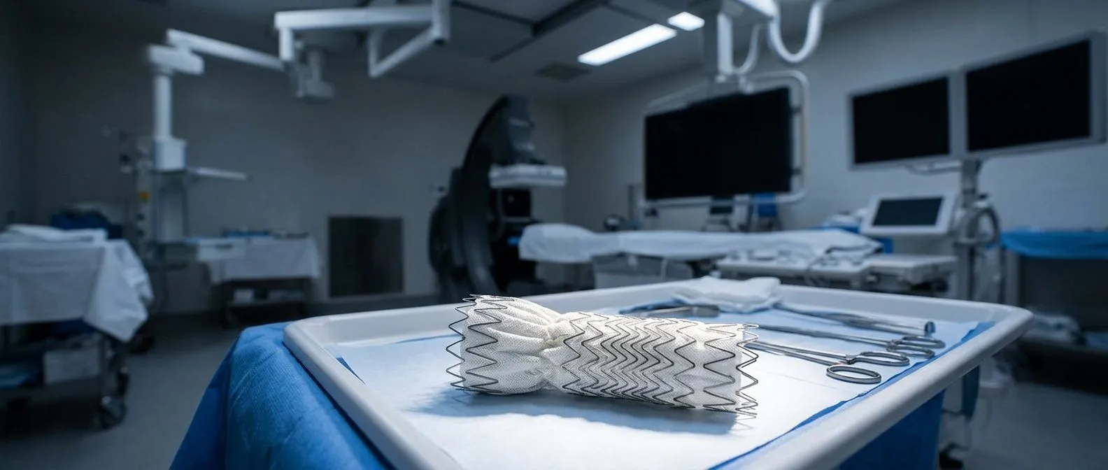
The two main treatment paths are medical management and surgical repair. Medical management relies on medications, especially beta blockers, to lower blood pressure and reduce the force of each heartbeat against the damaged wall. Surgical repair, most commonly performed as a thoracic endovascular aortic repair or TEVAR procedure, places a fabric-covered stent graft inside the aorta to seal the tear and redirect blood flow through a secure channel.
Choosing between these paths depends on whether the dissection is classified as complicated or uncomplicated. A complicated dissection shows signs that the aorta is rupturing, blood supply to organs is cut off, or pain cannot be controlled with medication. An uncomplicated dissection remains stable enough for careful monitoring and drug therapy. Both approaches require lifelong follow-up and imaging. According to Cleveland Clinic, prompt blood pressure control dramatically reduces early mortality in acute dissection, making the first hours of treatment critical.
How Does Type B Aortic Dissection Develop?
The aorta is the body's largest artery, and its wall has three distinct layers. A dissection begins when a small tear in the innermost layer — the intima — lets blood push between the layers, creating a false channel. In a type B dissection, this tear sits below the left subclavian artery, in the descending thoracic aorta, and does not involve the ascending aorta. That anatomical distinction matters because the ascending aorta requires open surgery, while the descending aorta can often be treated with less invasive endovascular techniques.
Several conditions raise the risk of developing a dissection. Long-standing high blood pressure weakens the aortic wall over time. Connective tissue disorders such as Marfan syndrome and Ehlers-Danlos syndrome make the wall structurally fragile. An atheromatous plaque — a fatty deposit in the artery wall — can also erode the intima and create a tear point. Patients who already have an aortic aneurysm face elevated risk because the dilated wall is thinner and under greater stress.
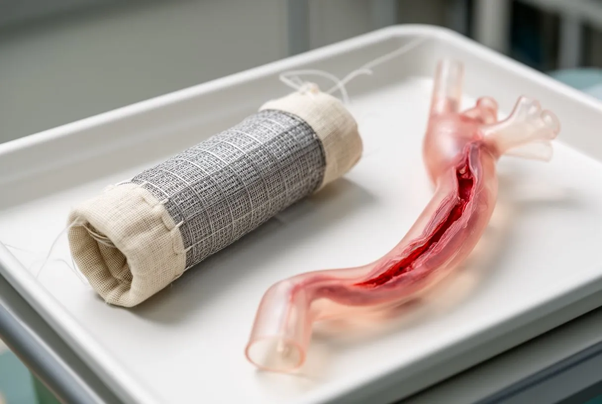
Where does the pain come from? The classic symptom is sudden, tearing chest or back pain that radiates downward. As the false channel extends, it can compress branch arteries feeding the kidneys, intestines, or legs, producing additional symptoms. When the dissection affects arteries supplying the arms, patients may experience arm pain or numbness. Some patients present with an atypical infarction — symptoms that mimic a heart attack but originate from the aortic tear rather than the coronary arteries. Auscultation with a stethoscope may reveal a difference in blood pressure between the left and right arm, a clinical clue that prompts urgent imaging.
When Is Surgery Needed for Aortic Dissection?
Not every type B dissection requires an operation. The decision hinges on whether complications are present at diagnosis or develop during initial medical management. Complicated dissections demand urgent intervention because they carry a high risk of death from rupture or organ failure. Signs of complication include ongoing chest pain despite medication, rapid expansion of the aortic diameter, evidence of rupture or leaking blood into the chest cavity, and compromised blood flow to the kidneys, legs, or intestines.
What makes a dissection uncomplicated? If blood pressure is controllable, pain resolves with medication, and imaging shows no organ malperfusion or aortic enlargement, the dissection is classified as uncomplicated. In these cases, the standard approach is aggressive medical therapy with serial imaging — typically CT angiography or MRI — to watch for changes. Many patients with uncomplicated type B dissection live for years on medication alone, though they need regular monitoring because the aorta can dilate over time.
Some centres now recommend earlier intervention for uncomplicated dissections when imaging predicts future complications. Guideline thresholds such as an aortic diameter greater than 5.5 cm or rapid growth exceeding 5 mm per year may prompt a discussion about elective TEVAR before an emergency arises. If you are weighing this decision, a thorough evaluation by a cardiac surgeon familiar with aortic disease is essential. Research such as the PREDICT PVL study illustrates how imaging predictors help clinicians anticipate which patients will benefit from proactive intervention.
What Is the TEVAR Procedure for Aortic Repair?
Thoracic endovascular aortic repair, known as TEVAR, is a minimally invasive aortic surgery that has transformed the treatment of complicated type B dissections. Instead of opening the chest, the surgeon accesses the aorta through small incisions in the groin, threading a catheter into the femoral artery. A compressed stent graft — a fabric tube reinforced with a metal mesh frame — is guided under X-ray imaging to the tear site. Once positioned, the graft expands against the aortic wall, sealing the tear and restoring normal blood flow through the true channel.
The stent graft acts as an internal bypass. Blood that previously leaked into the false channel now flows through the graft, and the false channel gradually shrinks as clot fills the space outside the graft. Compared with open surgery, TEVAR typically means shorter hospital stays, less blood loss, and faster recovery. The procedure does not require an autologous blood donation beforehand, and in many cases the arterial line used for monitoring is placed percutaneously rather than through a surgical cut-down.
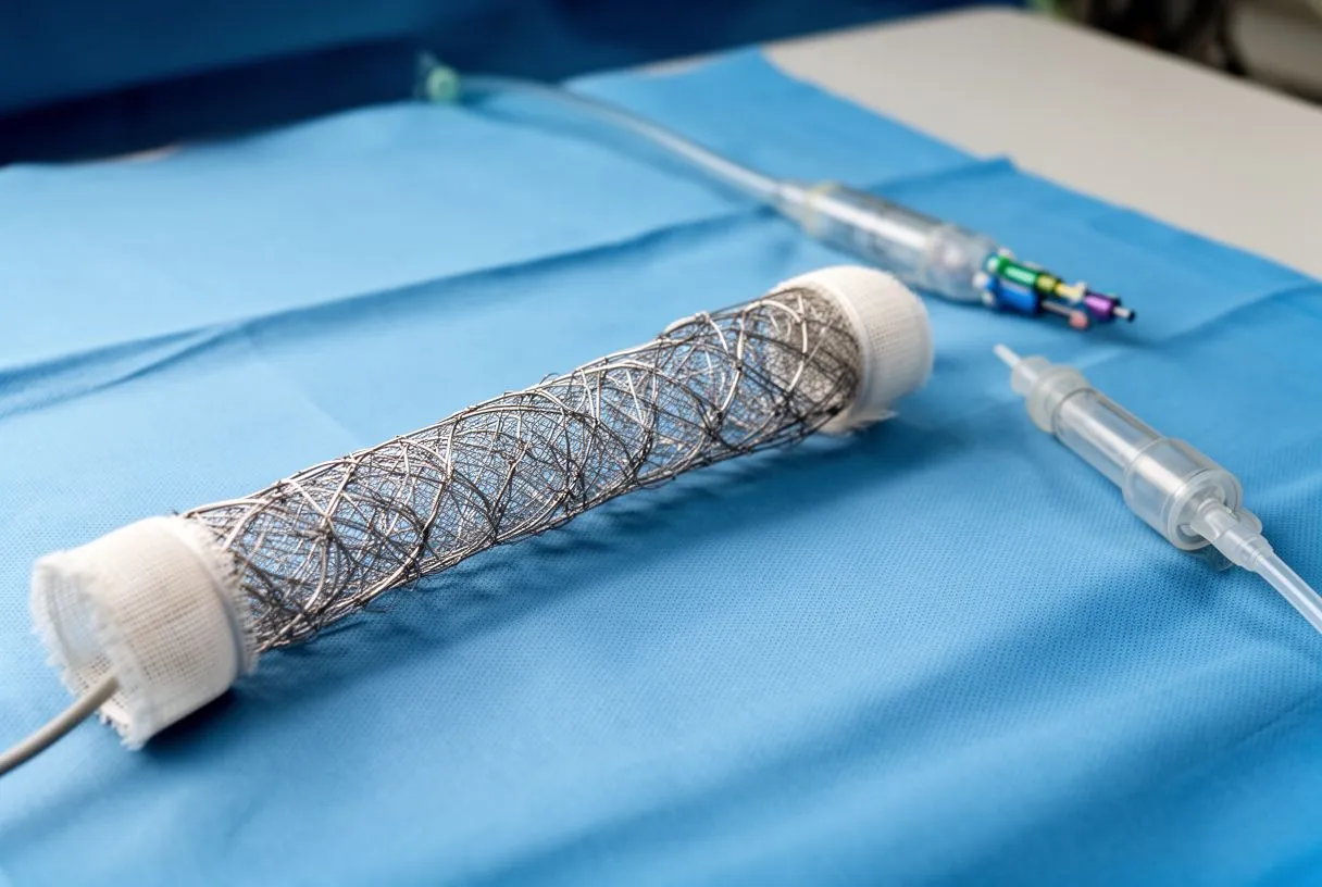
Who is a candidate for TEVAR? Suitable anatomy is the key factor. The dissection must have adequate "landing zones" — segments of healthy aorta above and below the tear where the stent graft can seal securely. If the tear extends too close to critical branch arteries, additional techniques or open surgery may be necessary. Patients with very tortuous or heavily calcified arteries may not be candidates because the graft cannot be safely delivered or seated. Your surgical team will use detailed CT imaging to determine whether your anatomy supports a TEVAR approach or whether an alternative strategy is more appropriate for your specific situation.
Aortic Dissection Medication and Blood Pressure Control
For both uncomplicated dissections managed medically and for patients recovering after stent graft placement, strict blood pressure control is the cornerstone of aortic dissection medication. The goal is to reduce the mechanical stress on the weakened aortic wall. Beta blockers are typically the first-line treatment because they lower both blood pressure and heart rate — the two forces that most directly stress the dissected wall. By slowing the heartbeat, beta blockers reduce the sheer force of each pulse against the torn layers.
When beta blockers alone do not achieve target blood pressure, additional medications are introduced. Angiotensin-converting enzyme inhibitors, calcium channel blockers, and diuretics may be combined to reach the recommended range — often a systolic pressure below 120 mmHg in the acute phase. Patients with an arterial graft from previous surgery need equally careful pressure management because their repaired aorta remains vulnerable. Some patients also take aspirin for cardiovascular protection, though this is prescribed based on individual cardiac risk assessment rather than as a dissection treatment itself.
Medication management is not a passive strategy. It requires regular dose adjustments, home blood pressure monitoring, and honest communication with your medical team about side effects. Fatigue, dizziness, and cold extremities are common with beta blockers and can often be managed by adjusting the dose or switching formulations. The principle is simple but powerful: every millimetre of mercury you lower your blood pressure reduces the force pushing against the damaged aortic wall, giving the body time to stabilise the dissection and reducing the chance of future complications.
Complicated vs Uncomplicated Type B Dissection
The distinction between complicated and uncomplicated type B dissection drives every treatment decision. An uncomplicated dissection is one where the tear is stable, blood pressure responds to medication, no organs are starved of blood, and imaging shows no imminent risk of rupture. These patients are typically admitted to an intensive care unit for pain control and blood pressure management, then transitioned to oral medications and outpatient imaging surveillance.
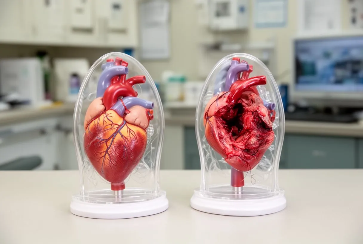
What turns a dissection complicated? Several scenarios shift the balance toward urgent intervention. Malperfusion syndrome occurs when the false channel compresses a branch artery, cutting blood supply to the kidneys, legs, intestines, or spinal cord. A patient may develop kidney failure, leg pain, or abdominal pain as the dissection progresses. Aortic rupture or contained leakage into the chest is the most dangerous complication and demands immediate repair. Rapid aortic expansion — growth exceeding 5 mm in six months — signals an unstable wall that may not hold much longer.
Sometimes a dissection begins as uncomplicated but becomes complicated hours or days later. This is why close monitoring in the acute phase is essential. Even after discharge, patients must watch for new symptoms and attend all follow-up imaging appointments. The following signs indicate that a previously stable dissection may be worsening and require urgent evaluation:
- New or returning chest or back pain: Pain that returns after initial control suggests the dissection is extending or the aorta is enlarging.
- Sudden leg weakness or numbness: Spinal cord or limb malperfusion can produce neurological symptoms rapidly.
- Abdominal pain or dark stools: Intestinal malperfusion can cause bowel ischaemia, a surgical emergency.
Aortic Stent Graft Recovery and Follow-Up
Recovery after a TEVAR procedure is generally faster than after open aortic surgery, but it still requires patience and discipline. Most patients spend one to three days in hospital, with the first night in intensive care for monitoring. The groin incisions heal within two weeks, and patients can usually walk within a day of the procedure. Full return to normal activities typically takes four to six weeks, though your surgeon will tailor this timeline to your overall health and the complexity of the repair.
Imaging before discharge confirms that the stent graft is seated correctly and that the false channel is beginning to shrink. A follow-up CT scan is usually performed at one month, then at six months, and annually thereafter. These scans are not optional — they are the only reliable way to detect endoleaks (persistent blood flow around the graft), graft migration, or new dissection. A small endoleak may be monitored, but a significant one requires a secondary procedure to seal it. The Contemporary Transcatheter Approaches to Mitral Regurgitation and related interventional cardiology literature underscore how imaging surveillance after structural heart and aortic interventions has become standard practice.
Blood pressure control remains critical even after a successful stent graft. The graft seals the primary tear, but the rest of the aorta is still vulnerable. Patients who had a dissection are at higher risk for new tears and aneurysm formation elsewhere in the aorta. This means beta blockers and other antihypertensive medications continue indefinitely, with doses reviewed at every follow-up visit. If you live far from your surgical centre — as many international patients do — coordinate with a local cardiologist who can manage your medications and communicate with your aortic team.
Aortic Dissection Survival Rate and Long-Term Outlook
Survival after type B aortic dissection has improved significantly over the past two decades, driven by better imaging, earlier diagnosis, and the availability of TEVAR. For uncomplicated dissections managed with medication, in-hospital mortality is relatively low when blood pressure is controlled promptly. For complicated dissections requiring emergency intervention, outcomes depend heavily on how quickly the repair is performed and whether organ damage has already occurred before surgery.
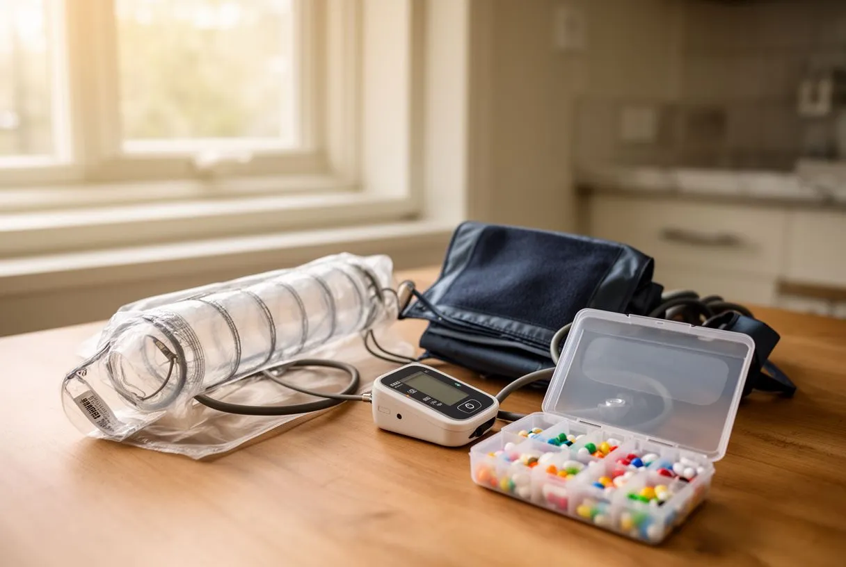
What happens years after a dissection? Long-term survival depends on sustained blood pressure control, regular imaging, and healthy lifestyle choices. The aorta that has dissected once is prone to further problems — progressive enlargement, new tears, or aneurysm formation at other levels. Some patients eventually need a second intervention, whether another stent graft or, in selected cases, open repair. The risk of late complications is highest in the first two years, which is why imaging frequency is greatest during that window and then tapers if the aorta remains stable.
It is important to be honest about the emotional impact. Many patients describe persistent anxiety after a dissection — fear that every new ache signals another tear. Cardiac rehabilitation programmes and peer support groups can help patients regain confidence in their bodies. Physical activity is encouraged once cleared, starting with walking and gradually increasing. Heavy lifting and contact sports are generally discouraged because they spike blood pressure. A well-managed aortic dissection is a chronic condition, much like diabetes — it requires daily attention but does not have to define your life.
Aortic Imaging and Monitoring After Dissection
Aortic imaging is the single most important tool in long-term aortic dissection follow-up. Computed tomography angiography, or CTA, provides detailed cross-sectional images of the entire aorta and is the most widely used modality. Magnetic resonance angiography offers similar detail without ionising radiation, making it attractive for younger patients who will need decades of surveillance. Both can detect changes as small as 2–3 millimetres in aortic diameter, which is the threshold that often triggers a change in management.
The recommended imaging schedule typically includes a scan before hospital discharge, another at one month, then at six months, and annually if the aorta remains stable. If the aorta shows enlargement, the interval shortens. If the aorta shrinks and stabilises after TEVAR, some centres extend the interval to every two years. Your imaging should be interpreted by a radiologist or cardiologist experienced in aortic disease, because subtle changes in the false channel or the stent graft can be easy to miss.
Patients who travel for their surgery face a practical question: where do I get my scans done locally? Work with your aortic team to establish a plan before you leave the surgical centre. Ask for a copy of your discharge imaging on a disc or secure portal, so your local radiologist has a baseline for comparison. Ensure the local facility uses the same modality — ideally CTA — at the same slice thickness, so measurements are consistent year over year. This coordination is essential for catching problems early and avoiding the stress of conflicting reports.
Finding the Right Cardiac Team for Your Dissection Care
Aortic dissection is a complex disease that benefits from a multidisciplinary team. You want a centre where cardiac surgeons, vascular surgeons, cardiologists, and imaging specialists work together rather than in isolation. The team should have experience with both open and endovascular techniques, because some patients need a hybrid approach that combines elements of both. A surgeon who performs TEVAR regularly will have lower complication rates than one who does the procedure occasionally, and this volume-outcome relationship is well documented in aortic surgery.
For patients in Mexico and those travelling internationally, access to experienced aortic care is a legitimate concern. Dr Alejandro Molina leads the cardiac surgery team at Cirugía de Corazón, with consulting rooms in Guadalajara, Querétaro, and Hermosillo. The practice focuses on patient education and shared decision-making, ensuring that you understand your diagnosis, your options, and the reasoning behind each recommendation before committing to a treatment path.
The most important step is a thorough consultation. Bring your imaging — CT scans, echocardiograms, and any prior surgical records — and be prepared to ask direct questions. What is my aortic diameter? Is my dissection complicated or uncomplicated? Am I a candidate for TEVAR or do I need open surgery? What is the expected recovery timeline? A qualified team will answer without rushing and will welcome a second opinion if you seek one. Do not self-diagnose or delay evaluation based on this article — contact a cardiac specialist to discuss your specific situation and book a consultation to review your case in detail.






