


Valve Annulus Dilation and Leaking Heart Valves
Valve annulus dilation causes leaking heart valves.
If you have been told that a heart valve is leaking, one phrase tends to appear in every report: the valve annulus. The annulus is the fibrous ring that anchors each valve, and when that ring stretches — a condition known as valve annulus dilation — the valve can no longer close tightly. Blood then flows backward with every heartbeat, a problem doctors call regurgitation. This guide explains what annulus dilation is, why it causes leaking valves, how the problem is diagnosed, and what repair options exist today, so you can walk into your next appointment understanding the words on your own echocardiogram.
What Does Annulus Mean in Cardiology?
Annulus is simply the Latin word for "ring." In cardiology, it describes the tough, saddle-shaped ring of connective tissue that forms the foundation of each heart valve. Your heart has four valves — the mitral, tricuspid, aortic and pulmonary — and each one sits in its own annulus. The valve leaflets, the thin flaps that actually open and close, are sewn by nature onto this ring the way fabric is attached to a frame.
The annulus does more than hold the valve in place. It flexes with every heartbeat, changing shape to help the valve close completely during contraction. When this ring is the right size and shape, the leaflets meet perfectly, like two doors sealing shut. When the ring enlarges or loses its normal shape, the doors no longer meet, and a gap opens through which blood leaks backward.
Understanding the National Institutes of Health describes the annulus as part of the heart's fibrous skeleton — the structural framework that also insulates the atria from the ventricles electrically. That is why annular problems rarely come alone: they often accompany leaflet disease, chamber enlargement or rhythm disturbances, and a complete evaluation always looks at the whole valve apparatus, not just the ring.
What Is Valve Annulus Dilation?
Valve annulus dilation means the ring supporting a valve has stretched beyond its normal diameter. Think of a collar that no longer fits a shirt: the leaflets are still healthy, but the frame they attach to has grown too wide for them to span. The result is a valve that closes incompletely, allowing blood to leak backward — the condition behind most leaking heart valve treatment decisions.
Why does dilation cause leaking? The leaflets have a fixed length. They cannot grow to match a larger ring, so as the annulus enlarges, the leaflets are pulled apart during closure and the valve becomes insufficient. Doctors call this "functional" regurgitation when the leaflets themselves are normal but the ring is the problem — a very common finding in enlarged hearts.
Where does it happen? Dilation can affect any annulus, but it behaves differently in each valve. Mitral valve annulus dilation is the most frequent cause of functional mitral regurgitation, while tricuspid valve annulus dilation almost always accompanies an enlarged right side of the heart. Aortic valve annulus dilation also occurs, usually in connection with aortic root disease.
Is it dangerous right away? Not necessarily. Mild dilation often produces no symptoms and may only need monitoring. But the leak itself slowly burdens the heart, so the timing of intervention matters. We recommend the less invasive route only where the evidence supports it for that patient, and every case is reviewed by the full multidisciplinary team before a treatment plan is proposed.
Where Is the Aortic Valve Located and What Size Is Normal?
The aortic valve sits between the left ventricle — the heart's main pumping chamber — and the aorta, the large artery that carries blood to the whole body. It is the last gate blood passes through before leaving the heart, which is why aortic valve annulus dilation matters so much: the entire circulation depends on that valve holding firm.
What is the normal size of the aortic valve annulus? In most adults, the aortic annulus measures roughly 2.0 to 2.6 centimetres in diameter, though the exact range varies with body size and sex. Precise Annulus Sizing is measured by CT scan or echocardiography before any procedure, because millimetres matter when choosing a valve or planning a repair.
What sits above the annulus? Immediately above the aortic annulus lies the aortic root, which widens slightly to hold the coronary arteries — the vessels that feed the heart itself. If the root dilates, it can pull the annulus outward and produce leaking even when the leaflets are structurally normal. This is why aortic valve annulus dilation is often assessed together with the aortic root, and why an aortic diameter above 5.5 centimetres is a recognised threshold for considering surgery.
What Causes Mitral Valve Annulus Dilation?
The mitral valve lies between the left atrium and the left ventricle, and its annulus is the most complex ring in the heart — it changes shape constantly with every beat. Valve annulus dilation in this location usually has one of several causes.
Leaflet prolapse. In Mitral Valve Prolapse, one or both leaflets flop backward into the atrium. Over the years, the extra motion stretches the annulus, so a leaflet problem becomes a ring problem too. This is why repair of a prolapsing valve almost always includes the annulus.
Chamber enlargement. When the left ventricle or left atrium enlarges — from heart muscle weakness, long-standing high blood pressure, or atrial fibrillation — it pulls the annulus outward with it. The leaflets stay normal, but the ring is too wide for them to close. This "functional" pattern is the most common form of secondary mitral regurgitation.
Age and calcification. Over decades, calcium deposits and tissue stiffening can remodel the annulus, flattening its natural saddle shape and widening its diameter.
The 60-to-40 rule is a useful surgical concept here: roughly 60% of the mitral valve's closing force comes from the posterior (back) leaflet's length, and about 40% of the annulus's circumference is also posterior. Surgeons planning repair focus especially on this posterior region because that is where the ring is most flexible and where most leaking begins. The practical takeaway for patients: annular dilation rarely exists alone, and the cause determines the repair.
What Are the Symptoms of a Leaking Heart Valve?
Early on, there may be none. A leaking valve can progress silently for years. Many patients first learn about regurgitation from a routine heart murmur heard with a stethoscope — a whooshing sound that suggests blood flowing backward. This is why heart murmur surgery options are only ever discussed after the leak has been measured and graded.
As the leak worsens, symptoms appear gradually. You may notice shortness of breath when climbing stairs, fatigue that sleep does not fix, a fluttering or irregular heartbeat, or swelling in the ankles and legs. Mitral leaks tend to cause breathlessness and palpitations; aortic leaks tend to cause fatigue and a pounding sensation in the chest. None of these symptoms is specific to valve disease, which is why proper testing is essential.
Symptoms are a signal, not a verdict. They tell you the heart is starting to struggle, and they are one of the clearest indications that valve heart valve regurgitation surgery should be considered rather than delayed. In our consultations we take the time to explain what the studies actually show, in plain words, and to answer every question before any decision — because a patient who understands their leak is far better equipped to choose the right moment for intervention.
How Is Mitral Valve Disease Diagnosed?
The diagnosis of valve disease follows a clear, logical sequence, and none of the steps hurts.
- Echocardiogram: the cornerstone test. Ultrasound waves create a moving picture of the heart, showing the leaflets, the annulus, and the direction and severity of the leak.
- Measurements that matter: the echo grades the leak from trace to severe, and measures the annulus diameter, chamber sizes and the pumping function of the heart.
Transesophageal echo. When the regular echo is not clear enough, a thin probe is passed gently down the oesophagus, giving sharp, close-up images of the mitral valve. This is a routine, well-tolerated test under light sedation, and it is often the key step before planning a repair.
Advanced imaging and exercise testing. A cardiac CT maps the annulus in three dimensions and is essential for Annulus Sizing before any valve procedure. Cardiac MRI quantifies the volume of blood leaking backward, and a stress echo can reveal whether the leak worsens with exercise. The first contact with our practice is a free video consultation: we get to know the patient, talk through the condition, and say which studies are still needed so no unnecessary ones are ordered — a face-to-face visit follows when the patient decides to go ahead.
What Is a Tricuspid Valve Annulus?
Location and role. The tricuspid valve sits between the right atrium and the right ventricle, the chambers that handle blood on its way to the lungs. Its annulus is the largest and least rigid ring in the heart, which makes it particularly prone to stretching whenever the right heart enlarges.
Why the right heart stretches it. Conditions that raise pressure in the lungs — long-standing left-sided valve disease, lung disease, or sleep apnea — force the right ventricle to work harder. The right atrium and ventricle balloon outward, and the tricuspid valve annulus dilation follows. The valve then leaks, and the leak adds to the right heart's burden, a self-reinforcing cycle.
What can be done. Tricuspid Valve Repair, most commonly an annuloplasty ring implanted around the stretched annulus, restores the ring to its natural diameter and allows the leaflets to meet again. Surgeons increasingly repair the tricuspid at the same time as a mitral operation, because a mildly leaking tricuspid valve left untreated can cause trouble years later.
Is Mild Calcification of the Aortic Valve Annulus Serious?
Usually, not yet. Mild calcification of the aortic valve annulus is a very common finding with age, and on its own it rarely causes symptoms. Calcium in the annulus often travels with calcium in the leaflets, so it can be an early sign of aortic valve stiffening — but a mildly calcified annulus by itself is usually a watch-and-measure situation.
When it matters. Calcification becomes important in three situations: when it progresses and narrows the valve (aortic stenosis), when it distorts the valve so much that it leaks, or when it changes treatment planning. A heavily calcified annulus can make valve repair more difficult and can influence whether a stented tissue valve or a mechanical valve is chosen, or whether a minimally invasive transcatheter approach is possible.
What monitoring looks like. An echocardiogram every one to two years, control of blood pressure and cholesterol, and prompt attention to new symptoms. If the leak or narrowing is significant, every case is reviewed by the multidisciplinary team against international practice guidelines before a recommendation is made, and you receive a clear proposal with the alternatives — the final decision is always yours.

What Are the Repair Options for Valve Annulus Dilation?
Valve annulus dilation treatment has one goal: restore the ring to the right diameter so the leaflets can close completely. There are several well-established ways to achieve this, and the right choice depends on the valve, the cause and your overall health.
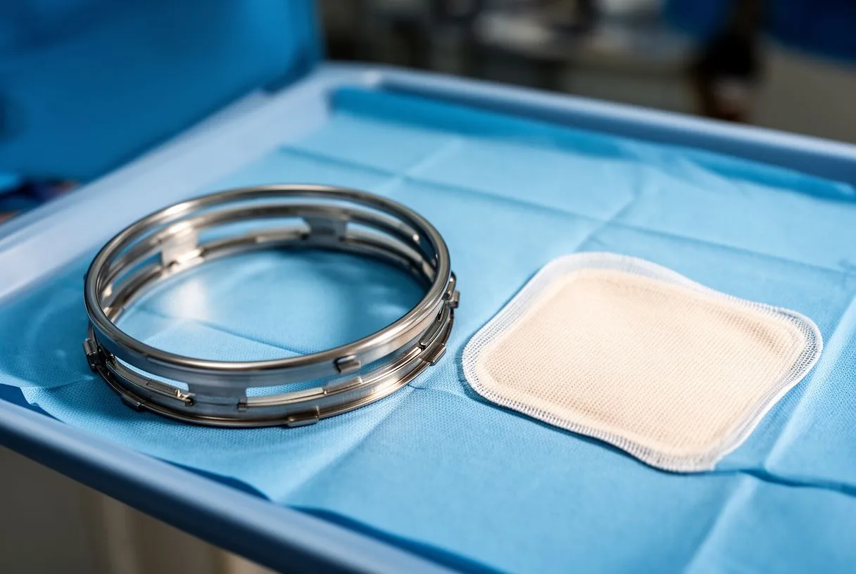
- Annuloplasty ring repair: the standard surgical answer. A firm ring — the valve annuloplasty ring — is sewn around the annulus to reshape and stabilise it, so the valve holds its repair for years.
- Leaflet repair plus annular support: for prolapse, the leaflet is refined and the ring is added, giving the most durable results.
- Valve replacement vs repair: when the annulus and leaflets are too damaged, the valve is replaced with a tissue or mechanical valve. This is a Stented Valve or, for some patients, a Stentless Valve — the trade-off between durability and blood-thinner requirements is discussed case by case.
Minimally invasive and transcatheter routes. Many annular repairs today are done through small incisions on the side of the chest — a minimally invasive valve repair that avoids opening the breastbone and shortens recovery. For aortic valves in patients at higher surgical risk, Transcatheter Aortic Valve Implantation — always called TAVR — delivers a Self-expanding Valve through a catheter in the groin, replacing the valve without open surgery. Published experience with these techniques includes specific refinements, such as studies describing single-dose lignocaine-based blood cardioplegia in valve replacement patients that help protect the heart during the operation (single-dose lignocaine-based blood cardioplegia study). We perform conventional, minimally invasive, robotic and transcatheter procedures, and in the consultation we explain which route the evidence supports for your specific anatomy.
What Is Recovery Like After Valve Annulus Dilation Surgery?
The first days. Most patients spend one to two nights in intensive care, where the heart's rhythm and function are watched closely, then move to a regular ward. Breathing tubes, drains and monitoring lines come out over the first 48 hours, and walking to a chair happens early — often the same day.
The first weeks. With a traditional sternotomy (breastbone incision), driving is usually possible after about four weeks and most daily activities by six. With minimally incisional or robotic approaches, patients typically regain comfort faster because the chest is not split. Cardiac rehabilitation — supervised, gradual exercise — is strongly recommended and makes a real difference to long-term recovery.
The first year and beyond. A repaired mitral or tricuspid valve needs periodic echocardiograms to confirm the annuloplasty ring is holding and the leak has not returned. Expect a follow-up echo within a few months, then yearly. Dr Molina performed the first robotic mitral valve repair in the Ángeles Health System and the first robotic heart surgery in Guadalajara.
When to seek help promptly. Call your cardiac team urgently for new or worsening breathlessness at rest, chest pain, fainting, a very rapid or irregular heartbeat, or sudden leg swelling. These can signal a change that needs assessment before it becomes an emergency.

Choosing your team and taking the next step. Valve annulus dilation is one of the most treatable problems in heart medicine: the ring can be reshaped, the leak can be stopped, and the results are durable when the repair is well chosen. The key is matching the procedure — repair, ring, minimally invasive route or TAVR — to your specific anatomy, because no single approach is right for everyone. If you have been told your valve is leaking or your annulus is enlarged, do not wait for symptoms to force the decision. We encourage second opinions and treat seeking one as a normal part of deciding about heart surgery. If you would like a video consultation with a heart surgeon to discuss your own case, Dr Alejandro Molina's multidisciplinary team in Guadalajara, Querétaro and Hermosillo is ready to review your studies with you, explain what they show in plain language, and help you decide with confidence.






