

Valve Migration After TAVR: Causes and Fixes
Valve migration TAVR causes and solutions explained by Guadalajara heart specialists.
Transcatheter aortic valve replacement, or TAVR, has changed the outlook for thousands of people with a narrowed aortic valve. Instead of open-heart surgery, a new valve is carried through an artery in the groin on a thin tube and set in place inside the old one. For most patients the valve settles exactly where it should and stays there. But in a small number of cases, the new valve can shift from its intended position — a complication known as valve migration after TAVR. This guide explains what that means, why it happens, how it is spotted, and what can be done about it.
What Is the TAVR Procedure and Why Have a Valve Replacement?
Why have a valve replacement at all? The aortic valve sits between the heart's main pumping chamber and the aorta, the large artery that supplies the whole body. When it becomes narrowed — a condition called aortic stenosis — the heart must push against a stubborn, stiff opening. Over time this causes breathlessness, fainting, chest discomfort, and eventually heart failure. Replacing the valve relieves that obstruction, and for severe stenosis it is often the treatment that changes the course of the disease.
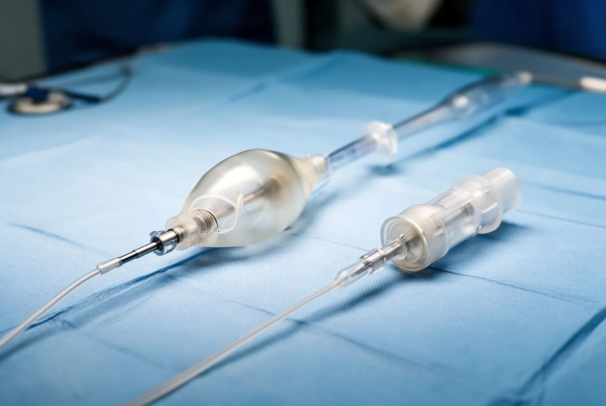
What is the TAVR procedure, step by step? The team threads a catheter — a long, thin tube — from an artery, usually in the groin, up to the heart. At its tip sits a replacement valve, compressed to the width of a pencil. When it reaches the right spot inside the diseased valve, a balloon inflates or the valve's own frame expands, and the new valve opens in place. No chest is opened, and the heart does not need to be stopped. Most patients walk within a day or two.
For many years TAVR was reserved for patients too frail for conventional surgery. That has changed. In a landmark randomized trial of intermediate-risk patients, transcatheter replacement produced outcomes comparable to surgical replacement, which is why the two approaches are now weighed side by side for many people (The Lancet). In our practice, every case is reviewed by the multidisciplinary team against international practice guidelines before a recommendation is given, and the patient receives a clear proposal with the alternatives — the final decision is always theirs.
What Is Valve Migration After TAVR?
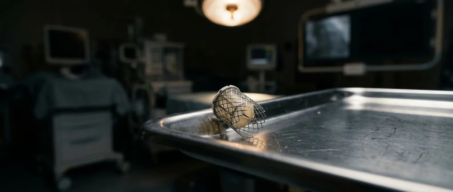
The simple definition. Valve migration after TAVR means the new valve moves from the position where it was implanted. A transcatheter valve is held in place by friction: its metal frame presses outward against the walls of the aortic root and the annulus, the ring of tissue where the native valve once sat. If that grip loosens, the frame can shift upward toward the aorta or downward into the left ventricle. The medical literature often calls the downward form valve embolization.
Transcatheter aortic valve migration is rare but real. It belongs to the family of complications surgeons group under valve malposition — the valve is present but not exactly where it needs to be. Migration matters because a displaced valve no longer sits between the correct structures. It can leak backward, block neighbouring pathways, or lose contact with the sealing skirt designed to stop paravalvular leaks.
The degree of displacement matters as much as the fact of it. A shift of a few millimetres may be watched safely. A valve that has embolized into the ventricle or the aortic arch needs prompt treatment. In our consultation we take the time to explain what the imaging studies actually show, in plain words, so the patient understands exactly where their valve sits before any decision about treatment.
What Causes Transcatheter Valve Migration?
Understanding transcatheter valve migration causes helps patients make sense of an unexpected event. Migration is almost never caused by anything the patient did. It is the result of anatomy, valve sizing, and sometimes technique. The common threads run through the list below:
- Undersizing: the chosen valve is slightly small for the annulus, leaving too little grip against the surrounding tissue.
- Severe calcium: heavy, uneven calcium on the native valve gives the new valve an irregular seat, so the frame cannot press evenly in every direction.
- Incorrect depth of implantation: a valve placed too high or too low has less overlap with the annulus and can slip.
- Rapid pacing issues: during deployment the heart is paced very fast to stop blood flow; if pacing fails or blood flow continues, the valve can be pushed off position.
- Fragile anatomy: a dilated aortic root or a very thin annulus offers less purchase for the frame.
Recognizing a TAVR complication where the valve slips. Operators recognize migration during the procedure itself: the valve moves on the imaging screen, the deployment balloon drifts, or the final angiogram shows the valve out of place. This is exactly why TAVR is performed in a hybrid operating room with live X-ray and echocardiography — the team can respond within seconds rather than discovering the problem days later.
Technique also plays a role. Balloon-expandable and self-expanding valve designs behave differently, and each requires its own deployment discipline. Dr Molina trained in structural cardiology and TAVR at CHU Bordeaux, and the team also trained at Jena, Bad Berka (Germany) and Buffalo (USA).
Early vs Late Valve Migration After TAVR: What Is the Difference?
Early valve migration in TAVR happens during or immediately after the procedure. Most episodes fall in this window. The valve shifts while it is being deployed or in the first hours on the intensive care unit. Because the patient is still in the catheterization suite or closely monitored, the team detects it at once and corrects it in the same setting, usually with excellent outcomes.
Late valve migration is much less common. Here the valve was well positioned at discharge but moves weeks, months, or occasionally years later. Reported causes include gradual frame expansion, changes in the annulus after infection, or degeneration of the leaflets that changes how forces pass through the valve. A documented pattern involves delayed migration of a valve placed inside an earlier surgical valve — a valve-in-valve situation — where the old valve's geometry makes a stable landing harder.
The timing changes the conversation. Early migration is an operator's problem, solved in the room. Late migration is a follow-up problem, found at an echocardiogram or during evaluation of new symptoms. Both are treatable, but late cases need a careful search for an underlying reason — particularly infection — before the valve itself is addressed.
What Are the Symptoms of TAVR Valve Instability?
TAVR valve instability symptoms overlap with the symptoms that led to the original procedure. The new valve is failing to do its job in its new position, so the body notices the same things it noticed before: breathlessness on exertion, dizziness, fatigue, or swelling of the ankles. The difference is the timeline — these symptoms return after a period of improvement, which is the signal that something has changed.
Warning signs that need same-day attention. Certain findings suggest the valve has moved into a dangerous position and affect the heart's conduction system or block blood flow:
- Fainting or near-fainting, especially with a very slow pulse — the displaced valve can injure the electrical pathway between the atria and ventricles.
- Sudden severe breathlessness or an irregular, racing heartbeat, which can signal the valve has embolized into the left ventricle.
- Chest pain with falling blood pressure, suggesting acute severe leakage around or through the displaced valve.
Most patients will never experience these events. But knowing them matters. A migration that is caught at a routine echocardiogram, before symptoms appear, is managed electively. One caught during an emergency admission is managed urgently. The gap between those two scenarios is exactly why structured follow-up after TAVR is not optional.
Who Is Most at Risk of Valve Migration After TAVR?
Valve migration risk factors in TAVR cluster around the anatomy of the landing zone. The phrase the team uses is the "landing zone" — the annulus and the few millimetres of tissue above and below it. Patients with a heavily calcified, asymmetric annulus, a large-difference between the annulus and the sinotubular junction, or a fragile aortic root all present a harder seat for the valve.
Imaging and sizing decisions carry the most weight. Before the procedure, a CT scan of the aortic root is measured in detail: annulus diameter, calcium distribution, and the angle of the aortic root. These measurements determine which valve size is chosen. When the CT is precise and the sizing is appropriate, the risk of migration drops sharply. Undersizing — deliberately or by error — is one of the most consistently reported factors.
Volume and experience matter too. Outcomes in both surgical and transcatheter aortic valve replacement improve with hospital volume, and complications such as malposition are less frequent in high-volume, experienced teams (Journal of the American Heart Association). Our team reviews every candidate's CT measurements together as a multidisciplinary group before choosing a valve size, because we believe the sizing decision is the single most important step in preventing this complication.
How Is Post TAVR Valve Movement Detected?
Post TAVR valve movement is usually found on echocardiography. The standard follow-up schedule after TAVR includes an echocardiogram at discharge, at around one month, and at one year, then periodically. The echocardiogram measures the position of the valve frame, the direction and severity of any leakage, the gradient across the valve, and the function of the heart muscle. A shift of a few millimetres between studies is the kind of finding that triggers a closer look.
CT imaging adds detail when needed. If echocardiography suggests the valve has moved or is not seated well, a CT scan of the aortic root can show the exact position of the frame, its relationship to the coronary arteries, and whether the frame has deformed. This information is essential when planning a repair or a second valve.
Sometimes the patient notices a change first — a return of breathlessness or a new murmur heard by their own doctor. That is enough to arrange the studies. When new symptoms appear after a TAVR, the usual first step is to repeat the echocardiogram and review the original imaging before proposing anything, so the problem is measured rather than assumed.
Can a Migrated TAVR Valve Be Repositioned or Removed?
Repositioning a migrated TAVR valve is often possible. If the valve is only slightly displaced and the frame is still intact, the team can thread a catheter to the valve, inflate a balloon inside it, and nudge it back into the annulus. Some self-expanding valve designs are deliberately built to be retrieved and repositioned before final release, which is one reason an operator may choose that platform for a patient with a difficult anatomy.
TAVR valve malposition treatment depends on where the valve went. When the valve has embolized downward into the left ventricular outflow tract, a catheter-based approach can often retrieve or stabilize it; published case series describe exactly this technique for outflow-tract-migrated valves. When the valve has moved into the ascending aorta and is causing no obstruction or leak, observation may be reasonable. If it is obstructing flow or interfering with future options, it usually needs to be addressed.
When retrieval fails, surgical removal follows. Valve migration that cannot be managed in the catheterization suite requires conventional open-heart surgery to remove the displaced device and implant either a new transcatheter valve or a surgical bioprosthesis. This sounds daunting, and it is a real operation — but it is performed in controlled conditions, and the need for it is uncommon relative to the total number of TAVRs performed worldwide.
What Are the TAVR Revision Surgery Options?
TAVR revision surgery options fall into three broad families. The right choice depends on the valve's position, the patient's anatomy, and what the first valve did wrong.
- Valve-in-valve implantation: a new transcatheter valve is placed inside the displaced or dysfunctional one, using it as the landing platform — the same principle used when a transcatheter valve degenerates years later.
- Catheter retrieval and repositioning: for valves still reachable with a catheter and not yet firmly lodged, the original device is pulled back or nudged into place, sometimes followed by a second valve if the first cannot be salvaged.
- Surgical explanation and replacement: open surgery to remove the migrated valve entirely — the option reserved for valves that have embolized into the ventricle, become infected, or sit where no catheter can safely reach. When the whole device must be removed, this is called a TAVR valve explantation.
Choosing between them is a team decision. A TAVR failure from valve migration is treated very differently from a failure from leaflet degeneration or infection, and the workup has to distinguish among them. Every revision case is reviewed by the team against international guidelines, and the less invasive route is recommended only where the evidence supports it for that specific patient. We also encourage patients to seek a second opinion — we treat that as a normal part of deciding about heart surgery, not as a challenge.
Recovering After TAVR Through the Groin Artery
How long does it take to recover from a heart valve replacement through the groin artery? Far faster than from open surgery. Most patients go home within two to five days. The puncture site in the groin needs a few days of care — keep it clean, avoid heavy lifting for about a week — and driving is usually permitted once the site is healed and medications allow it. Many people return to normal daily activities within two weeks.
What should you expect six months after an aortic valve replacement? By six months, most TAVR patients feel markedly better than before the procedure: less breathlessness, more stamina, better sleep. The heart muscle is still remodelling — the muscle thickened by years of pushing against a narrowed valve takes time to relax back toward normal. Regular follow-up continues, and the echocardiogram at six months to a year confirms the valve is stable. A general guide to life after valve procedures and heart-healthy habits can be found at Harvard Health Publishing.
Follow-up is where problems are prevented. The schedule exists precisely so that a valve that is starting to move, leak, or fail is caught while the fix is simple. Keeping every scheduled echocardiogram after TAVR matters, because a gap in follow-up is when problems are missed. If you are considering TAVR, or you have had one and new symptoms have appeared, talk to a qualified cardiac team rather than waiting to see if things settle on their own. The first contact with our practice is a free video consultation: we get to know you, talk through your condition, and tell you which studies are actually needed — so no unnecessary ones are ordered — before any face-to-face visit at our consulting rooms in Guadalajara, Querétaro or Hermosillo.
In summary, valve migration after TAVR is a recognized but uncommon complication: it arises mainly from sizing and anatomy, is usually detected early through structured follow-up, and has a clear treatment pathway ranging from simple repositioning to valve-in-valve implantation or surgical explanation. The best protection is careful pre-procedure planning by an experienced multidisciplinary team and honest, regular follow-up afterwards — and the final decision about any procedure always belongs to the patient.






