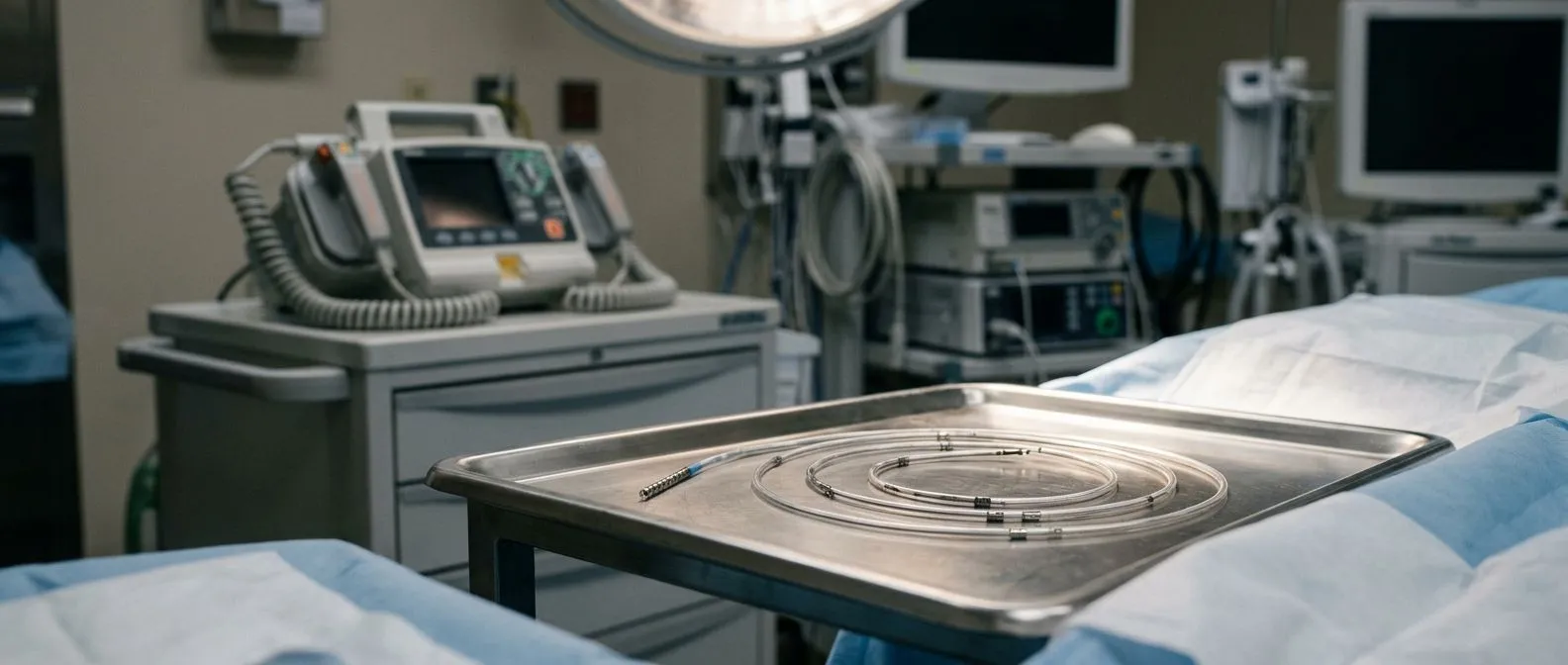
Ventricular Tachycardia Treatment: Emergency Signs and Care
Ventricular tachycardia treatment saves lives when every second counts.
What Is Ventricular Tachycardia?
Ventricular tachycardia is a dangerously fast heart rhythm that originates in the ventricles — the two lower chambers responsible for pumping blood to the lungs and the rest of the body. In a normal heartbeat, an electrical signal begins in the atrium, the upper collecting chamber, and travels down to the ventricles in a coordinated fashion, producing a resting rate of 60 to 100 beats per minute. In ventricular tachycardia, abnormal electrical circuits fire from within the ventricular muscle itself, driving the heart to 150 or even 250 beats per minute. At those speeds the ventricles cannot fill properly between beats, cardiac output drops sharply, and the brain and other organs receive inadequate blood flow.
Cardiologists classify ventricular tachycardia as sustained if it lasts more than 30 seconds and non-sustained if it self-terminates before that threshold. Sustained VT is the more urgent concern because it rarely stops on its own and can degenerate into ventricular fibrillation — a chaotic rhythm that produces no meaningful pumping at all and causes sudden cardiac arrest within minutes. Non-sustained episodes may still signal serious underlying disease, but they are typically discovered during routine monitoring rather than during an emergency room visit.
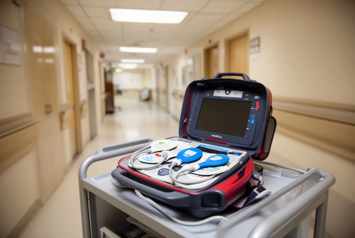
The distinction between VT with a pulse and pulseless VT is critical for treatment. When a patient still has a measurable pulse, medications and controlled electrical cardioversion can restore normal rhythm. When there is no pulse, the situation is treated identically to cardiac arrest: immediate defibrillation and cardiopulmonary resuscitation are required. An Automated External Defibrillator can be life-saving in these moments, and every minute of delay reduces survival significantly, which is why public-access defibrillators and prompt bystander CPR are so strongly emphasised in cardiac arrest education.
Ventricular Tachycardia Symptoms and Emergency Warning Signs
Many patients describe the first sign of ventricular tachycardia as a sudden, pounding sensation in the chest — the heart feels as though it is racing or fluttering violently. Palpitations may be accompanied by dizziness, lightheadedness, or a vague sense that something is profoundly wrong. Shortness of breath and chest pressure or pain are also common, particularly in patients who have underlying coronary artery disease. Some people experience a brief warning — a moment of nausea or visual disturbance — before losing consciousness, while others collapse without any prodrome at all.
Syncope, the medical term for fainting, is one of the most alarming presentations of sustained VT. When the heart cannot maintain blood pressure because it is beating too fast to fill effectively, the brain is starved of oxygen and consciousness is lost. Any patient who experiences unexplained syncope should be evaluated urgently by a cardiac team, because ventricular tachycardia is one of several life-threatening rhythms that can cause it. Arm pain radiating from the chest, profuse sweating, and a grey or ashen complexion are additional red flags that the episode may be progressing toward cardiac arrest.
- Sudden collapse or loss of consciousness: call emergency services immediately and begin CPR if trained.
- Rapid heartbeat with chest pain or pressure: do not drive yourself; wait for an ambulance with monitoring capability.
- Dizziness, severe shortness of breath, or cold sweating: these suggest the heart is not maintaining adequate blood pressure.
Not every fast heartbeat is an emergency, but the line between benign palpitations and ventricular tachycardia cannot be drawn without an electrocardiogram. If a rapid heartbeat lasts more than a few seconds, is accompanied by dizziness or chest pain, or results in fainting, call emergency services immediately. Have someone take you or wait for an ambulance equipped with monitoring and defibrillation capability rather than attempting to drive yourself to the hospital.
What Causes Ventricular Tachycardia?
The most common cause of sustained ventricular tachycardia in adults is scar tissue left behind by a previous heart attack. When a coronary artery is blocked and heart muscle dies, the body heals the area with a dense, fibrous scar. This scar cannot conduct electrical impulses the way healthy muscle does, so signals loop around it in abnormal circuits — re-entry pathways that fire repeatedly and generate the rapid rhythm of VT. Patients who have had large infarctions, particularly those with significantly reduced pumping function, carry the highest risk of developing this arrhythmia over time.
Ventricular tachycardia can also arise in hearts that have never had a heart attack. Structural conditions such as hypertrophic or dilated cardiomyopathy stretch and distort the ventricular walls, creating zones where electrical conduction slows and re-entrant circuits form. Inflammatory diseases, infiltrative disorders, and valve abnormalities can produce similar substrate changes over time. An atheromatous plaque that ruptures and triggers acute ischaemia can also provoke VT even before permanent scar forms, which is why unstable patients presenting with chest pain and rapid rhythms are treated as emergencies from the moment they arrive.
Less commonly, VT originates without any structural heart disease at all. Genetic ion channelopathies — conditions such as long QT syndrome, Brugada syndrome, and catecholaminergic polymorphic VT — alter the microscopic flow of ions across heart muscle cells and make the ventricles electrically unstable. Severe electrolyte disturbances, particularly low potassium and magnesium, can trigger VT in otherwise healthy hearts. Certain medications that prolong the QT interval, recreational stimulants, and even profound emotional stress are additional recognised triggers that an electrophysiologist will investigate during a thorough evaluation.

How Is Ventricular Tachycardia Diagnosed?
The single most important diagnostic test for ventricular tachycardia is the 12-lead electrocardiogram. During an active episode, the ECG shows a wide-complex tachycardia — the QRS complex, which represents ventricular electrical activation, is broader than normal because the impulse is travelling through abnormal pathways rather than the heart's specialised conduction system. A skilled electrophysiologist can often distinguish VT from supraventricular rhythms with aberrant conduction by examining specific features, but when the diagnosis is uncertain, treating the rhythm as VT is the safer clinical default.
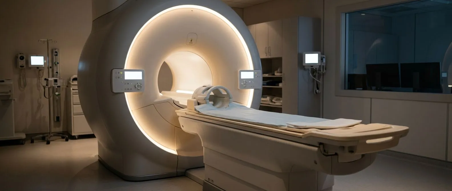
When episodes are intermittent, a resting ECG may be normal between attacks. In these cases ambulatory monitoring provides the answer. A Holter monitor records every heartbeat for 24 to 48 hours, while an event recorder or implantable loop recorder can capture rhythm for weeks or months. Cardiac imaging is essential to identify the underlying structural substrate. Echocardiography assesses pumping function and wall-motion abnormalities, and cardiac magnetic resonance imaging reveals scar tissue and subtle structural changes with remarkable detail, as demonstrated in studies on magnetic resonance imaging in the evaluation of pectus excavatum and related thoracic conditions.
Blood tests help identify reversible triggers. Electrolyte panels detect low potassium or magnesium, thyroid function tests rule out hyperthyroidism, and drug screens can reveal medication-induced QT prolongation. In patients with established coronary disease, the presence of Apolipoprotein B elevation helps characterise residual cardiovascular risk. During an electrophysiology study, catheters are threaded into the heart to map the exact location of abnormal circuits and provoke VT in a controlled setting, providing both diagnostic confirmation and a roadmap for ablation if that treatment is planned.
Ventricular Tachycardia Treatment Options
Ventricular tachycardia treatment falls into two broad categories: emergency management of an active episode and long-term prevention of recurrence. In the acute setting, the patient's haemodynamic stability determines everything. If blood pressure is maintained and the patient is conscious, intravenous antiarrhythmic medication is the first step. If the patient is unstable — hypotensive, confused, or in shock — synchronized electrical cardioversion is performed immediately to reset the rhythm before it deteriorates into full cardiac arrest.
For long-term prevention, the treatment strategy depends on the underlying cause. Patients with scar-related VT from a previous heart attack often benefit from an implantable cardioverter-defibrillator, a device that monitors the heartbeat continuously and delivers a shock to terminate dangerous rhythms. Catheter ablation targets the abnormal circuits directly, burning or freezing the tissue responsible for the re-entry pathway. Medication with antiarrhythmic drugs reduces episode frequency in many patients, and optimising treatment of the underlying condition — coronary artery disease, heart failure, or electrolyte imbalance — is fundamental to reducing overall risk.
Choosing among these options is rarely simple, and the decision should be individualised by a cardiac electrophysiologist who understands the patient's full clinical picture. Factors include the type and frequency of VT, the heart's pumping function, prior procedures, medication tolerance, and the patient's overall health and preferences. A patient who has had a single brief non-sustained episode with normal heart function faces a very different risk profile from one who experiences repeated sustained episodes with a weakened left ventricle, and the treatment plans must reflect that distinction rather than applying a one-size-fits-all approach.
Antiarrhythmic Drugs for VT Management
Antiarrhythmic medications work by altering the electrical properties of heart muscle cells — slowing conduction, extending the refractory period, or suppressing the abnormal automaticity that drives ventricular tachycardia. Amiodarone is the most widely used agent for VT, particularly in patients with structural heart disease, because it is effective across multiple mechanisms and can be administered intravenously during an acute episode or taken orally for long-term suppression. Sotalol and mexiletine are alternatives, each with distinct pharmacological profiles and side-effect considerations that the prescribing physician weighs carefully.
Despite their benefit, antiarrhythmic drugs are not without risk. Amiodarone, for example, can affect thyroid function, liver enzymes, lung tissue, and sun sensitivity, requiring regular monitoring of blood tests and organ function over the course of treatment. Proarrhythmia — the paradoxical worsening of arrhythmia by the very drug meant to treat it — is a concern with many agents, which is why drug selection is typically guided by an electrophysiologist rather than a general practitioner. Dosing is adjusted for kidney and liver function, and interactions with other medications must be reviewed at every visit.
In the emergency setting, lidocaine is sometimes used for suspected ischaemia-related VT, though amiodarone remains the evidence-supported first choice in most resuscitation protocols. For patients who also have atrial fibrillation or atrial flutter, certain antiarrhythmics can address both supraventricular and ventricular rhythms, simplifying the medication regimen. The published clinical evidence supporting these choices continues to evolve, and research appearing in journals such as JAMA Network informs ongoing guideline updates that shape clinical practice and drug selection recommendations.
Catheter Ablation for Ventricular Tachycardia
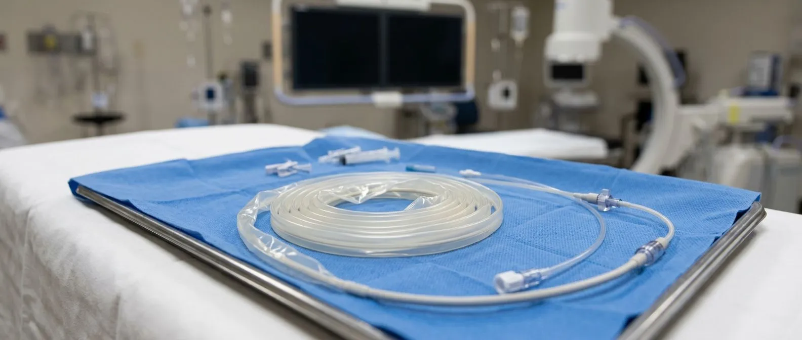
Catheter ablation is a minimally invasive procedure that targets the precise region of heart muscle generating ventricular tachycardia. During the procedure, thin flexible catheters are inserted through veins in the groin and advanced into the heart under X-ray guidance. Once positioned, the catheter tip delivers radiofrequency energy — controlled heat — to the abnormal tissue, creating small scars that block the re-entry circuit and prevent the rhythm from recurring. In selected cases, the catheter is advanced through the arterial line approach to reach certain ventricular sites that are difficult to access from the venous side.
Ablation is most commonly recommended for patients with scar-related VT who continue to experience episodes despite medication, or for those who receive frequent shocks from an implantable defibrillator. It can also be the preferred approach for patients with idiopathic VT — rhythms that arise from structurally normal hearts, often originating in the right ventricular outflow tract. The goal is not always complete elimination of VT but a meaningful reduction in episode frequency, ICD shocks, and symptoms, which dramatically improves quality of life and reduces hospital admissions.
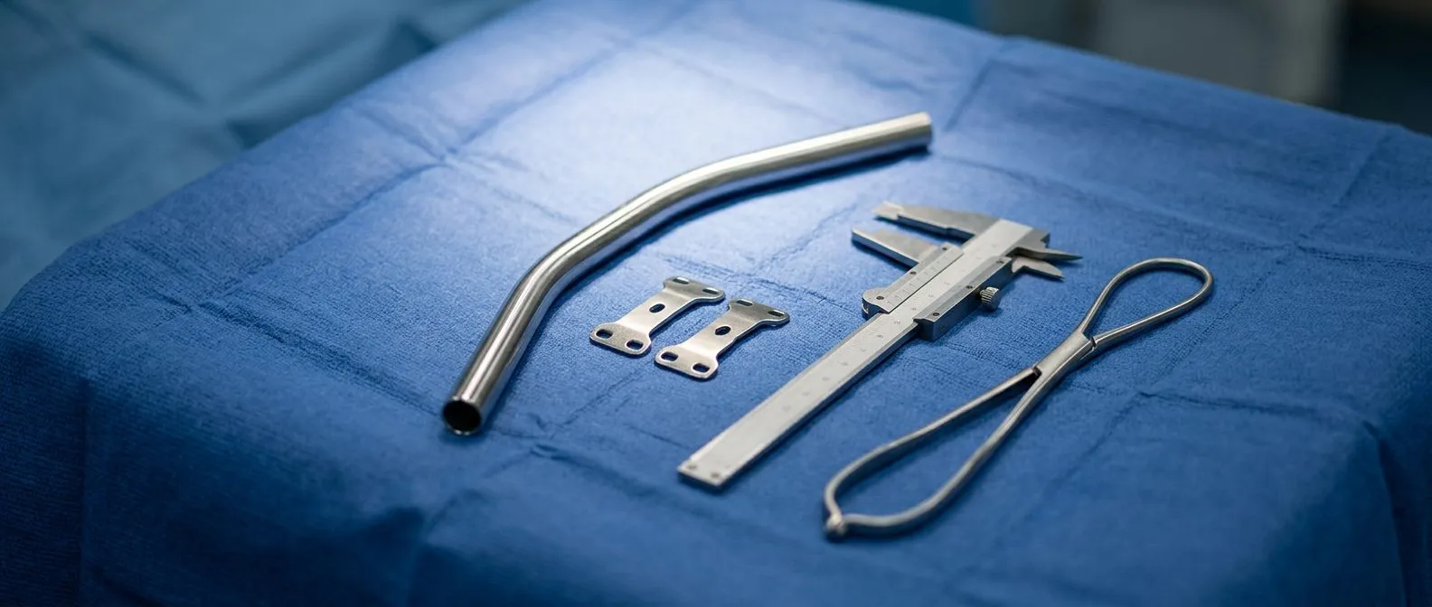
The procedure carries real risks, including bleeding, infection, damage to blood vessels or heart valves, and the possibility of creating new arrhythmia circuits. Success depends on the complexity of the substrate and the skill of the operator, which is why ablation is performed in specialised electrophysiology laboratories by experienced teams. For patients who also require other cardiac surgery — for instance, those needing repair of the ascending aorta or valve replacement — combined approaches have been described in the surgical literature, including simultaneous pectus excavatum repair using sternal plate in a patient undergoing the Bentall procedure, illustrating how complex structural repair and arrhythmia management can sometimes be coordinated in a single operative plan.
Electrical Cardioversion and Emergency VT Treatment
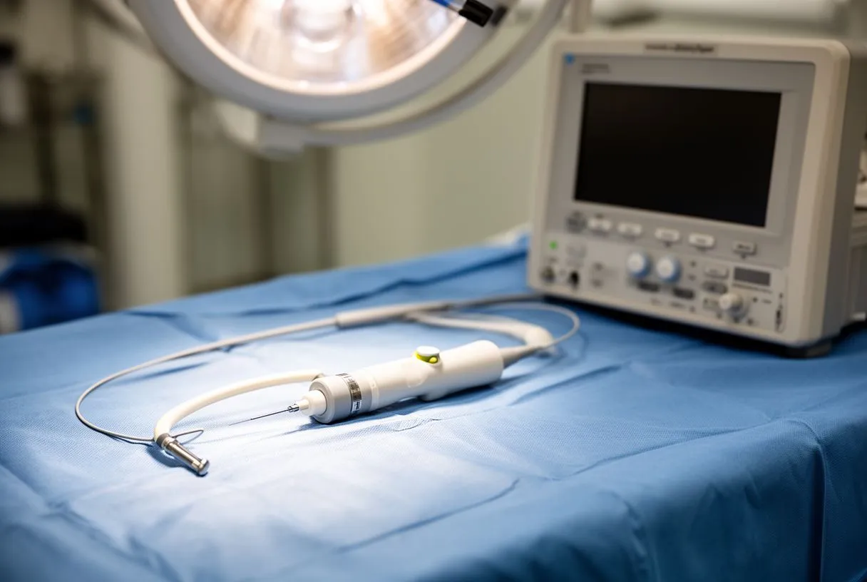
When ventricular tachycardia produces haemodynamic compromise — falling blood pressure, altered consciousness, chest pain, or signs of shock — the fastest way to restore a normal rhythm is synchronised electrical cardioversion. Pads are placed on the chest, and a brief, controlled electric shock is delivered timed to the QRS complex, instantly depolarising the entire heart and allowing the normal pacemaker to resume control. The patient is sedated or anaesthetised for comfort, and the team stands ready to escalate to full defibrillation if the rhythm degenerates into ventricular fibrillation.
In pulseless VT, the algorithm changes entirely. The patient is in cardiac arrest, and the priority is immediate unsynchronised defibrillation — a high-energy shock not timed to any specific cardiac event. CPR is performed between shocks, and intravenous medications such as amiodarone or lidocaine are administered according to established resuscitation protocols. Aspirin is not a treatment for active VT, though it is often part of the broader cardiac care plan for patients who also have coronary artery disease and are at risk of further ischaemic events. The goal of the resuscitation team is to restore a perfusing rhythm as quickly as possible and then identify and treat the underlying trigger.
After successful cardioversion or resuscitation, the patient is admitted for continuous monitoring, blood work, and cardiac imaging to determine why the episode occurred. This evaluation is essential because VT is often a symptom of another problem — ischaemia, electrolyte disturbance, drug effect, or progressive structural disease — and unless that root cause is addressed, recurrence is likely. The inpatient stay also provides time to initiate or adjust medications and, where appropriate, plan for catheter ablation or defibrillator implantation before discharge.
Recovery and Long-Term Management After VT
Surviving an episode of ventricular tachycardia marks the beginning, not the end, of a patient's cardiac journey. The first weeks after discharge focus on medication stabilisation, wound care if a defibrillator was implanted, and gradual return to daily activity. Patients are typically advised to avoid driving for a defined period — guidelines often recommend several months — because the risk of recurrent VT with syncope poses a danger behind the wheel. Cardiac rehabilitation, supervised exercise, and lifestyle modification form the foundation of secondary prevention during this critical recovery window.
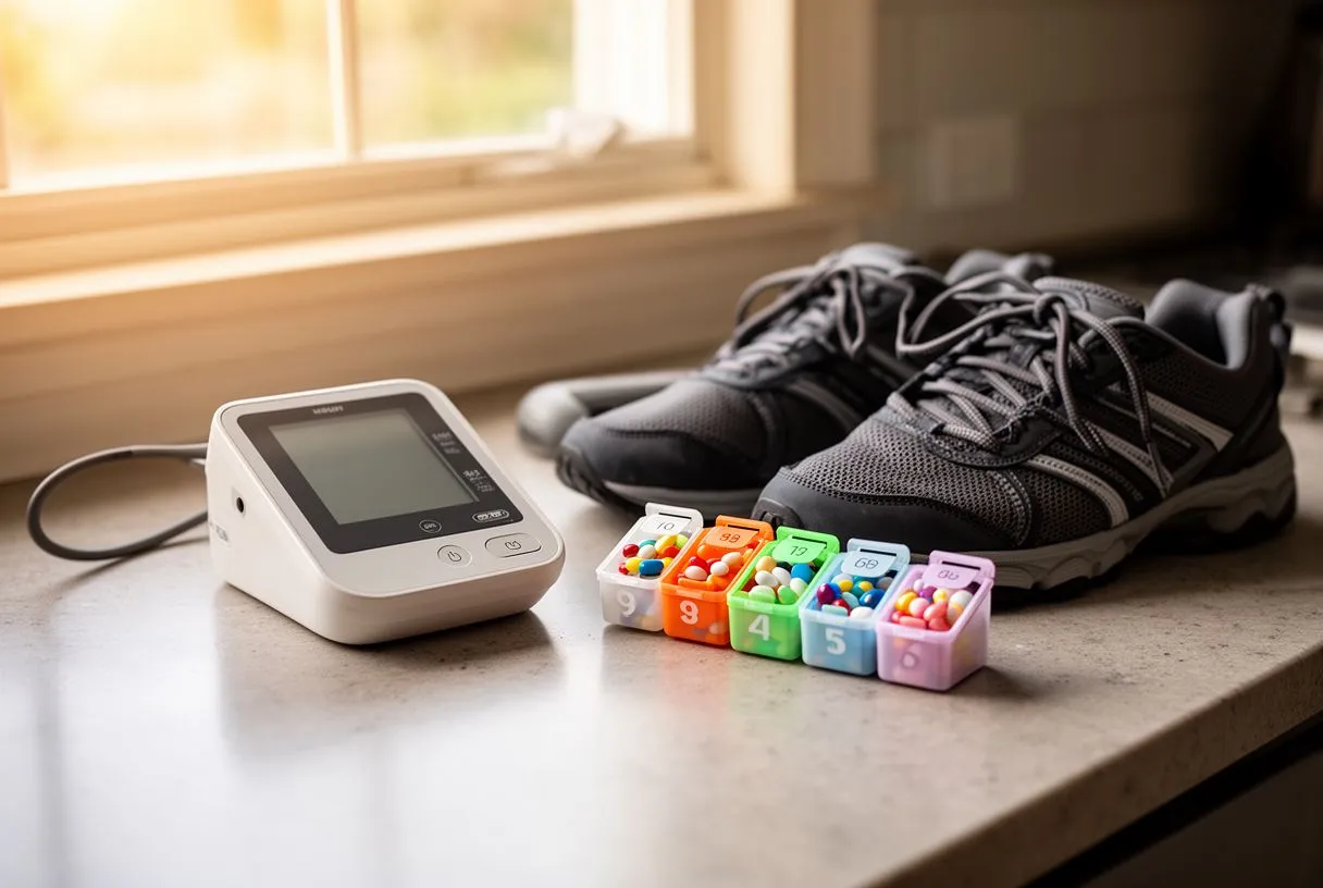
Long-term management is a partnership between the patient and the cardiac team. Regular follow-up visits include device interrogation for ICD patients, medication review, and assessment of heart function. Auscultation with a stethoscope, blood tests, and periodic imaging help detect subtle changes before they become problems. Patients are taught to recognise warning symptoms and understand what their device does, so they can respond appropriately if they feel a shock or experience palpitations. Education about medication adherence is critical because missed doses are among the most common causes of VT recurrence.
Quality of life after VT depends heavily on the underlying condition and the success of treatment. Many patients return to full, active lives with appropriate therapy, while others live with the knowledge that their heart is vulnerable and that further intervention may be needed. Psychological support is an under-recognised but important component of recovery, as living with an arrhythmia — especially one that has caused syncope or required a shock — can produce anxiety that itself affects quality of life. An honest conversation with the cardiac team about expectations, limitations, and prognosis is one of the most valuable tools a patient has.
When to Seek Urgent Care for Ventricular Arrhythmia
The clearest signal that ventricular tachycardia requires emergency attention is the combination of a rapid heartbeat with any sign of poor blood flow: dizziness, chest pain, shortness of breath, sweating, or loss of consciousness. These symptoms mean the heart is not pumping effectively, and the rhythm may be on the edge of degenerating into cardiac arrest. Do not wait to see if it resolves on its own. Call emergency services, sit or lie down to prevent injury if you faint, and unlock the door so paramedics can reach you quickly.
Patients with known VT or an implanted defibrillator should have a clear action plan developed with their electrophysiologist. This plan specifies which symptoms warrant an emergency call, which warrant a same-day clinic visit, and which can wait for a scheduled appointment. If a defibrillator delivers a shock, the patient should contact the cardiac team promptly, and more than one shock in 24 hours is universally treated as a medical emergency. The action plan should be shared with family members, because they are often the ones who recognise the severity of an episode and act first.
- Call emergency services: if you experience a rapid heartbeat with dizziness, chest pain, or fainting.
- Contact your cardiac team the same day: if your defibrillator delivers a single shock and you feel otherwise stable.
- Schedule a routine follow-up: for new palpitations without other symptoms, to arrange monitoring and evaluation.
Ultimately, ventricular tachycardia is a condition that demands expert, individualised care. The information in this guide is intended to help patients and families understand the condition, recognise the danger signs, and participate meaningfully in treatment decisions — not to replace a clinical evaluation. If you or a loved one has experienced symptoms described here, or if you have been diagnosed with VT and have questions about your treatment plan, a cardiac electrophysiologist can discuss your specific situation and the options best suited to your needs.