


Abdominal Aorta Screening: Checking for an Aneurysm
Abdominal aorta screening detects aneurysms before they rupture.
What Is Abdominal Aorta Screening?
Understanding the aorta and why it matters: The aorta is the largest artery in your body, a muscular tube about the diameter of a garden hose that carries oxygen-rich blood from your heart down through your chest and into your abdomen. The segment that passes through the belly is called the abdominal aorta. Over time, the wall of this vessel can weaken and bulge outward, forming a balloon-like swelling called an abdominal aortic aneurysm. If it grows large enough, it can rupture, causing life-threatening internal bleeding. Abdominal aorta screening is a simple, painless ultrasound test designed to catch this silent threat before it reaches that danger point.
Why screening rather than waiting for symptoms: Most aneurysms develop slowly over years without producing any warning signs. By the time a person feels pain or notices a pulsing sensation in the abdomen, the aneurysm may already be dangerously large. Screening flips this timeline: it finds the aneurysm while it is still small, monitorable, and treatable with a planned elective repair rather than emergency surgery. According to WebMD, early detection through targeted ultrasound programmes has measurably reduced rupture-related deaths in the populations studied.
What screening does not do: It is important to understand that an abdominal aorta screening ultrasound is not a general heart check or a full vascular examination. It looks specifically at one vessel and measures its diameter. It does not evaluate the heart valves, the coronary arteries on the heart's surface, or smaller branch vessels in the legs. If your doctor suspects broader vascular disease, additional studies such as CT angiography or a complete echocardiogram may be recommended alongside or instead of the focused screening scan.
Who Needs an Aorta Screening Test?
Age and sex thresholds that drive screening: Clinical guidelines generally recommend abdominal aorta screening for men aged 65 to 75 who have ever smoked, because this group carries the highest baseline risk. The evidence for universal screening in women is less robust, but women with a family history of aneurysm, a history of smoking, or other significant cardiovascular risk factors may also benefit from a one-time scan. Some international programmes invite all men at age 65 for a single ultrasound, regardless of smoking history, because the cost per rupture prevented remains favourable.
Family history as a decisive factor: If a first-degree relative — a parent, sibling, or child — has had an aortic aneurysm or a sudden aortic dissection, your own risk is substantially higher regardless of sex. In these families, screening is often recommended earlier, sometimes beginning at age 50 or even 45, and may be repeated every few years rather than performed only once. Genetic conditions such as Marfan syndrome, vascular Ehlers-Danlos syndrome, and bicuspid aortic valve disease also warrant dedicated surveillance imaging of the entire aorta, not just the abdominal segment.
Patients with existing cardiovascular disease: If you already carry a diagnosis of coronary artery disease, peripheral arterial disease, or have had a previous vascular procedure such as a balloon angioplasty, your vascular system has already demonstrated a tendency toward wall pathology. An atheromatous plaque — a fatty deposit in the artery wall — can weaken the underlying tissue and contribute to aneurysm formation. Your cardiologist may therefore include an abdominal aorta measurement as part of a broader vascular assessment, even if you fall outside the standard screening age band.
How Does Abdominal Aorta Screening Work?
The screening test uses standard ultrasound technology, the same safe imaging method employed during pregnancy. A technician places a small handheld probe, called a transducer, against the skin of your abdomen after applying a warm gel. The probe emits high-frequency sound waves that bounce off the aortic wall and the blood inside it, creating a real-time grey-scale image on a nearby monitor. No needles, no contrast dye, and no radiation are involved. The entire scan typically takes ten to fifteen minutes.

What the technician measures: The key measurement is the maximum anteroposterior diameter of the abdominal aorta, usually taken at the level of the renal arteries where the aneurysm most commonly develops. A normal diameter is roughly 2 centimetres or less. A reading between 3 and 4.9 centimetres is considered a small or moderate aneurysm that warrants surveillance. A diameter of 5.5 centimetres or greater is the threshold at which elective surgical repair is typically recommended, because the annual rupture risk rises sharply above that size.
Interpreting the results with your doctor: The ultrasound report will include the measured diameter, the location of any widening, and sometimes a description of the aortic wall. Your physician then contextualises this single number against your overall health picture. A 4-centimetre aneurysm in a frail 80-year-old with limited life expectancy may be managed conservatively with regular monitoring, while the same measurement in a healthy 60-year-old might prompt a more proactive discussion. Research such as the study on Prevalence and Outcomes of Concomitant Aortic Stenosis and Cardiac Amyloidosis illustrates how overlapping cardiac conditions can complicate aneurysm management decisions, underscoring the value of a personalised clinical conversation.
What Are the Symptoms of an Aortic Aneurysm?
The problem with relying on symptoms: The abdominal aortic aneurysm earned its grim nickname, the "silent killer," precisely because it produces no symptoms in the majority of cases until the moment of rupture. People walk around with a 5-centimetre bulge in their aorta feeling completely normal. This is precisely why abdominal aorta screening exists: to find the problem before the body sends any distress signal at all.
When symptoms do appear, they tend to be vague and easily mistaken for other conditions. Some patients report a deep, constant ache in the lower back or the side of the abdomen that does not change with position or movement. Others notice a pulsing sensation near the navel, as though they can feel their heartbeat through the belly wall. Occasionally, a physician may detect an unusually prominent pulse during a routine physical examination using a stethoscope in a process called auscultation, sometimes hearing a bruit — a whooshing sound created by turbulent blood flow within the dilated segment.
Rupture symptoms are a medical emergency: If the aneurysm tears, the symptoms shift dramatically. Sudden, severe, tearing pain in the abdomen or lower back, a drop in blood pressure, dizziness, and even loss of consciousness signal that blood is escaping the aorta into the abdominal cavity. At this stage, survival depends on immediate emergency surgery. Unlike an atypical infarction — a heart attack with unusual symptoms — a ruptured aneurysm offers almost no window for second-guessing. The key takeaway is simple: never wait for symptoms to justify a screening test, because the symptom that matters most may be the one that arrives too late.
Abdominal Aorta Ultrasound: What to Expect During the Procedure
Knowing what happens during the scan helps reduce anxiety, especially for patients who have never had an ultrasound before. The test takes place in a dedicated imaging room or a vascular laboratory. You will be asked to lie flat on an examination table, typically on your back. The sonographer may ask you to roll slightly onto one side to get a clearer view of the aorta, which sits just in front of the spine, deep behind the intestines.

Preparation and comfort: You will usually be asked to fast for several hours before the appointment, because gas in the bowel can block the ultrasound beam and obscure the aortic image. Wearing loose, comfortable clothing makes it easy to expose the abdomen. The gel applied to the skin feels cool and slightly sticky but does not sting or irritate. The probe presses firmly against the belly — enough to obtain a clear image — but should not be painful. If you experience discomfort, tell the technician immediately, as they can adjust the pressure.
After the scan: There is no recovery period or downtime. You wipe off the gel, dress, and resume normal activities immediately. The images are reviewed by a qualified vascular specialist or radiologist, and a formal report is sent to your referring physician. Results are typically available within a few days. If the scan shows a critically large aneurysm, the imaging team will contact your doctor urgently, and you may be referred for same-day assessment. For the vast majority of screening patients, however, the result is reassuring — either a normal aortic diameter or a small aneurysm that simply needs periodic rechecking.
Aortic Aneurysm Risk Factors and Prevention
Modifiable and non-modifiable risks: Some risk factors cannot be changed. Age is the most significant: the incidence of abdominal aortic aneurysm rises steadily after 60. Male sex is another fixed risk, as men develop aneurysms roughly four times more often than women. Family history, as discussed earlier, reflects an inherited tendency toward connective tissue weakness in the arterial wall. These factors determine who should be screened, but they are not directly actionable.
The modifiable risks are where prevention makes a measurable difference. Smoking is the single most powerful controllable factor — it damages the arterial wall by promoting inflammation and degrading the structural proteins that keep the aorta resilient. Quitting smoking slows aneurysm growth and reduces rupture risk even in patients who already have a diagnosed aneurysm. Chronic high blood pressure forces the aortic wall to withstand higher pressures with every heartbeat, accelerating any weakness. Controlling hypertension with medication and lifestyle changes therefore plays a direct role in aneurysm prevention. High cholesterol contributes to atheromatous plaque buildup that can further weaken the wall; managing lipid levels — sometimes guided by markers such as Apolipoprotein B — helps protect the entire vascular tree.

Can lifestyle changes shrink an existing aneurysm? No. Once the aorta has dilated, it does not return to its original diameter through diet, exercise, or medication. Lifestyle changes and blood pressure control can slow the rate of growth, but they cannot reverse the structural damage. This is why surveillance imaging is essential once an aneurysm is found. Research into the molecular drivers of vascular remodelling, including pathways such as Wnt/β-catenin signalling explored in a detailed review published by Nature, may one day yield therapies that directly target aneurysm progression, but for now, prevention means risk-factor management and early detection.
Abdominal Aorta Screening Guidelines for Men and Women
Current screening recommendations at a glance: Different countries and professional societies publish slightly varying guidelines, but the common threads are clear and worth summarising for quick reference.
- Men aged 65–75 who have ever smoked: One-time abdominal ultrasound screening is strongly recommended.
- Men aged 65–75 who have never smoked: A one-time screen is still reasonable and offered in many national programmes.
- Women aged 65–75 who have ever smoked: Selective screening may be considered, especially with additional risk factors; evidence is less definitive than for men.
- First-degree relatives of aneurysm patients: Screening is recommended, often starting at age 50, with periodic repeats depending on the initial result.
- Patients with genetic connective tissue disorders: Comprehensive aortic surveillance begins earlier and covers the entire aorta, not just the abdominal segment.
How often should screening be repeated? If your initial ultrasound shows a normal aortic diameter (under 3 centimetres), a single screen is generally considered sufficient for men in the standard age group, because the likelihood of a new aneurysm developing later is low. If a small aneurysm (3.0–3.9 cm) is found, repeat ultrasound is typically scheduled every two to three years. For a moderate aneurysm (4.0–4.4 cm), the interval shortens to roughly 12 months. For a larger aneurysm approaching 4.5 cm, surveillance may move to every six months while the surgical team prepares for possible intervention.
These intervals are guidelines, not rigid rules. Your cardiologist or vascular surgeon will tailor the surveillance schedule to your specific anatomy, overall health, and rate of growth. An aneurysm that grows faster than 0.5 centimetres per year may trigger earlier surgical referral even if the absolute diameter has not yet reached 5.5 centimetres. The key is that someone is watching the measurement trend over time, which is impossible without first having that baseline screening scan.
What Happens If an Aneurysm Is Detected?
Small aneurysms — watchful waiting: If screening reveals an aneurysm under 4.5 centimetres, the standard approach is active surveillance rather than immediate surgery. This means periodic ultrasound measurements to track the growth rate, blood pressure control to reduce stress on the aortic wall, and smoking cessation counselling if applicable. Many small aneurysms grow very slowly and never reach a size requiring intervention. The goal during this phase is to detect any acceleration in growth before it becomes dangerous.
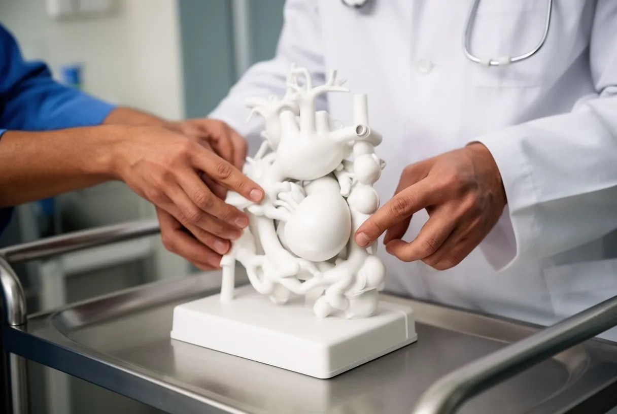
Surgical thresholds and options: When the aneurysm reaches approximately 5.5 centimetres in diameter, or grows rapidly between scans, the balance of risk shifts: the danger of rupture begins to outweigh the risks of planned surgical repair. Two main surgical approaches exist. Open surgical repair involves replacing the weakened aortic segment with a synthetic tube called a graft through a large abdominal incision. This is a major operation but offers a durable, long-lasting result. Endovascular aneurysm repair, or EVAR, is a minimally invasive alternative in which a fabric-covered stent graft is threaded into the aorta through small incisions in the groin arteries and deployed inside the aneurysm to reinforce the wall. EVAR generally involves a shorter hospital stay and faster initial recovery, but it requires specific aneurysm anatomy to be suitable and involves lifelong imaging follow-up to ensure the stent remains properly seated.
Shared decision-making: The choice between open repair and EVAR is not simply a technical question for the surgeon; it depends on your age, fitness for surgery, life expectancy, and the shape of your aneurysm. A younger, very fit patient may benefit from the long-term durability of open repair. An older patient with other medical conditions may face a lower overall risk with EVAR. In complex cases — for example, when aneurysm disease coexists with other cardiac problems requiring procedures such as balloon aortic valvuloplasty or the placement of a balloon-expandable valve — the cardiac and vascular teams coordinate to plan the safest combined strategy. Your surgical risk is often estimated using scoring systems such as EuroSCORE II, which helps frame an honest discussion about likely outcomes rather than offering false reassurance.
Understanding cost structures: The cost of an abdominal aorta screening ultrasound varies widely depending on the country, the healthcare setting, and whether the scan is performed as part of an organised screening programme or as an individual private appointment. In countries with national screening programmes, the test may be free at the point of service for eligible age groups. In private or fee-for-service settings, the cost can range from a modest outpatient fee to a more significant charge if the scan is bundled with a full vascular assessment. For patients travelling internationally for cardiac care, factoring in the cost of a screening ultrasound as part of a broader evaluation package is often sensible.
Insurance coverage for screening: Many insurance plans cover one-time screening ultrasound for men aged 65 to 75 with a smoking history, reflecting established guideline recommendations. Coverage for women, for younger patients, or for repeat surveillance scans may require additional justification from your physician. If the scan is performed for diagnostic reasons — for example, because your doctor felt a pulsatile mass on examination or because you reported abdominal pain — it is more likely to be covered as a medically necessary study rather than a preventive screening test. Patients should verify coverage details with their insurer before the appointment to avoid unexpected charges.
Value beyond the price tag: While cost is a legitimate concern, it is worth considering the value proposition of screening in straightforward terms. An abdominal aorta screening ultrasound is a brief, non-invasive, low-cost test that can identify a potentially fatal condition years before it becomes an emergency. The alternative — discovering an aneurysm only at the moment of rupture — involves emergency surgery, intensive care, and far greater expense with significantly worse outcomes. For patients with relevant risk factors, the screening scan represents one of the most cost-effective preventive investments available in cardiovascular medicine.
Aortic aneurysm screening cost and insurance considerations
Understanding the costs: The cost of an abdominal aortic aneurysm screening ultrasound varies widely by country, healthcare setting and whether the scan is done as part of an organised screening programme or at an individual private appointment. In countries with national screening programmes, the scan may be free for the age groups that qualify. In private or fee-for-service settings, the cost can range from a modest outpatient fee to a considerable charge if the scan is part of a full vascular assessment. If you travel abroad for cardiac care, it is usually worth including the cost of a screening ultrasound in a broader assessment package.
Insurance coverage for screening: Many insurance plans cover a one-time screening ultrasound for men aged 65 to 75 with a history of smoking, in line with established guideline recommendations. To obtain coverage if you are a woman, are younger or need repeat surveillance scans, your doctor may have to provide additional justification. If the scan is done for diagnostic purposes, for example because your doctor felt a pulsating mass during the examination or because you reported abdominal pain, it is more likely to be covered as a medically necessary study rather than a preventive screening test. Check the details of your coverage with your insurer before the appointment to avoid unexpected charges.
The value beyond the price: Although cost is a legitimate concern, it is worth weighing the value of screening in plain terms. An abdominal aortic aneurysm screening ultrasound is a short, non-invasive, low-cost test that can identify a potentially life-threatening condition years before it becomes an emergency. The alternative, discovering an aneurysm only when it ruptures, means emergency surgery, intensive care and a far greater expense, as well as considerably worse outcomes. If you have relevant risk factors, screening is one of the most cost-effective preventive investments available in cardiovascular medicine. We recommend discussing the specific cost and logistics of screening with your cardiology team during a consultation, since the options vary between our Guadalajara, Querétaro and Hermosillo locations.
When to Seek Urgent Medical Attention for Aortic Health
Screening is a preventive measure, but it is equally important to recognise the warning signs that demand immediate emergency care. If you have a known aneurysm or carry significant risk factors, certain symptoms should never be managed at home or delayed until the next clinic appointment.
- Sudden severe abdominal or back pain: A tearing or ripping sensation that appears without warning and does not ease with rest or position change may indicate aneurysm expansion or rupture.
- Dizziness, fainting, or collapse: A rapid drop in blood pressure from internal bleeding can cause sudden light-headedness or loss of consciousness.
- A rapidly expanding pulsing sensation in the abdomen: If a previously subtle pulse becomes prominent and painful, the aneurysm may be growing quickly.
- New, severe pain after a known aneurysm diagnosis: Any new and unexplained pain in the abdomen or lower back in someone under surveillance should be treated as urgent until proven otherwise.
Acting on warning signs: If any of these symptoms occur, go to the nearest emergency department immediately or call your local emergency number. Do not drive yourself if you are dizzy or in severe pain. In the emergency setting, a rapid CT scan can confirm whether the aneurysm has ruptured or is leaking, and the surgical team can mobilise for emergency repair. Time is the single most important variable: the faster a ruptured aneurysm is identified and treated, the better the chance of survival. This is not a moment to call the clinic for a routine appointment — it is a reason to activate the emergency system directly.
For patients without a known aneurysm but with significant risk factors, this information is not meant to cause fear but to build awareness. The purpose of abdominal aorta screening is precisely to prevent you from ever facing an emergency like this. By identifying an aneurysm while it is still small and stable, screening transforms a potentially catastrophic event into a planned, managed, and highly treatable condition. If you are in a high-risk group, we encourage you to book a consultation with our cardiac team to discuss whether a screening ultrasound is appropriate for your individual situation. A simple conversation today can provide both reassurance and a clear plan for protecting your vascular health in the years ahead.






