

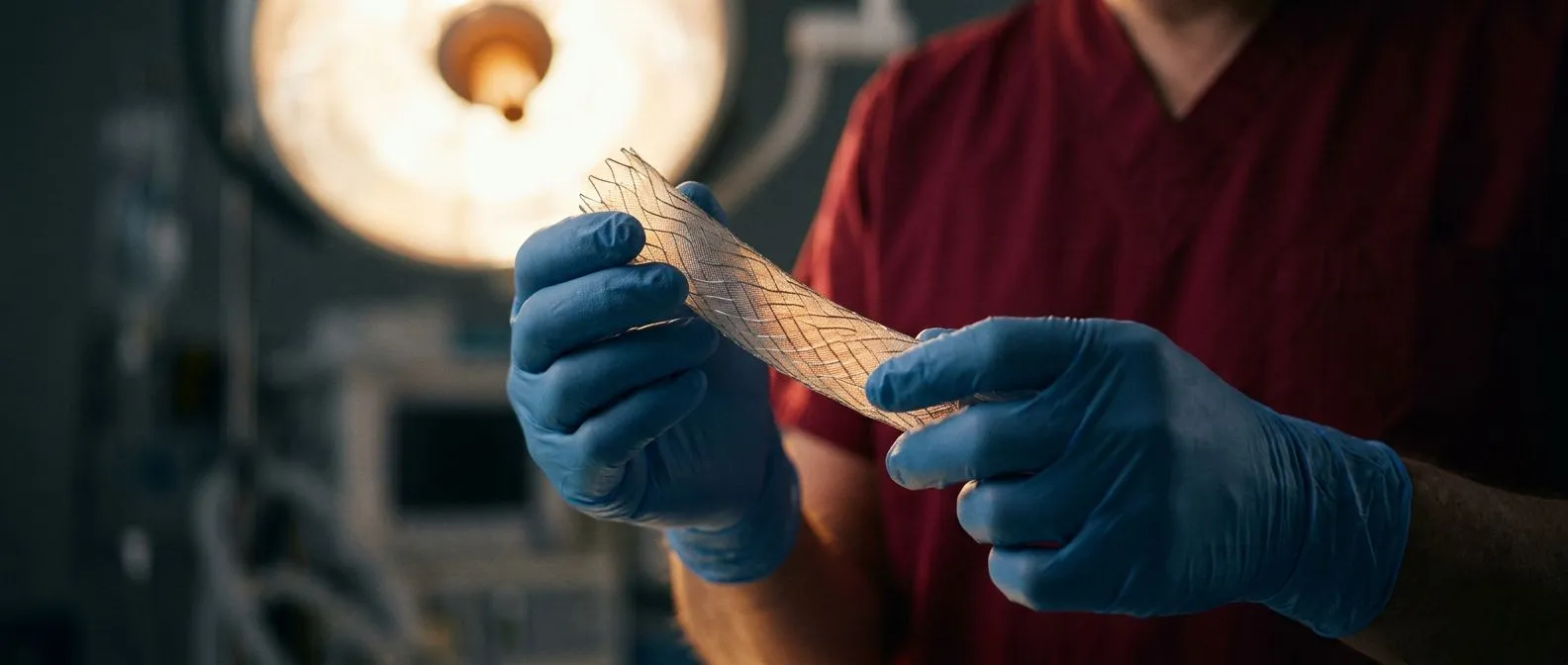
Abdominal Aortic Aneurysm: Warning Signs and Repair
Abdominal aortic aneurysm repair options, warning signs, and when to seek urgent care from heart surgery specialists in Guadalajara.
What Is an Abdominal Aortic Aneurysm and Why Does It Form?
An abdominal aortic aneurysm is a weakened, bulging area in the wall of the aorta, the large blood vessel that carries blood from your heart down through your abdomen. Over time, the constant pressure of blood flow stretches the damaged wall outward, much like a worn spot on an inner tube. Most people with this condition feel nothing at all until the aneurysm grows large or threatens to rupture, which is exactly why understanding the underlying causes matters so much for early detection and timely abdominal aortic aneurysm repair.
What weakens the aortic wall? The most common driver is atherosclerosis, a gradual buildup of fatty deposits called atheromatous plaque along the inner lining of arteries. These plaques narrow and stiffen the vessel, altering blood flow dynamics and concentrating stress on specific segments. When the aortic wall can no longer withstand that stress, it begins to dilate. Research published in the Journal of the American Heart Association has shown that aging-related oxidative damage in vascular cells contributes to progressive remodeling of the aortic wall, especially in the abdominal segment where blood pressure forces are highest.
Who is most at risk? Risk factors cluster in predictable ways. Men over sixty-five, long-term smokers, and people with a family history of aortic disease face the highest likelihood. Chronic high blood pressure accelerates wall damage by exposing the aorta to elevated force with every heartbeat. Connective tissue disorders such as Marfan syndrome can also undermine structural integrity at younger ages. If you carry several of these risk factors, a targeted screening conversation with a cardiac specialist is one of the most practical steps you can take to protect yourself.
Abdominal Aortic Aneurysm Symptoms You Should Never Ignore
Most abdominal aortic aneurysms develop silently. In the early stages, the vessel may expand to a significant size without producing any noticeable sensation. This silent progression is the greatest danger, because the first symptom many patients experience is the aneurysm beginning to leak or rupture. That said, some people do notice subtle warning signs as the aneurysm enlarges and presses on nearby structures, and recognizing these clues can make the difference between planned abdominal aortic aneurysm repair and an emergency operation.
What does a growing aneurysm feel like? As the bulge expands, it may press against the spine or surrounding nerves, producing a deep, pulsating ache in the lower back or the side of the abdomen. Some patients describe a persistent throbbing sensation near the navel that syncs with their heartbeat. Unlike muscular back pain, this discomfort does not improve with rest or position changes. A thorough physical examination that includes careful auscultation, the clinical term for listening to body sounds with a stethoscope, may reveal a bruit, an abnormal whooshing sound over the aneurysm caused by turbulent blood flow.
Can symptoms masquerade as other problems? Yes, and that overlap frequently delays diagnosis. Pain radiating from the abdomen into the groin can mimic kidney stones or hernia pain. Numbness or coolness in the legs may suggest a vascular cause but be attributed to a pinched nerve. Because these symptoms are vague and intermittent, patients and primary-care providers alike may chase other explanations first. If you are in a high-risk group and experience any of these patterns persistently, ask your physician specifically whether an aortic ultrasound is appropriate.
Aortic Aneurysm Warning Signs Before Rupture
When an abdominal aortic aneurysm approaches rupture, the clinical picture changes dramatically. The slow, silent growth gives way to acute, unmistakable symptoms that demand immediate emergency attention. Understanding these late-stage abdominal aortic aneurysm symptoms can save your life or the life of someone you love, because every minute matters once the vessel wall begins to tear.
Sudden, severe pain is the hallmark. A rupturing aneurysm typically causes abrupt, excruciating pain in the abdomen or lower back, often described as tearing or ripping in character. The pain may radiate to the flank, groin, or legs. Patients frequently experience a rapid drop in blood pressure, dizziness, clammy skin, and a racing heart as the body struggles to compensate for internal blood loss. This cascade reflects blood escaping from the aorta into the abdominal cavity, a life-threatening situation that requires surgical intervention within hours.
Internal bleeding from a ruptured aneurysm can also produce arm pain or generalized numbness if blood loss is severe enough to compromise circulation throughout the body. If the aneurysm leaks slowly rather than bursting all at once, there may be a brief window of consciousness during which the patient can seek help. Recognizing the signs of impending rupture matters because emergency open abdominal aortic surgery carries significantly higher mortality than a planned procedure performed before complications arise, a reality well documented in major cardiovascular research registries.
If you or someone near you experiences sudden, intense abdominal or back pain accompanied by fainting or a visible pulsating mass in the abdomen, call emergency services immediately. Do not wait for the pain to pass, and do not attempt to drive yourself to the hospital. Rapid transport to a facility capable of vascular or cardiac surgery offers the best chance of survival.
How Doctors Diagnose an Abdominal Aortic Aneurysm
Diagnosis begins with a focused conversation about risk factors and a physical examination, but imaging studies confirm the aneurysm and determine its size. Because abdominal aortic aneurysm symptoms overlap with so many other conditions, imaging is essential rather than optional. The diagnostic pathway is straightforward, and most patients can complete the evaluation within a single outpatient visit.
Ultrasound is the first-line tool. Abdominal ultrasound is quick, painless, uses no radiation, and provides accurate measurements of the aortic diameter. For screening purposes, a one-time ultrasound in men aged sixty-five to seventy-five who have ever smoked is recommended by major cardiovascular societies. If the ultrasound reveals an aneurysm, the physician will measure its widest dimension and compare it against established abdominal aortic aneurysm size criteria to decide whether surveillance or repair is appropriate. This measurement becomes the reference point for all future monitoring.
CT angiography adds detail when needed. If the aneurysm is large enough to warrant surgical planning, a computed tomography angiogram produces detailed cross-sectional images that map the exact anatomy of the aneurysm, its relationship to branching vessels such as the renal arteries, and the quality of surrounding tissue. This study is critical for determining whether a patient is a candidate for an endovascular stent graft or whether open surgery is necessary. In some complex cases, an arterial line, a thin catheter inserted into an artery for continuous blood pressure monitoring, may be used during preoperative assessment to precisely characterize cardiovascular stability.
Abdominal Aortic Aneurysm Size Criteria for Surgery
Not every aneurysm requires immediate repair. The decision to operate hinges on the aneurysm's diameter, its rate of growth, and the patient's overall health. Established guidelines provide clear thresholds that help surgeons and patients weigh the risk of surgery against the risk of rupture, and these thresholds form the backbone of abdominal aortic aneurysm treatment planning.
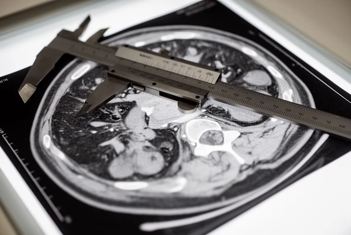
What size triggers repair? The widely accepted threshold for surgical intervention is an aortic diameter greater than 5.5 centimeters in men, with some guidelines suggesting a slightly smaller cutoff for women due to differences in average aortic size and rupture risk. Below this threshold, most aneurysms are monitored with regular ultrasound, typically every six to twelve months depending on size. An aneurysm that grows more than 0.5 centimeters in six months also warrants surgical consideration regardless of absolute size, because rapid expansion signals accelerating wall weakness. These evidence-based thresholds are reviewed in detail by the New England Journal of Medicine and other major medical literature.
For aneurysms below the surgical threshold, blood pressure management and smoking cessation are the cornerstones of conservative care. Medications such as Aspirin may be recommended to reduce cardiovascular risk overall, though it does not directly shrink the aneurysm. The goal during surveillance is to intervene at the optimal moment, before the rupture risk exceeds the surgical risk, and to avoid unnecessary operations on aneurysms that may never cause harm.
Open Abdominal Aortic Surgery vs Aortic Stent Graft Procedure
When the time comes for abdominal aortic aneurysm repair, two primary surgical options exist. Each has distinct advantages, risks, and recovery profiles, and the choice between them depends on the patient's anatomy, age, comorbidities, and surgical risk score. Understanding both approaches helps you participate meaningfully in the decision-making conversation with your cardiac team.
Open surgery is the traditional, durable approach. In open abdominal aortic surgery, the surgeon makes a long incision in the abdomen, clamps the aorta above and below the aneurysm, opens the weakened segment, and sews a synthetic arterial graft in its place. This graft becomes a permanent, strong conduit for blood flow. Open repair has decades of outcome data behind it and remains the gold standard for long-term durability. However, it is a major operation requiring general anesthesia, several days in the intensive care unit, and weeks of recovery at home. Surgeons may use autologous blood, the patient's own blood collected before or during surgery, to minimize transfusion risks from donor blood.
Endovascular repair is less invasive but not for everyone. The aortic stent graft procedure, also called endovascular aneurysm repair or EVAR, involves threading a fabric-and-metal tube through a small incision in the groin, guiding it into the aneurysm under X-ray, and expanding it to reinforce the aortic wall from the inside. Because it avoids a large abdominal incision, recovery is faster and hospital stay is shorter. However, not all aneurysms have the right anatomy for a stent graft. The aneurysm neck, the segment of healthy aorta above the bulge, must be wide enough and straight enough to create a seal. In rare anatomical challenges, such as an ectopic or abnormally positioned kidney, endovascular repair may still be feasible, as described in a case published by the Turkish Journal of Vascular Surgery.
Aortic Stent Graft Procedure Step by Step
For patients who qualify, the endovascular approach has transformed abdominal aortic aneurysm repair from a major open operation to a targeted, minimally invasive procedure. Understanding what happens during the operation reduces anxiety and helps families prepare for what to expect on the day of surgery.
Step 1: Access and imaging. The procedure begins with small incisions in the groin to expose the femoral arteries. The surgeon inserts guide wires and catheters, advancing them under real-time X-ray fluoroscopy toward the aneurysm. Contrast dye is injected to create a detailed roadmap of the aorta and confirm the aneurysm's dimensions. This step ensures the stent graft will be positioned precisely, with its upper and lower ends landing on healthy aortic wall to create a secure seal.
Step 2: Graft deployment. The stent graft, compressed inside a delivery catheter, is carefully advanced to the aneurysm site. Once positioned, the surgeon withdraws the outer sheath, allowing the graft to expand and press against the aortic wall. Some systems use balloon angioplasty, inflating a small balloon inside the graft to ensure full expansion and contact. The graft now forms a new, reinforced channel for blood flow, bypassing the weakened aneurysm sac, which gradually shrinks as pressure is redirected.
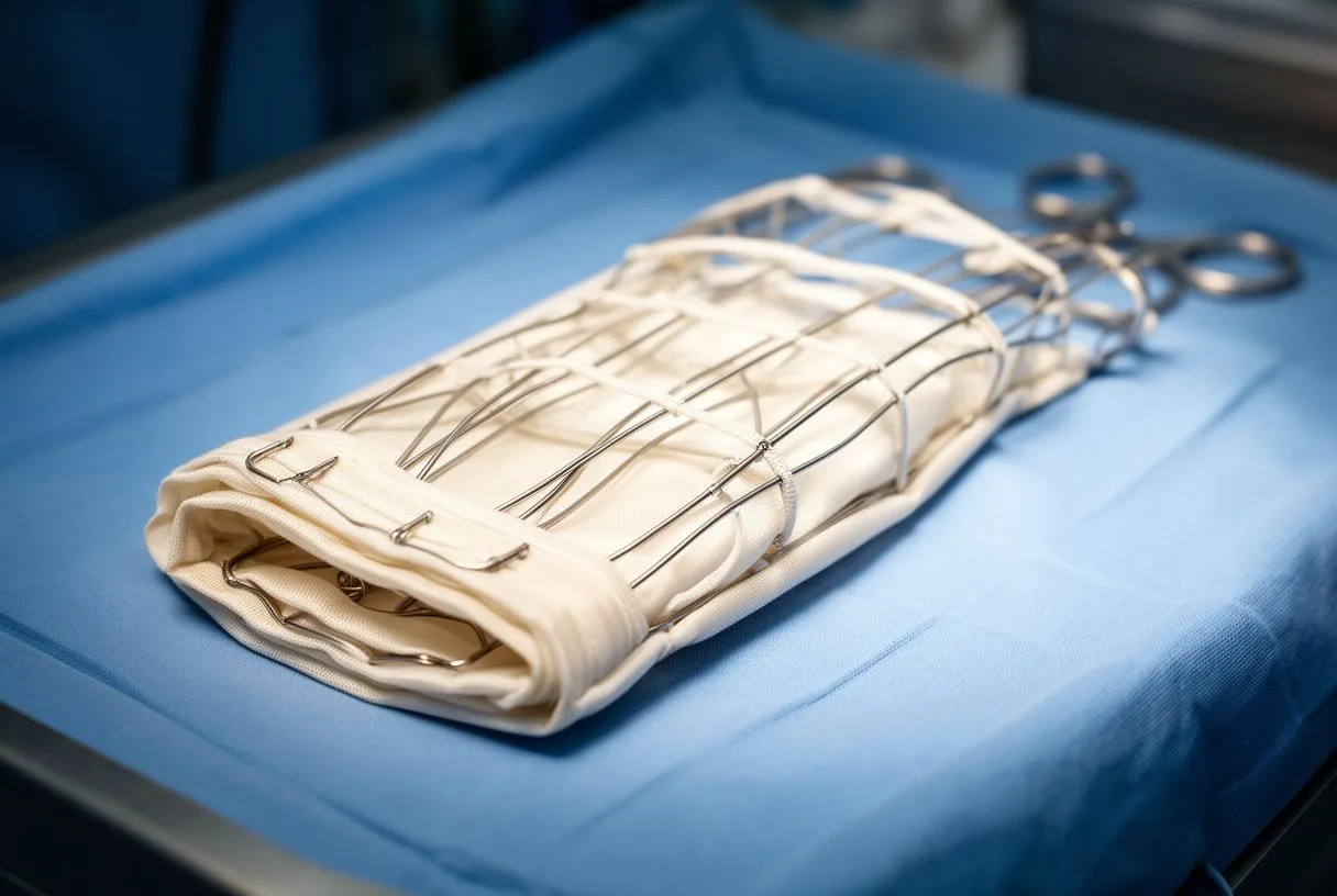
Step 3: Confirmation and closure. Final angiography images verify that the graft is properly seated with no leaks around the edges, a complication called endoleak. Once the surgical team is satisfied, the catheters are removed, the groin incisions are closed, and the patient is transferred to recovery. Most patients spend one to two nights in the hospital and can return to light activity within two weeks. Follow-up CT scans at one month, six months, and annually are essential to confirm the graft remains sealed and the aneurysm is shrinking.
Abdominal Aortic Aneurysm Recovery Timeline
Recovery from abdominal aortic aneurysm repair varies significantly depending on whether you underwent open surgery or endovascular repair. Setting realistic expectations helps you plan for time off work, arrange household help, and recognize when healing is progressing normally versus when something needs medical attention.
Endovascular repair recovery. After an aortic stent graft procedure, most patients walk the same day and leave the hospital within forty-eight hours. You can expect mild groin discomfort and bruising for about a week. Light activities resume in one to two weeks, and most people return to work in two to three weeks. Full lifting restrictions lift after four to six weeks. The long-term commitment is surveillance imaging, because stent grafts require lifelong monitoring to detect endoleaks or graft migration that could necessitate a secondary intervention.
Open surgery recovery. Open abdominal aortic surgery involves a longer hospital stay, typically five to seven days, including one to two days in intensive care. An arterial cannula may remain in place for continuous blood pressure monitoring during the early postoperative period. Pain from the abdominal incision is managed with medication and gradually improves over two to three weeks. Walking is encouraged early to prevent blood clots, but heavy lifting and strenuous activity are restricted for six to eight weeks to allow the abdominal wall and graft to heal securely. Full recovery generally takes two to three months.
In both approaches, cardiac rehabilitation and gradual return to exercise improve outcomes. Your surgical team will tailor recommendations to your specific situation, comorbidities, and the demands of your daily life. The goal is not a generic timeline but a safe, individualized plan that respects the seriousness of the operation while moving you steadily back to independence.
Aortic Aneurysm Repair Complications and Risks
No surgical procedure is without risk, and abdominal aortic aneurysm repair is no exception. Candid discussion of potential complications is a hallmark of high-quality patient education. Understanding the risks empowers you to weigh options, prepare for recovery, and recognize warning signs early.
Open surgery risks. Because open repair is a major abdominal operation, potential complications include bleeding requiring transfusion, infection at the incision site, bowel injury, kidney impairment from clamping the aorta near the renal arteries, and cardiovascular events such as heart attack or stroke. Surgeons calculate individual risk using tools like EuroSCORE II, which incorporates age, cardiac history, lung function, and other factors to estimate operative mortality. The presence of an atheromatous plaque in the aortic arch or neck vessels can increase stroke risk during clamping and should be assessed preoperatively. In some centers, advanced techniques such as awake ECMO, a form of temporary heart-lung support that avoids general anesthesia, are explored for high-risk patients, though this remains uncommon.

Endovascular repair risks. The most common procedure-specific complication is endoleak, where blood continues to flow into the aneurysm sac outside the graft. Many endoleaks resolve spontaneously or are managed with a minor secondary procedure, but persistent leaks can allow the aneurysm to grow and eventually rupture. Other risks include graft infection, migration of the stent over time, and access-site complications in the groin. Because these issues can develop silently, lifelong imaging follow-up is non-negotiable.
- Endoleak: blood leaking around the graft, the most common endovascular complication requiring monitoring.
- Cardiac events: heart attack or arrhythmia such as atrial flutter can occur during or after major vascular surgery.
- Limb ischemia: reduced blood flow to the legs if graft limbs narrow or kink, requiring prompt correction.
When to Seek Urgent Care for an Aortic Aneurysm
Whether you are under surveillance for a small aneurysm or have already undergone abdominal aortic aneurysm repair, knowing when to seek emergency care is one of the most important pieces of information you can carry. A clear action plan reduces hesitation in moments when speed matters most.
Before repair: signs of rupture. If you have a known aneurysm and experience sudden, severe abdominal or back pain, dizziness, fainting, or a rapid heartbeat, treat it as a rupture. Call emergency services immediately. Do not take pain medication and wait. Do not schedule a routine appointment. The window for life-saving surgery is narrow, and transport to a vascular surgery center must happen as fast as possible. Inform the emergency responders that you have a known aortic aneurysm so they can prioritize the right destination hospital.
After repair: signs of graft problems. If you have undergone endovascular repair, new abdominal or back pain, fever, or a pulsating sensation near the original aneurysm site may indicate an endoleak, graft infection, or other complication. Redness, swelling, or drainage from surgical incisions also warrants prompt evaluation. For open repair patients, persistent fever, wound separation, or increasing abdominal swelling should not be dismissed as normal healing. Contact your surgical team or go to the nearest emergency department without delay.
- Sudden tearing pain: call emergency services immediately, do not wait or self-medicate.
- Fever after surgery: may signal graft infection, requires same-day evaluation.
- New pulsating mass: possible graft or aneurysm expansion, seek urgent imaging.
Abdominal aortic aneurysm repair, whether through open surgery or a stent graft procedure, is one of the most well-studied interventions in vascular medicine. The key to a good outcome lies in early detection, accurate sizing, and choosing the right procedure for your anatomy and health profile. If you or a loved one has risk factors or a known aneurysm, we encourage you to discuss your specific situation with a qualified cardiac and vascular surgery team. Dr Alejandro Molina and the team at Cirugía de Corazón in Guadalajara, Querétaro, and Hermosillo are available for consultation to help you understand your options and plan the safest path forward. Please book a consultation rather than relying on this information for self-diagnosis.






