Partial anomalous pulmonary venous drainage
Learn about partial anomalous pulmonary venous drainage, its symptoms, diagnosis, and surgical treatment options for this congenital heart condition.
Not everyone goes looking for information about anomalous venous drainage, so if you are reading this, you or someone you love has probably been diagnosed with it. I understand: it is not easy to find reliable information on this subject written for patients and their families. So relax, you have come to the right place.
In this article we will look at an important but perhaps little-known subject: partial anomalous pulmonary venous drainage. We will cover what the condition is, the treatments available, the tests needed, possible complications, the benefits of treatment and, most importantly, how to keep a good quality of life in the long term. By the end you will have a clear idea of what you are facing.
Let's start with the anatomy and circulation of a normal heart
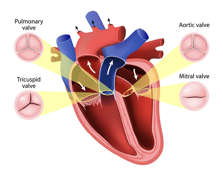
The heart's normal circulation is a complex, essential system that moves blood through the heart and the rest of the body, delivering oxygen and nutrients to the tissues and removing waste products. These are its main steps:
- Right-sided or pulmonary circulation: this begins in the right atrium, which receives oxygen-poor blood from the body through the superior and inferior venae cavae. From there, blood flows through the tricuspid valve into the right ventricle. Once in the right ventricle, the heart contracts and pumps the blood into the pulmonary artery, which carries it to the lungs.
- The lungs: the pulmonary artery carries oxygen-poor blood to the lungs, where gas exchange takes place. In the lungs, the blood picks up oxygen and releases carbon dioxide, a process known as oxygenation.
- Left-sided or systemic circulation: once the blood has been oxygenated in the lungs, it returns to the heart through the pulmonary veins into the left atrium. Normally there are 4 pulmonary veins draining into the left atrium. From the left atrium, blood flows through the mitral valve into the left ventricle.
- The body: from the left ventricle, the heart contracts again and pumps the oxygenated blood into the aorta, the body's main artery. The aorta branches into smaller arteries that carry blood to every organ and tissue. Once the oxygen-rich blood has been delivered and the tissues have taken up the oxygen and nutrients, the oxygen-poor blood makes its way back to the heart to repeat the cycle without interruption.
This process keeps a constant flow of blood throughout the body. The heart, like an efficient pump, works in step with the valves and arteries to maintain proper circulation and make sure every tissue receives the oxygen and nutrients it needs to work properly.
What is partial anomalous pulmonary venous drainage?
Partial anomalous pulmonary venous drainage is a condition in which one or more of the veins that carry blood from the lungs to the heart are not connected correctly. In other words, instead of emptying into the left atrium as they should, they “send back” their blood to the right side of the heart, even though it is already oxygenated.
As a result, some of the oxygenated blood does not flow properly to the left side of the heart, which can affect heart function and circulation. It also makes the right side of the heart work “double”, because part of the blood it has already sent to the lungs comes straight back to where it started.
How is it different from total anomalous venous drainage?
In total anomalous pulmonary venous drainage, unlike the partial form, all of the pulmonary veins are abnormally connected to the venous system, which means none of them drains correctly into the left atrium. The oxygenated blood returning from the lungs does not reach the left atrium to be pumped to the rest of the body; instead, it is diverted to the right atrium or another systemic vein, causing a mixing of oxygen-rich and oxygen-poor blood and a significant disturbance of the circulation.
The key differences between partial and total anomalous venous drainage are:
- Venous connections: in the partial form, only some of the pulmonary veins are abnormally connected, whereas in the total form all of them are.
- Blood flow: in the partial form, part of the oxygenated blood flows correctly to the left atrium, whereas in the total form all of it is diverted to the right atrium or the systemic venous system.
- Severity: the total form is generally more serious, with more obvious and potentially more dangerous symptoms than the partial form.
The diagnosis, symptoms and treatment of total anomalous venous drainage differ from those of the partial form. As with any congenital (present from birth) heart condition, it is essential that a medical team specialised in paediatric or adult cardiology handles the assessment and treatment of these patients. Treatment usually involves early surgical correction to restore normal circulation and prevent long-term complications.
What are the symptoms, and how is anomalous venous drainage diagnosed?
The symptoms can vary with the severity of the condition and the patient's age. Some patients have more obvious symptoms, while others may have none or only mild ones. The most common symptoms include:
- Cyanosis: a bluish colour of the lips, nails and skin due to a lack of oxygen in the blood.
- Breathing difficulty: a feeling of breathlessness or difficulty breathing, especially during exercise or physical activity.
- Tiredness and weakness: getting tired and weak easily.
- Feeding difficulties in babies: babies may have trouble feeding and grow slowly.
- Recurrent chest infections: greater susceptibility to respiratory infections such as pneumonia and bronchitis.
- Abnormal heart rhythm: arrhythmias or irregular heartbeats may occur.
Anomalous venous drainage is usually diagnosed with a combination of medical tests and imaging. Some of the methods used are:
- Physical examination: when examining you with a stethoscope, your doctor may hear a heart murmur or abnormal heartbeats, although this is not always the case, because the signs can be very subtle or even absent.
- Echocardiogram: an ultrasound test that shows real-time images of the heart and its structures. It can identify the abnormal venous drainage and assess blood flow in the heart.
- Chest X-ray: can show changes in the size and shape of the heart and lungs.
- Electrocardiogram (ECG): records the heart's electrical activity to detect arrhythmias or other rhythm abnormalities.
- Cardiac MRI or CT: these give detailed images of the heart and its blood vessels, allowing a more precise assessment of the anatomy and the abnormalities.
- Cardiac catheterisation: sometimes needed to obtain more detailed information about the vascular abnormalities and the pressures inside the heart.
Once the diagnosis is made, it is important that you or your relative are assessed and treated by a medical team specialised in paediatric or adult cardiology with experience in managing anomalous venous drainage. Treatment will depend on the severity of the condition and may include medical and surgical options to correct the abnormality and improve the circulation.
What happens if the condition is not treated?
In patients with partial anomalous venous drainage, the disturbed circulation causes physiological changes in the cardiovascular system. These changes are directly related to the abnormal flow of blood from the lungs to the heart, and vary with the severity and the specific type of venous malformation. Some of the most common are:
- Volume overload of the right atrium.
- Dilation of the right ventricle.
- Reduced systemic blood flow.
- Pressure overload in the pulmonary artery.
- Tissue hypoxia (lack of oxygen).
- Arrhythmias or heart failure.
Is there a treatment for partial anomalous venous drainage?
The approach to treatment depends on the severity of the condition and the symptoms. Because it may be obvious from birth and need immediate intervention in some cases, or remain completely silent and only be found in adulthood, after the age of 30, 40 or even 50, treatment must be individualised according to its severity and its effects on the cardiovascular system.
Treatments and possible interventions
- Medical management: for mild cases, the doctor may recommend medical management, which can include medicines to help improve heart function and relieve symptoms.
- Surgery: in more serious cases, surgery may be needed to correct the anomalous drainage. Cardiovascular surgeons can repair the incorrect venous connections and restore proper blood flow, either with minimally invasive procedures or with open-heart surgery.
- Catheterisation: in some cases, doctors may choose cardiac catheterisation procedures. This less invasive approach uses catheters to correct the abnormal connections and improve circulation.
Possible complications
It is important to bear in mind that, as with any medical procedure, complications can occur. However, advances in technology and the experience of medical professionals have significantly reduced the risks of these interventions. Your medical team will work closely with you and your family to minimise any potential risk and ensure the best possible outcome.
Benefits of treatment
Correcting partial anomalous venous drainage can bring several significant benefits for health and quality of life. Potential benefits include:
- Better heart function: restoring proper blood flow can help improve how the heart works and reduce the associated symptoms. Essentially, we return to a normal circulation and, little by little, the heart resumes its normal physiological function.
- Relief of symptoms: many patients find their symptoms ease once the drainage problem has been corrected.
- Prevention of complications: we avoid complications that could otherwise develop, such as arrhythmias, pulmonary hypertension, heart failure, right ventricular hypertrophy or death.
The take-home message
We can conclude that partial anomalous venous drainage is a condition that affects blood flow and heart function. However, there are effective and safe treatments that can improve quality of life and offer the prospect of a healthy life.
The exact timing of surgical correction will depend on many factors and on each patient's personal circumstances. It is an important conversation to have with your doctor before making a decision. Mutual trust and respect between doctor and patient are essential to move forward, as is the right care, to achieve the best possible results. Remember: you and your family are not alone on this journey, and we are here to help you every step of the way.

About the author
Dr Alejandro Molina is a cardiovascular surgeon practising in Guadalajara, Querétaro and Hermosillo. He trained in cardiothoracic surgery at the Universidad de Guadalajara, completed advanced training in cardiac surgery, minimally invasive surgery, robotic surgery and structural interventions at the University of Montreal, and a fellowship in structural cardiology and TAVR at CHU Bordeaux, France. He performed the first robotic mitral valve repair in the Ángeles Health System and the first robotic heart surgery in Guadalajara.
Medical degree: Universidad Autónoma de Guadalajara, Cédula Profesional 5757260 · Cardiothoracic surgery: Universidad de Guadalajara, Cédula de Especialidad 10768871 · Current certification, Consejo Nacional de Cirugía del Tórax, no. 447