Bradycardia: When a Slow Heart Rate Needs a Pacemaker
Discover when a bradycardia pacemaker becomes essential for slow heart rate.
A resting pulse that dips well below the expected range often raises immediate questions for patients and their families. When a patient experiences persistent fatigue, lightheadedness, or sudden fainting spells, clinical evaluation may point toward a diagnosis requiring a bradycardia pacemaker to maintain a safe, physiological rhythm. Understanding how the heart's natural electrical wiring functions—and what happens when those impulses falter—allows individuals to make grounded, confident decisions alongside their cardiovascular specialists.
What Is a Bradycardia Pacemaker?
Understanding bradycardia fundamentals begins with defining standard cardiac pacing. In clinical terms, bradycardia refers to an adult resting heart rate that drops below 60 beats per minute. While a low pulse is common and entirely benign among highly trained athletes or during deep sleep, pathological bradycardia occurs when the heart cannot pump an adequate supply of oxygenated blood to satisfy the body's metabolic demands. When intrinsic electrical impulses slow down or encounter physical blocks, a small, surgically implanted electronic device—a bradycardia pacemaker—can monitor and actively stimulate the myocardium to beat at a reliable rate.
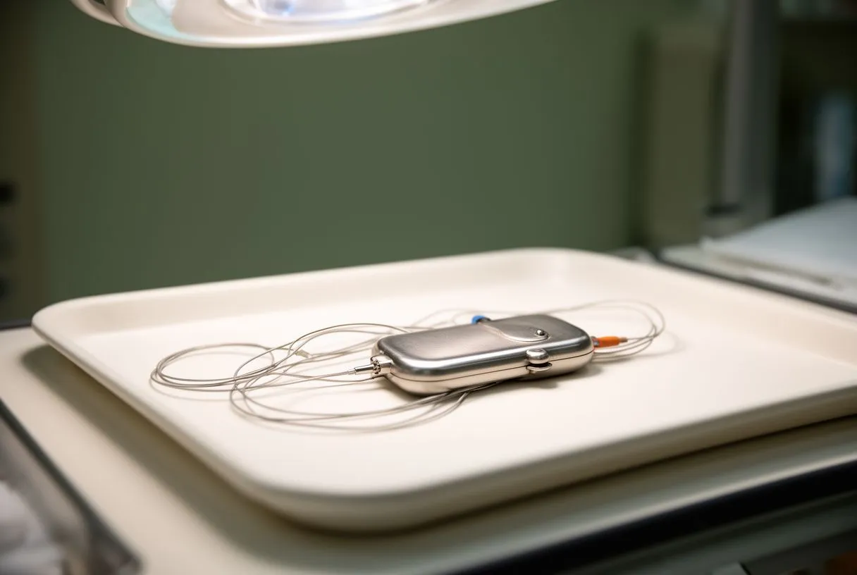
According to health resources such as WebMD, a permanent pacing system consists of an insulated pulse generator containing a miniature battery and computerized circuitry, alongside one or more flexible electrical leads placed inside the cardiac chambers. By continuously tracking the intrinsic beat-to-beat activity of the tissue, the device delivers microscopic, painless electrical discharges only when the native pulse falters. This demand-based pacing ensures that normal heartbeats pass uninterrupted while preventing dangerous pauses.
For international patients seeking treatment through cross-border healthcare or traveling to centers of excellence in Guadalajara, Querétaro, or Hermosillo, understanding device mechanics helps alleviate anxiety. Modern systems do not replace the natural heart; rather, they serve as a vigilant safety net. Whether treating sinus node dysfunction or high-grade atrioventricular block, the modern bradycardia pacemaker is tailored to restore energy levels and prevent rhythm-induced syncope.
How Bradycardia Affects the Heart
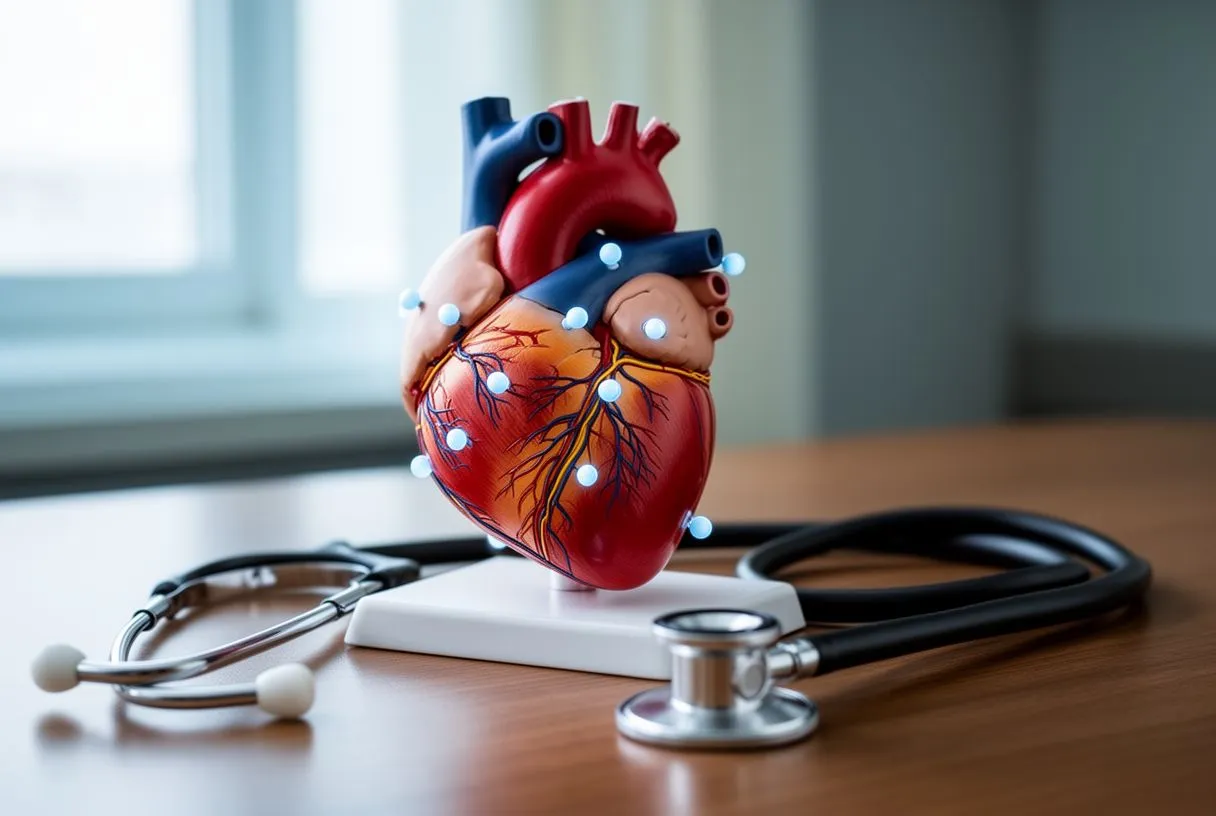
The electrical conduction pathway governs how blood travels through each chamber. In a healthy heart, an electrical trigger originates in the sinoatrial node located in the upper right atrium. This pulse sweeps across the atria, prompting them to contract, before reaching the atrioventricular node. From there, the current travels down the bundle of His and through the Purkinje fibers, causing the ventricles to squeeze blood outward to the lungs and systemic circulation. When fibrosis, degenerative wear, or ischemic injury interrupts this pathway, the rate drops significantly.
Broad epidemiological research underscores how cardiovascular conditions contribute to functional impairment. As shown in the Global, regional, and national disability-adjusted life-years (DALYs) for 359 diseases and injuries and healthy life expectancy (HALE) for 195 countries and territories, 1990-2017: a systematic analysis for the Global Burden of Disease Study 2017, heart diseases remain a leading driver of reduced quality of life globally. A chronically depressed rate reduces total cardiac output, which can eventually lead to exercise intolerance, fluid retention, and secondary strain on the left ventricle.
Sinus node dysfunction and heart block represent the two principal pathophysiological mechanisms behind symptomatic slow rates. Sick sinus syndrome encompasses sinus pauses, sinus arrest, and alternating fast-slow rhythms (tachy-brady syndrome). In contrast, atrioventricular block involves delayed or completely severed communication between the upper and lower chambers. In complete heart block, the ventricles must rely on an unreliable intrinsic escape rhythm, often dropping below 30 to 40 beats per minute, which creates severe hemodynamic instability.
Common Symptoms of Slow Heart Rate
Recognizing low cardiac output signs early can prevent traumatic injuries associated with falls. When systemic blood pressure drops due to prolonged pauses between contractions, the brain is the first organ to register the deficit. Patients frequently report moments of sudden unsteadiness, visual graying, or profound weakness during routine physical efforts like climbing stairs or carrying groceries.
Differentiating normal resting pulse variations is essential because a low number alone does not automatically necessitate surgical intervention. The critical distinction lies in whether the slow rhythm impairs end-organ perfusion. If a patient remains entirely energetic with normal cognitive clarity and exercise capacity, watchful waiting is generally appropriate. Conversely, when slowness coincides with tangible functional limitation, a thorough cardiovascular workup is imperative.
- Presyncope and Syncope: Sudden, transient loss of consciousness or near-fainting episodes resulting from brief pauses in blood flow to cerebral vessels.
- Unexplained Chronic Fatigue: Persistent exhaustion that fails to improve with sleep, caused by diminished oxygen transport throughout major organ systems.
- Exertional Shortness of Breath: Inability of the pulse to rise appropriately during physical activity, leading to immediate breathlessness and muscle fatigue.
- Cognitive Fog and Confusion: Memory lapses, difficulty concentrating, or sudden disorientation linked to reduced cerebral blood delivery.
Diagnosing Bradycardia and Conduction Disorders
Initial clinical assessment begins with a detailed medical history and a standard resting 12-lead electrocardiogram. The surface tracing provides an instantaneous snapshot of rhythm, allowing electrophysiologists to inspect the PR interval, QRS duration, and morphology of P waves. However, because electrical pauses often occur unpredictably, a normal resting baseline tracing does not exclude intermittent conduction failure.
When transient episodes evade stationary detection, extended ambulatory monitoring becomes necessary. Clinicians utilize 24- to 48-hour Holter monitors, multi-week patch event recorders, or insertable cardiac monitors to continuously record rhythm dynamics. Pairing patient-logged symptom timestamps with electrocardiographic strips allows the medical team to verify whether a dizzy spell corresponds to a documented pause or advanced heart block.
Advanced diagnostic imaging techniques often complement rhythmic evaluations to determine whether underlying myocardial disease is present. As outlined in the Clinical recommendations for cardiovascular magnetic resonance mapping of T1, T2, T2* and extracellular volume: A consensus statement by the Society for Cardiovascular Magnetic Resonance (SCMR) endorsed by the European Association for Cardiovascular Imaging (EACVI), mapping myocardial tissue helps identify infiltrative processes, structural scarring, or active inflammation. Ruling out reversible triggers, such as medication side effects or severe thyroid deficiencies, ensures that device therapy is deployed only when truly indicated.
When Bradycardia Requires a Pacemaker
Clinical criteria for device therapy follow clear international guidelines established by major cardiovascular societies. Implantation of a bradycardia pacemaker is definitively recommended when documented symptomatic bradycardia directly produces syncope, presyncope, or heart failure symptoms. It is also strongly indicated in asymptomatic patients who present with high-grade or third-degree atrioventricular block, where the risk of sudden cardiac arrest or profound hemodynamic collapse is elevated.
Distinguishing reversible and structural causes protects patients from undergoing unneeded surgical procedures. Before scheduling an implantation, the clinical team meticulously checks for secondary culprits, such as supratherapeutic beta-blocker or calcium channel blocker dosing, severe electrolyte derangements, or acute Lyme carditis. If rhythm normalization occurs once an offending drug is tapered under controlled supervision, permanent device therapy can frequently be avoided.
When structural degeneration of the conduction bundle is irreversible, delaying intervention can prove dangerous. An unexpected cardiac pause lasting several seconds can precipitate falls, bone fractures, or motor vehicle collisions. In these scenarios, the consensus of the surgical team focuses on establishing a steady baseline rate to protect both longevity and independent daily functioning.
Types of Pacemaker Devices Available
Single-chamber and dual-chamber units remain the historical mainstays of clinical rhythm management. A single-chamber device utilizes a solitary lead placed either in the right atrium or the right ventricle. Dual-chamber systems feature two separate leads—one terminating in the right atrium and the other in the right ventricle. This allows the generator to coordinate atrial and ventricular contractions sequentially, closely preserving the natural timing of the heart.
Biventricular devices and leadless pacing innovations cater to distinct specialized clinical profiles. Biventricular pacemakers, commonly used in cardiac resynchronization therapy, employ three leads to coordinate both ventricles in patients who also suffer from heart failure and conduction delay. In recent years, miniaturized leadless pacemakers have also emerged, resting entirely inside the right ventricle without requiring transvenous leads or a subcutaneous chest pocket.
- Single-Chamber Systems: Paces either the atrium or ventricle; frequently used for chronic atrial fibrillation accompanied by slow ventricular response.
- Dual-Chamber Systems: Paces both upper and lower chambers; ideal for sick sinus syndrome and standard complete heart block to restore synchrony.
- Biventricular Systems: Coordinates both ventricles simultaneously; suited for patients experiencing dyssynchronous heart failure alongside conduction abnormalities.
- Leadless Miniaturized Devices: Self-contained capsules placed via catheter; eliminates lead-related complications for select single-chamber candidates.
The Pacemaker Implantation Procedure Explained
Preoperative preparation and anesthesia prioritize patient comfort and absolute surgical sterility. Most procedures take place in an advanced electrophysiology laboratory or a specialized hybrid operating room under local anesthesia combined with conscious intravenous sedation. This ensures the patient remains relaxed, pain-free, and breathing spontaneously throughout the intervention without the deeper systemic demands of general anesthesia.
The surgical technique step-by-step involves creating a small incision, typically two inches in length, just beneath the collarbone. The cardiac surgeon gains access to the cephalic, axillary, or subclavian vein to navigate the insulated leads under continuous fluoroscopic X-ray visualization into the heart. Once the lead tips are secured into the cardiac tissue, electrical testing confirms appropriate sensing and pacing thresholds.
The leads are then connected to the small pulse generator, which is nestled into a carefully fashioned subcutaneous pocket beneath the skin or over the pectoral muscle. The surgical team closes the incision using layered absorbable sutures and applies a sterile, protective dressing. In standard, uncomplicated scenarios, the entire implantation procedure requires approximately one to two hours to complete.
Recovery and Living with a Pacemaker
Immediate post-implantation care focuses on wound healing and lead stabilization. Patients typically spend several hours in a monitored recovery suite, and many are discharged home the same afternoon or following a single overnight observation. For the first four to six weeks, individuals are advised to avoid lifting heavy objects or raising the arm on the operated side above shoulder height to allow the lead anchors to fibrose securely into the heart wall.
Resuming daily activities and exercise occurs gradually under medical guidance. Light walking is encouraged almost immediately after discharge to promote circulation and prevent stiffness. Once cleared by their cardiologist, patients can return to swimming, golfing, gardening, and light recreational activities, experiencing substantially improved stamina now that their heart maintains an adequate rate.
Modern devices feature robust shielding against everyday electromagnetic fields. Cell phones, microwaves, computers, and household appliances can be used safely without disrupting device function, provided phones are kept a few inches away from the generator site. Patients are issued a medical device identification card, which should be presented at airport security checkpoints to bypass magnetic wands directly over the generator.
Evaluating Surgical Risks and Long-Term Care
Procedural complications and mitigation strategies are rare when procedures are carried out by experienced cardiovascular surgical teams. Potential risks include pocket hematoma, localized infection, lead dislodgement, or inadvertent pneumothorax during venous puncture. Rigorous sterile protocols, prophylactic antibiotics, and precise fluoroscopic guidance minimize these possibilities to low single-digit percentages.
Battery longevity and device replacement are predictable aspects of lifelong pacing. Today's lithium-iodine batteries generally function between 7 and 15 years, depending on pacing frequency and programmed output. Routine in-clinic or remote telemetry checks continuously monitor battery voltage. When the device approaches its elective replacement indicator, an outpatient procedure is scheduled to exchange the generator while preserving the established leads.
Routine follow-up appointments allow electrophysiologists to fine-tune pacing settings non-invasively using an external programmer. Settings can be tailored to match the patient's individual lifestyle, ensuring maximum physiological responsiveness during movement while extending overall battery life.
Seeking Expert Cardiac Care in Mexico
World-class infrastructure and surgical expertise in Mexico attract both domestic residents and traveling international patients seeking high-caliber cardiac care. Modern private surgical hospitals in major hubs such as Guadalajara, Querétaro, and Hermosillo house advanced electrophysiology suites equipped with state-of-the-art diagnostic imaging and monitoring technologies.
A cardiologist can carry out a thorough evaluation to identify the exact mechanism behind a slow pulse. Whether an individual requires specialized rhythm diagnostic testing, medical management, or a modern pacing implant, care plans are executed with meticulous clinical precision and empathetic communication.
If you or a loved one is navigating episodes of unexplained fainting, chronic exhaustion, or a newly diagnosed low heart rate, do not rely on self-diagnosis. Scheduling a comprehensive consultation with a qualified cardiac team provides the diagnostic clarity and tailored treatment necessary to protect your heart health and restore your active quality of life.