Chest X-Ray Before and After Heart Surgery
Chest x-ray heart surgery guide shows what imaging reveals before and after cardiac procedures.
What Does a Chest X-Ray Show Before Heart Surgery
When you or a loved one are preparing for heart surgery, a chest x-ray is one of the first imaging tests your cardiac team will request. This simple, painless picture of your chest gives the surgeon a baseline view of your heart's size, the position of major blood vessels, and the condition of your lungs before any intervention takes place. Think of it as the "before" photograph in a before-and-after comparison — it establishes what is normal for you so that any change after surgery can be measured against it. A preoperative chest x-ray also helps your surgeon confirm that there are no hidden problems, such as a lung infection or fluid accumulation, that could complicate your procedure.
What your surgeon is actually looking for: The heart sits in the middle of the chest between the lungs, and its silhouette on an x-ray tells a trained eye a great deal. The cardiac silhouette — the shadow the heart casts on the image — reveals whether the heart is enlarged, which can suggest long-standing valve disease or heart failure. The ascending aorta, the first segment of the body's largest artery as it leaves the heart, may appear widened if an aneurysm is present. Your surgeon measures these structures precisely, because decisions about surgical approach and risk often hinge on whether the heart and great vessels fall within normal dimensions or show signs of chronic disease.
The lungs are equally important on this baseline image. Your team checks for signs of fluid overload, chronic lung disease, or any opacity that might represent infection or scarring. If you have a history of smoking or long-standing cardiac symptoms, the lungs may show changes that influence how you will be managed during and after surgery. For example, evidence of chronic obstructive lung disease might prompt the anaesthesiologist to adjust ventilation settings. Everything the team learns from this single image feeds into the surgical plan, ensuring that when you arrive in the operating room, your team already knows the landscape of your chest.
Why Do I Need a Preoperative Chest X-Ray for Cardiac Surgery
Many patients wonder why they need another x-ray when they have already had a CT scan or echocardiogram. The answer is that each imaging test answers a different question. An echocardiogram shows how the heart valves move and how well the heart pumps. A CT scan provides detailed cross-sectional anatomy. A chest x-ray, by contrast, gives a fast, low-radiation overview that confirms the position of any existing hardware — such as a pacemaker — and screens for acute lung problems that other tests might miss. According to WebMD, chest x-rays remain one of the most commonly ordered preoperative tests because they are quick, widely available, and reveal information that changes surgical management in a meaningful percentage of patients.
Screening for hidden lung and vascular problems: Some patients arrive for heart surgery with a cough they have attributed to a cold, only for the x-ray to reveal a pleural effusion — fluid between the lung and the chest wall — that needs drainage before surgery is safe. Others may have an atheromatous plaque, a fatty deposit in the wall of a blood vessel, that has calcified enough to be visible on x-ray and signals widespread vascular disease. These findings do not always cancel surgery, but they reshape the plan. The surgeon may adjust the incision site, order additional blood tests, or coordinate with a lung specialist to optimise lung function before the operation.
The preoperative x-ray also documents the position of the trachea and main bronchi, the airways that branch from the windpipe into the lungs. If the heart or a great vessel is enlarged enough to push an airway out of its normal position, the anaesthesia team needs to know in advance so they can choose the right breathing tube and ventilation strategy. This is especially relevant for patients undergoing surgery on the aortic valve or the ascending aorta, where enlargement can shift the airway anatomy. Every detail captured before surgery becomes a reference point for the images taken afterward, making the preoperative chest x-ray an indispensable part of your cardiac surgical record.
What Changes Appear on a Chest X-Ray After Heart Surgery
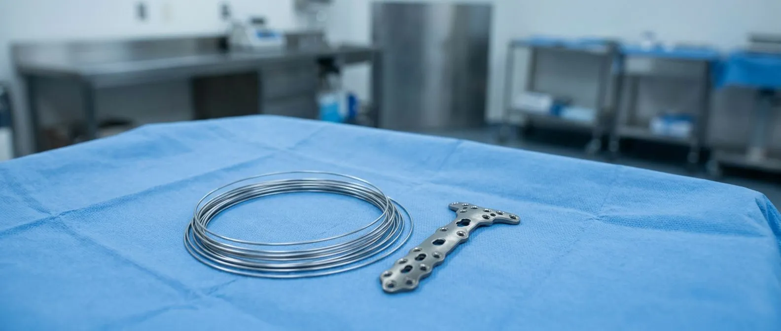
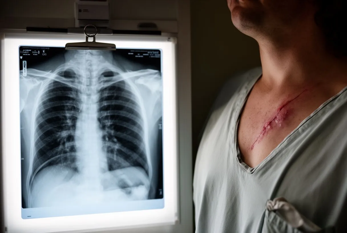
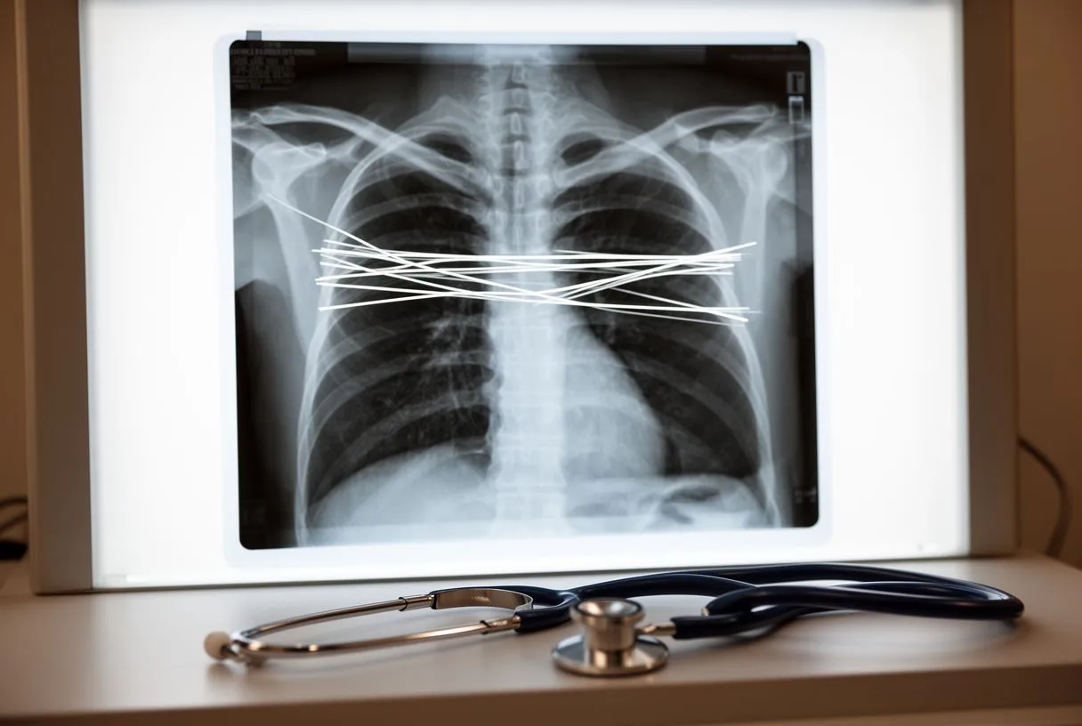
After heart surgery, the first chest x-ray is usually taken in the intensive care unit within hours of your arrival, often while you are still waking from anaesthesia. This image looks dramatically different from your preoperative one. The most visible change is the presence of sternotomy wires — thin metal loops the surgeon uses to close the breastbone (sternum) after a midline chest incision. These wires appear as bright white lines running vertically down the centre of the chest and remain visible on every future x-ray for the rest of your life. They are a normal and expected finding, not a cause for alarm.
Lines, tubes, and hardware you will see: In the immediate postoperative period, the x-ray shows several temporary items that your care team uses to monitor and support you. An arterial line, a thin catheter placed in a wrist or groin artery, provides continuous blood pressure readings and appears as a faint shadow near the wrist or in the upper chest. An arterial cannula, a larger tube used during cardiopulmonary bypass to connect your circulation to the heart-lung machine, may still be visible if it has not yet been removed. Chest tubes placed to drain blood and air from around the heart and lungs show as rounded or tubular densities at the lower edges of the chest. All of these are routine, and your team will remove them as you recover.
The cardiac silhouette itself often looks wider in the first few days after surgery. This widening reflects temporary swelling of the tissues around the heart and is not necessarily a sign of a problem. The mediastinum — the central compartment of the chest that contains the heart, great vessels, and thymus — typically appears broader because of postoperative fluid and surgical oedema. If you had coronary artery bypass surgery, small surgical clips may be visible along the path of the bypass grafts. If you had valve surgery, the prosthetic valve may appear as a dense, ring-shaped shadow that replaces the natural valve on the image. Your surgeon compares these findings against the preoperative x-ray to confirm that everything is in the expected position.
How Long Until Your Chest X-Ray Returns to Normal After Surgery
The timeline for x-ray normalisation depends on the type of surgery, your overall health, and whether any complications arise. In the first week, the chest x-ray typically still shows widening of the mediastinum, small amounts of pleural fluid, and the gradual removal of lines and tubes. By the end of the first week, most chest tubes have been removed, and the mediastinal width begins to narrow as swelling subsides. The sternotomy wires remain, but the surrounding tissue inflammation that made them look prominent starts to fade. Your team takes serial x-rays during this period to track these changes and confirm that the heart and lungs are recovering as expected.
Weeks to months — gradual resolution: By two to three weeks after surgery, many patients' x-rays show a cardiac silhouette closer to its preoperative size, though subtle widening may persist. Small pleural effusions, especially on the left side where the surgery was performed, can linger for weeks and are common after valve surgery or bypass using an internal mammary artery graft. These effusions usually shrink without intervention, but if they grow or cause breathlessness, your surgeon may drain them. A rare but important consideration is an atypical infarction — a heart attack that presents with unusual symptoms — which can sometimes be suspected if the x-ray shows new pulmonary congestion in the absence of obvious surgical causes. Such findings prompt urgent evaluation with blood tests and echocardiography.
By three months, the x-ray in most patients shows a stable cardiac silhouette, resolving or resolved pleural fluid, and clear lung fields. The sternotomy wires are the only permanent visible change. In a small number of patients, chronic changes such as pleural thickening or a slightly widened mediastinum persist indefinitely and are simply noted as post-surgical findings. Research into unusual postoperative imaging presentations continues to expand our understanding; for instance, a recent case report documented a rare vascular anomaly discovered through postoperative imaging follow-up (Case Report: A rare case of multi-vessel coronary artery-coronary sinus fistula combined with multiple coronary aneurysms initially presenting with frequent premature ventricular complexes). This underscores why your cardiac team values follow-up imaging even when you feel well.
What Are Common Sternotomy X-Ray Findings
Patients who have had a median sternotomy — the vertical cut through the breastbone used in most open-heart surgeries — often ask whether the metal wires will set off airport security alarms or cause problems later. On the x-ray, these wires appear as a series of bright loops arranged in a vertical line down the sternum. Over time, they may develop a slightly irregular appearance as bone remodels around them, but this is normal. The wires rarely cause problems, though in some cases a wire may irritate surrounding tissue and need removal years later. On a chest x-ray heart surgery follow-up, your surgeon checks wire alignment to ensure the sternum is healing in proper alignment.
Sternum healing and what the wires tell your surgeon: The pattern and spacing of the wires reveal whether the bone is healing symmetrically. If the wires are pulling apart or bent, the surgeon may suspect sternal instability — a condition where the two halves of the breastbone are not fusing properly. This can happen in patients with osteoporosis, diabetes, or those who cough forcefully in the early recovery period. Sternal instability may require a supportive brace or, in severe cases, surgical re-fixation. The x-ray is one of several tools — alongside physical examination and sometimes a CT scan — used to assess bone healing.
Beyond the wires, the postoperative x-ray reveals other common findings that are expected rather than alarming. Here is a summary of what patients and families typically see:
- Sternotomy wires: Permanent metal loops that close the breastbone; they remain visible on all future x-rays.
- Pleural effusion: Fluid between the lung and chest wall, common on the left side, usually resolves within weeks.
- Mediastinal widening: Central chest fullness from postoperative swelling; normal in the first one to two weeks.
- Surgical clips: Small metal markers along bypass grafts or valve sutures, harmless and permanent.
- Atelectasis: Partial lung collapse at the bases, very common after general anaesthesia, improves with deep breathing.
Can a Chest X-Ray Detect Complications After Cardiac Surgery
One of the most important roles of the postoperative chest x-ray is catching complications early, often before symptoms become severe. A chest x-ray can reveal a pneumothorax — air trapped between the lung and the chest wall — which may cause sudden breathlessness and requires urgent drainage. It can show mediastinal widening beyond what is expected from normal swelling, which may signal bleeding around the heart and warrants immediate investigation. If a patient develops atrial flutter, a rapid irregular rhythm originating in the upper chambers of the heart, the x-ray may show pulmonary congestion as a secondary sign because the irregular rhythm reduces the heart's pumping efficiency. While the diagnosis of atrial flutter relies on an electrocardiogram rather than an x-ray, the imaging findings often prompt the cardiac team to investigate further.
Recognising warning signs on imaging: Auscultation — listening to the chest with a stethoscope — combined with a chest x-ray helps your team distinguish between ordinary postoperative changes and genuine complications. For example, crackles heard on auscultation together with fluffy white shadows on the x-ray suggest pulmonary oedema, which means fluid is building up in the lungs. This can result from heart strain, kidney dysfunction, or simply fluid overload in the early recovery period. The treatment depends on the cause: diuretics to remove excess fluid, adjustment of intravenous fluids, or in some cases further cardiac imaging to check valve function. Rare congenital conditions can also complicate the picture; for instance, Williams syndrome, which affects blood vessel and heart development, may present with unusual vascular patterns that complicate surgery and recovery (A Case of William's Syndrome in a Ugandan Child: A Feasible Diagnosis Even in a Low-Resource Setting). Such cases remind us that every patient's anatomy is unique.
The chest x-ray also helps detect less common but serious problems. A mediastinal abscess, a collection of infected fluid in the central chest, may appear as an abnormal widening or air-fluid level and requires prompt surgical drainage. Pericardial effusion, fluid around the heart, can compress the heart and impair filling, and the x-ray may show an enlarged, globular cardiac silhouette. While echocardiography is more sensitive for pericardial effusion, the chest x-ray often provides the first clue. If the x-ray shows a significant change from the previous day, your surgeon will act quickly to determine the cause, because early intervention dramatically improves outcomes after cardiac surgery.
What Does Pulmonary Edema Look Like on a Post-Surgery X-Ray
Pulmonary oedema — fluid that leaks into the air sacs of the lungs — is one of the most common findings your team watches for after heart surgery. On a chest x-ray, it appears as diffuse, fluffy white shadows that spread outward from the centre of the chest, often described as a "bat-wing" pattern when both sides are affected. The lungs, which should look dark and clear on a healthy x-ray, take on a hazy, ground-glass quality. In the immediate postoperative period, mild pulmonary congestion is common because the body retains fluid after surgery and the heart needs time to adjust to its new workload. Your team manages this with careful fluid balance and sometimes diuretics, medications that help the kidneys remove excess water.
Distinguishing normal from concerning fluid: The key question your cardiac team asks is whether the fluid on the x-ray is improving or worsening over successive images. If the haziness is clearing each day as tubes are removed and the patient begins to walk, it is part of normal recovery. If the shadows are expanding or the patient is becoming more breathless, the team investigates further. An arterial graft, a blood vessel used to bypass a blocked coronary artery, may not function as expected, and the resulting heart strain can manifest as worsening pulmonary oedema. In such cases, the surgeon may order an urgent echocardiogram or even a return to the operating room. Autologous blood — blood collected from the patient's own drainage and reinfused — is sometimes used to avoid transfusion reactions, and your team monitors the x-ray to ensure reinfusion does not contribute to fluid overload.
Patients who have undergone valve replacement are particularly susceptible to transient pulmonary congestion, because the newly placed prosthetic valve changes the pressure dynamics inside the heart. The left atrium, the upper chamber that receives blood from the lungs, may take days to weeks to adapt to the new flow pattern. During this adjustment, a small amount of back-pressure can push fluid into the lungs. This usually resolves as the heart remodels. Your team uses the chest x-ray alongside blood tests and clinical examination to determine whether the congestion is part of expected adaptation or a sign that the valve is not seating properly. In most cases, the images improve steadily and reassure both patient and surgeon that recovery is on track.
How Does a Chest X-Ray Guide Your Heart Surgery Recovery
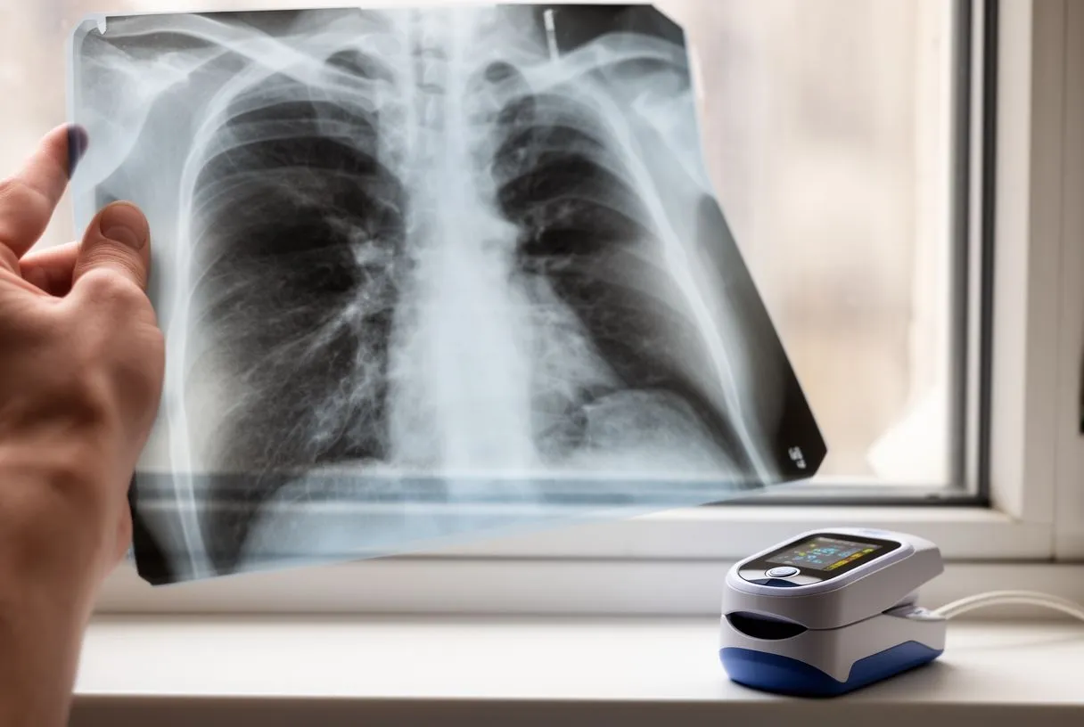
Every chest x-ray taken during your recovery serves as a checkpoint, confirming that your heart and lungs are healing and helping your team decide when to remove tubes, adjust medications, or advance your activity level. In the intensive care unit, x-rays are often taken daily for the first few days. As you transfer to a regular ward and then to home, the frequency decreases. By the time you attend your first outpatient follow-up — typically two to four weeks after discharge — a chest x-ray heart surgery comparison with your preoperative image helps your surgeon confirm that the heart size has returned toward baseline and that any pleural fluid has resolved or is shrinking.
Tracking the removal of tubes and lines: One practical way the x-ray guides recovery is by confirming the safe removal of hardware. When a chest tube is pulled, a follow-up x-ray checks that no air has entered the pleural space as a result. When a central venous catheter — a line placed in a large neck or chest vein — is removed, the x-ray confirms there is no residual pneumothorax. If a patient has had an arterial graft placed during bypass, the x-ray helps monitor for any new fluid that might suggest graft-related issues. The team also checks the position of a temporary pacemaker wire, which is often left in place for several days after surgery in case the heart rhythm needs support. An automated external defibrillator may be stationed nearby in the early recovery period for high-risk patients, though it is rarely needed.
The x-ray also helps your team decide when it is safe to increase your activity. If lung fields remain clear and the cardiac silhouette is stable, the physiotherapy team will encourage more walking and breathing exercises. Here is a quick summary of what each recovery stage typically shows on the x-ray:
- Day 1–3 (ICU): Tubes, lines, mild mediastinal widening, possible atelectasis at lung bases.
- Day 4–7 (ward): Most tubes removed, decreasing effusion, improving lung expansion.
- Week 2–4 (outpatient): Near-normal cardiac silhouette, small or resolved effusion, clear sternotomy wires.
- Month 3 and beyond: Stable findings; wires and clips are the only permanent visible changes.
For patients travelling to Mexico for cardiac surgery — whether from within the country or from abroad — understanding the practical aspects of imaging is part of preparing for the journey. If additional x-rays are needed because of a longer hospital stay or a complication, the per-image cost is modest. Patients should ask their surgical coordinator for a written breakdown of what imaging is included and what might incur an additional charge.
Insurance, travel, and practical considerations: International patients often ask whether their insurance will cover imaging performed in Mexico. This depends on the insurer and the policy; some plans reimburse out-of-country medical costs, while others do not. Patients should obtain a detailed invoice that includes procedure codes, as these are often needed for reimbursement claims. For patients who have risk factors such as elevated apolipoprotein B — a protein carried on cholesterol particles that signals higher cardiovascular risk — the preoperative workup may include additional blood tests alongside the chest x-ray, and the combined results help the surgeon assess overall operative risk. The preoperative x-ray also checks for any calcification in the aorta or other vessels that could complicate surgery, which is particularly relevant for older patients or those with long-standing hypertension.
Travelling home after surgery raises another practical question: when is it safe to fly? Most cardiac surgeons recommend waiting at least two to four weeks after heart surgery before air travel, partly because cabin pressure changes can affect lung function and fluid balance. A chest x-ray taken shortly before travel confirms that the lungs are clear and that any pleural fluid has resolved enough to make flying safe. Patients who had balloon angioplasty — a procedure that uses a small inflated balloon to open a narrowed artery — as part of their treatment, or who are taking aspirin to prevent clot formation, should discuss their travel plans with their surgeon. The x-ray is one piece of the puzzle; your overall clinical status, including wound healing and heart rhythm stability, determines the final clearance to travel.
What a chest X-ray costs in Mexico for heart patients
If you are travelling to Mexico for heart surgery, whether from another part of the country or from abroad, knowing the practical side of imaging is part of preparing for the trip. The usual pattern is at least one chest X-ray before surgery and another afterwards. If you need additional X-rays because of a longer hospital stay or a complication, the cost per image is moderate. Ask the surgical coordinator which imaging studies are included and which could carry an additional charge, and ask for a written breakdown. Dr Alejandro Molina and the Cirugía de Corazón team provide transparent cost estimates that cover the whole surgical process, imaging included.
Insurance, travel and practical considerations: If you are an international patient, you may want to know whether your insurance will cover imaging done in Mexico. That depends on the insurer and the policy: some plans reimburse medical expenses outside the country and others do not. Get an itemised invoice that includes the procedure codes, as these are usually needed to claim reimbursement. If you have risk factors such as raised apolipoprotein B levels, a protein carried in cholesterol particles that signals higher cardiovascular risk, the preoperative assessment may include additional blood tests alongside the chest X-ray; the combined results help the surgeon assess overall operative risk. The preoperative X-ray can also reveal calcification in the aorta or other blood vessels that could complicate surgery, which is especially relevant if you are older or have had high blood pressure for a long time.
If you are travelling home after surgery, another practical question arises: when is it safe to fly? Most cardiac surgeons recommend waiting at least two to four weeks after heart surgery before air travel, partly because changes in cabin pressure can affect lung function and fluid balance. A chest X-ray shortly before the trip confirms that your lungs are clear and that any pleural fluid has reduced enough for you to fly safely. If you had a balloon angioplasty, a procedure that uses a small inflated balloon to open a narrowed artery, as part of your treatment, or you take aspirin to prevent clots, discuss your travel plans with your surgeon. The X-ray is only one piece of the puzzle; your overall clinical condition, including wound healing and the stability of your heart rhythm, will determine the final clearance to travel.
Key Takeaways on Chest X-Ray Before and After Heart Surgery
A chest x-ray is far more than a routine box to tick before and after heart surgery. It is a dynamic tool that shapes your surgical plan, monitors your recovery in real time, and catches complications before they become dangerous. The preoperative image establishes your baseline — the size of your heart, the clarity of your lungs, and the position of any existing hardware. The postoperative images track the removal of tubes, the resolution of fluid, the healing of your sternum, and the position of any new valves or grafts. Together, these images form a visual narrative of your journey through cardiac surgery.
What to remember as a patient or family member: The most important message is that seeing something unusual on your postoperative x-ray does not always mean something is wrong. Sternotomy wires, surgical clips, mild mediastinal widening, and small pleural effusions are expected findings that resolve with time. What matters is the trend — whether each image shows improvement compared to the one before. If your surgeon is concerned, they will explain what they see and what the next step is. Never hesitate to ask your cardiac team to show you your x-ray and explain it in plain terms; this is a reasonable and encouraged part of your recovery. Understanding your own imaging builds confidence and helps you participate actively in decisions about your care.
Every patient's recovery is unique, and imaging is only one component of a comprehensive cardiac assessment. If you are preparing for heart surgery or recovering from one, the most valuable step you can take is to discuss your specific situation with a qualified cardiac team. Dr Alejandro Molina and the specialists at Cirugía de Corazón, with practices in Guadalajara, Querétaro, and Hermosillo, are available to review your case, explain your imaging in clear language, and guide you through every stage of the surgical journey. Booking a consultation is the first step toward understanding what your own chest x-ray reveals and what it means for your heart health going forward.