Coronary Arteries Blocked: Symptoms, Tests and Treatment
Blocked coronary arteries treatment starts with knowing the symptoms and tests.
What Are Blocked Coronary Arteries?
Blocked coronary arteries occur when the vessels that supply oxygen-rich blood to your heart muscle become narrowed or fully obstructed by fatty deposits called atheromatous plaque. The coronary arteries wrap around the outside of the heart, and when their inner walls accumulate cholesterol, calcium, and inflammatory cells, the channel for blood flow shrinks. This process develops over years — often silently — and the first sign many patients notice is chest discomfort during exertion. Understanding what is happening inside those vessels is the first step toward choosing the right blocked coronary arteries treatment with your cardiac team.
The heart muscle, or myocardium, demands a constant supply of oxygen. When coronary artery stenosis — a medical term for narrowing — reduces that supply, the heart cannot pump efficiently. A partial blockage may cause symptoms only under stress, such as walking up stairs or carrying groceries. A complete blockage, however, starves a portion of the heart muscle and causes a heart attack, known medically as a myocardial infarction. The location and severity of the blockage determine how urgently intervention is needed and which treatment option offers the lowest risk for your situation.
For patients in Mexico and across Latin America, coronary artery disease remains one of the most common reasons people seek cardiac care. Many arrive at consultation having lived with mild symptoms for months or even years, attributing chest tightness to indigestion or aging. The reality is that coronary blockages do not improve on their own. They either stabilize with medication and lifestyle changes, or they progress. Early diagnosis and an honest conversation about treatment options — from medication to coronary angioplasty Mexico to heart bypass surgery — give you the best chance of a durable outcome.
Blocked Coronary Arteries Symptoms to Watch For
The most recognized symptom of blocked coronary arteries is angina — a pressure, squeezing, or heaviness in the center of the chest that typically appears during physical activity and eases with rest. This discomfort may radiate to the left arm, jaw, neck, or back, and some patients describe it as feeling like a heavy weight sitting on their chest. Angina that follows a predictable pattern — brought on by effort, relieved by rest — is called stable angina and usually indicates a partial blockage. When chest pain occurs at rest, lasts longer than twenty minutes, or grows more severe, it may signal an unstable plaque and requires emergency evaluation.
Atypical symptoms in women, older adults, and people with diabetes: Not everyone with coronary artery blockage experiences textbook chest pain. Women more often report nausea, shortness of breath, profound fatigue, or discomfort between the shoulder blades rather than crushing chest pressure. Older patients and those with long-standing diabetes may have nerve damage that blunts pain signals, leading to what clinicians call a silent or atypical infarction — a heart attack that happens without the expected warning. This is why relying on symptoms alone is unreliable, and why diagnostic testing plays such a central role in evaluation.
Some patients first learn they have blocked heart arteries only after a routine electrocardiogram shows evidence of a previous, unrecognized heart attack. Others notice gradually decreasing exercise tolerance — they cannot walk as far or climb as many stairs as they once did without feeling winded. A careful physical examination, including auscultation — listening to the heart with a stethoscope — may reveal murmurs or irregular rhythms that prompt further investigation. If you or a family member experience any combination of chest discomfort, unexplained breathlessness, or sudden fatigue, it is worth discussing with a qualified cardiac specialist rather than waiting for symptoms to resolve on their own. Research published in The Lancet has consistently shown that earlier recognition of coronary symptoms correlates with better long-term outcomes.
How Do Coronary Arteries Become Blocked?
The underlying process behind blocked coronary arteries is atherosclerosis, a chronic condition in which cholesterol deposits accumulate within the walls of arteries. It begins when low-density lipoprotein (LDL) cholesterol particles penetrate the inner lining of the vessel wall, drawn in by elevated blood levels of a protein called apolipoprotein B. Once inside, these particles oxidize, triggering an inflammatory response. White blood cells arrive to clean up the oxidized cholesterol but instead become trapped, forming a fatty streak that gradually enlarges into an atheromatous plaque. Over years, that plaque calcifies and narrows the artery channel.

Several factors accelerate plaque formation. High blood pressure damages the artery lining and makes it more permeable to cholesterol. Smoking introduces chemicals that injure vessel walls and promote clot formation. Diabetes raises blood sugar levels that glycate — stiffen — arterial tissue and accelerate inflammation. A family history of early coronary disease, obesity, and a sedentary lifestyle all increase risk. Importantly, these risk factors tend to cluster: a patient with hypertension and diabetes faces a significantly higher chance of developing coronary artery stenosis than someone with only one risk factor. Identifying and addressing each one is central to any treatment plan.
Plaque does not grow at a steady, predictable rate. Some plaques remain stable for decades and never cause symptoms, while others rupture suddenly. When a plaque's fibrous cap breaks open, its cholesterol-rich core contacts the bloodstream, and the body responds by forming a clot — much like it would on a skin cut. If that clot is large enough, it occludes the artery entirely, causing a myocardial infarction. This is why a person who felt well yesterday can present with a heart attack today. The risk of rupture depends more on plaque composition than size, which is why imaging and functional tests matter as much as measuring how narrow the artery appears.
Diagnostic Tests for Blocked Coronary Arteries
When blocked coronary arteries are suspected, the evaluation begins with non-invasive tests. An electrocardiogram (ECG) records the heart's electrical activity and can reveal signs of reduced blood flow or previous damage. A stress test — usually a treadmill or bicycle exercise test combined with imaging — shows how the heart performs under exertion, when oxygen demand rises. Echocardiography uses sound waves to create images of the heart muscle and valves, revealing whether any wall segments move abnormally. These tests are safe, widely available, and help the cardiologist decide whether more invasive investigation is warranted.
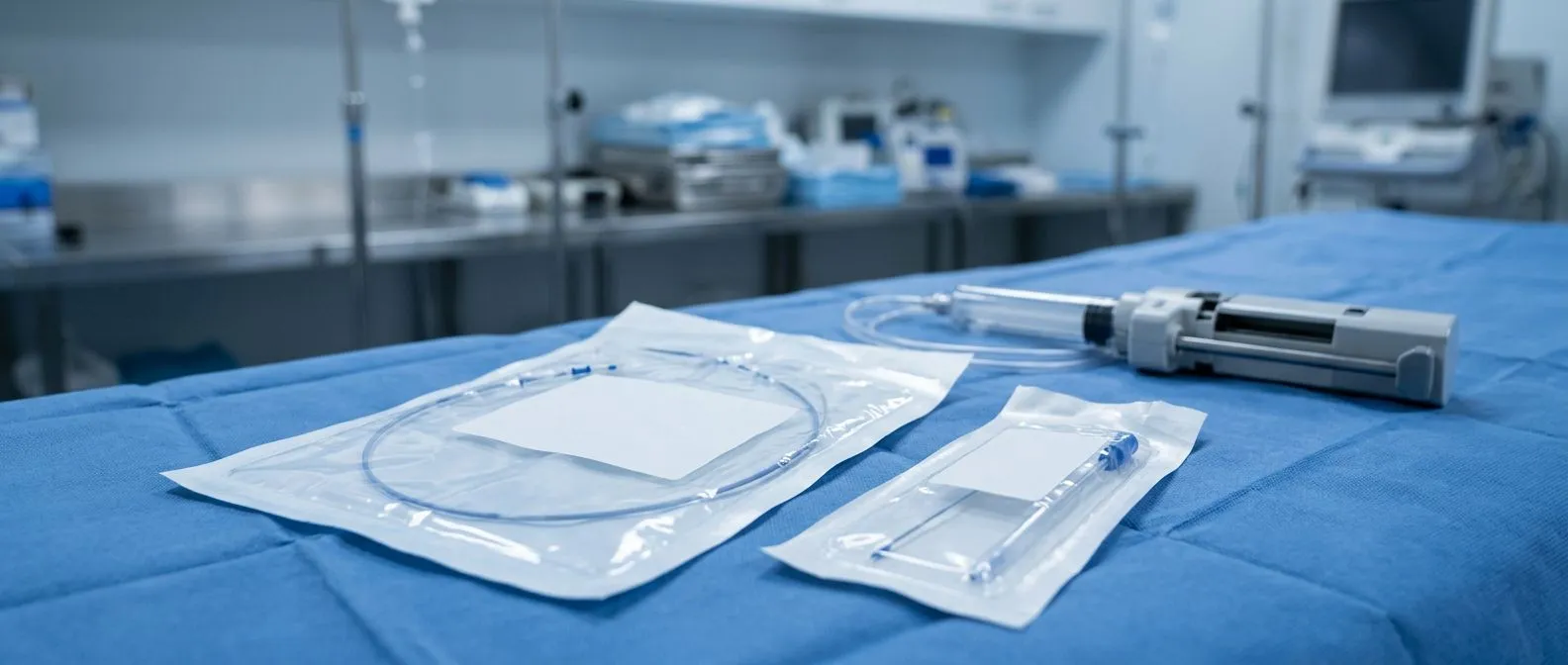
If non-invasive tests suggest significant blockage, the next step is often a cardiac catheterization procedure, also called coronary angiography. A thin tube called a catheter is threaded — usually through the wrist or groin — into the coronary arteries, and a contrast dye is injected while X-ray images are captured. This allows the cardiologist to see exactly where and how severely each artery is narrowed. In some cases, additional tools such as intravascular ultrasound provide detailed pictures of the plaque from inside the vessel. The procedure is both diagnostic and potentially therapeutic: if a treatable blockage is found, the same access can be used to perform an intervention.
Other tests may be used depending on the clinical picture. A CT coronary angiogram offers a non-invasive alternative for patients at intermediate risk, producing detailed three-dimensional images of the coronary arteries without catheter insertion. Blood tests measure cardiac enzymes — proteins released when heart muscle is injured — and help distinguish unstable angina from an actual heart attack. Updated clinical classifications of pulmonary and cardiac hemodynamic definitions, as outlined in research such as the Haemodynamic definitions and updated clinical classification of pulmonary hypertension, also inform how cardiologists interpret right-heart catheterization data in complex cases. The goal of testing is not simply to find a blockage but to understand its functional impact on your heart.
Blocked Coronary Arteries Treatment: Medication First
For many patients, blocked coronary arteries treatment begins with medication rather than a procedure. The goals are threefold: relieve symptoms, prevent plaque progression, and reduce the risk of heart attack. Antiplatelet therapy — most commonly aspirin — helps prevent clot formation on existing plaques. Statins lower LDL cholesterol and stabilize plaque, making rupture less likely. Beta-blockers reduce the heart's workload by slowing its rate and lowering blood pressure. Nitrates dilate blood vessels and ease angina. Calcium channel blockers and a newer class called ranolazine can also reduce chest pain. For most patients with stable coronary disease, a well-designed medication regimen provides meaningful symptom relief and a good quality of life.

When medication is enough, and when it is not: If your angina occurs predictably and responds well to medication, and if stress testing shows no large area of heart muscle at risk, your cardiologist may recommend continuing medical therapy with regular monitoring. This approach is supported by clinical evidence and avoids the risks inherent in any procedure. However, medication alone may be insufficient when blockages are severe, when symptoms escalate despite optimal drug therapy, or when imaging reveals that a large territory of heart muscle depends on a critically narrowed artery. In those situations, revascularization — restoring blood flow through angioplasty or bypass — becomes the recommended path.
Medication also plays a crucial role after any intervention. Stents and bypass grafts address the most severe blockages, but they do not cure atherosclerosis. Without ongoing statin therapy, blood pressure control, and antiplatelet medication, new blockages can form — in stented segments, in bypass grafts, or in previously uninvolved arteries. This is why cardiac teams emphasize that a stent or bypass is one chapter in a lifelong management plan, not a cure. Lifestyle modifications — stopping smoking, adopting a Mediterranean-style diet, regular moderate exercise, and stress reduction — are not optional add-ons; they are as important as any prescription. Managing conditions like atrial flutter, an irregular heart rhythm that can accompany coronary disease, also requires careful coordination with your cardiologist.
Coronary Angioplasty and Coronary Stent Procedure
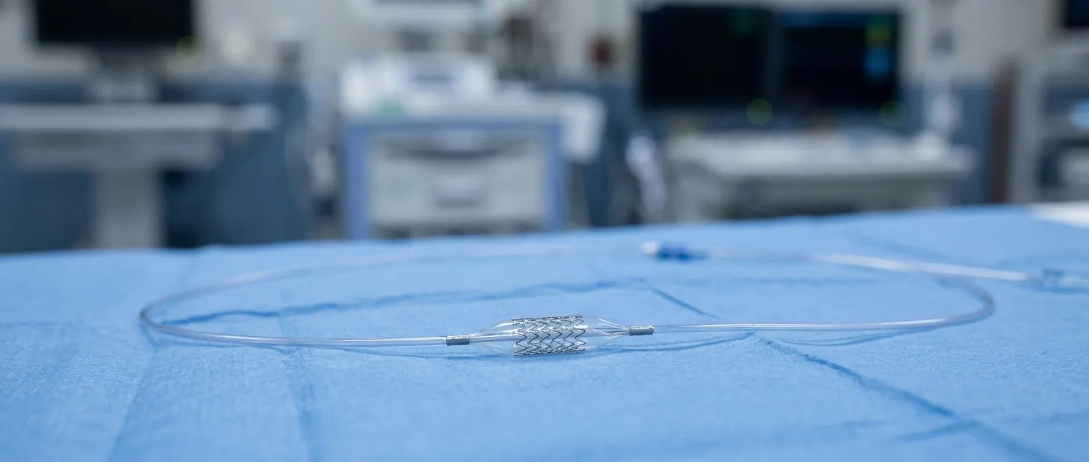
Coronary angioplasty, also called percutaneous coronary intervention, is a minimally invasive procedure that opens narrowed arteries from within. After numbing the access site — typically the wrist — the cardiologist guides a catheter to the coronary artery blockage. A tiny balloon at the catheter tip is inflated, compressing the plaque against the artery wall and widening the channel. This balloon angioplasty technique is almost always combined with stent placement: a small mesh tube is positioned in the newly opened segment and expanded to hold the artery open. Most stents used today are drug-eluting, meaning they slowly release medication that prevents scar tissue from re-narrowing the vessel.
The coronary stent procedure usually takes between thirty minutes and two hours, depending on the number and location of blockages. Patients are typically awake but sedated, and many go home the next morning. Discomfort is minimal — most patients feel only mild pressure at the access site. For patients with a single or double vessel blockage that is accessible by catheter, angioplasty with stenting offers a faster recovery than open surgery and excellent symptom relief. However, stents are not permanent guarantees: a small percentage of stented arteries re-narrow over months, a process called restenosis, and blood-thinning medication must be taken as prescribed to prevent clot formation inside the stent.
Deciding between angioplasty and bypass surgery depends on several factors: the number of arteries involved, the anatomy of the blockages, whether the left main coronary artery is affected, and the patient's overall health. Patients with diabetes and multiple vessel disease often have better long-term outcomes with bypass surgery, according to major studies. Those with isolated blockages in one or two accessible arteries may be excellent candidates for stenting. Your cardiac team will weigh these factors using established tools — including the EuroSCORE II, which estimates surgical risk — and present you with an honest comparison. The goal is not to sell a procedure but to find the approach that offers the lowest risk and most durable benefit for your individual situation.
Heart Bypass Surgery for Severe Blockages
When multiple coronary arteries are severely blocked, or when the left main artery — the vessel that supplies a large portion of the heart — is narrowed, coronary artery bypass grafting (CABG) may be the recommended treatment. In this operation, a cardiac surgeon creates new routes for blood to reach the heart muscle, bypassing the blocked segments. The grafts are constructed from blood vessels taken from elsewhere in the body. The internal mammary artery, which runs behind the chest wall, is the most commonly used arterial graft because it remains open longer than vein grafts and resists atherosclerosis. Surgeons may also use the radial artery from the forearm or the saphenous vein from the leg.

During conventional bypass surgery, the heart is stopped and a heart-lung machine takes over circulation — a technique called cardiopulmonary bypass. This allows the surgeon to work on a still, bloodless field. In selected patients, an off-pump technique may be used, where the heart continues beating while the surgeon places grafts using specialized stabilization tools. The choice depends on the number of grafts needed, the surgeon's experience, and the patient's anatomy. The ascending aorta — the large artery emerging from the heart — is typically where vein or arterial grafts are connected to receive blood. The other end is attached to the coronary artery downstream of the blockage, restoring flow to the heart muscle.
Bypass surgery is a major operation, and the decision to proceed is made after careful risk assessment. Surgeons evaluate factors such as age, kidney function, lung disease, previous strokes, and the condition of the heart muscle — particularly whether it has been weakened by previous heart attacks. The EuroSCORE II calculator combines these variables to estimate the risk of mortality, giving patients and families a realistic basis for informed consent. Large-scale European surveys, including the Euro Heart Survey on Valvular Heart Disease and related registries, have contributed to the evidence base that guides these decisions. While bypass surgery carries higher upfront risk than stenting, it often provides more complete and durable revascularization for patients with complex disease.
Recovery After Blocked Coronary Arteries Treatment
Recovery timelines differ substantially depending on whether you had a stent or bypass surgery. After coronary angioplasty and stenting, most patients spend one night in the hospital and resume light activities within a week. The access site in the wrist heals quickly, and you will be prescribed antiplatelet medication — often a combination of aspirin and a second drug — to prevent clot formation inside the stent. Your cardiologist will schedule a follow-up visit to review symptoms, adjust medications, and confirm that blood pressure and cholesterol are at target levels. Most people return to work within one to two weeks and feel markedly better in terms of exercise tolerance and chest pain relief.
After heart bypass surgery, recovery is longer but follows a predictable trajectory. Patients typically spend four to six days in the hospital, including one or two days in the intensive care unit for close monitoring. An arterial line — a thin catheter placed in an artery for real-time blood pressure monitoring — and an arterial cannula used during the heart-lung bypass are removed once stable. The chest incision takes six to eight weeks to heal fully, and during that time patients are advised to avoid heavy lifting and strenuous upper-body activity. Cardiac rehabilitation — a supervised program of exercise, education, and emotional support — typically begins four to six weeks after surgery and continues for several months.
- Week 1–2: Rest, gentle walking, wound care, pain management, and close follow-up with your surgical team.
- Week 3–6: Gradual increase in walking distance, return to light daily activities, begin outpatient cardiac rehabilitation.
- Week 6–12: Most patients resume full activity, including exercise and travel, pending clearance from their cardiologist.
Long-term follow-up is essential regardless of which treatment you received. Blockages can recur, and new ones can develop in previously uninvolved arteries. Your cardiologist will monitor cholesterol, blood pressure, and blood sugar, and may recommend periodic stress testing or imaging. If you were placed on anticoagulation medication — for example, if you also have a condition requiring the type of monitoring studied in the ARISTOTLE trial — your dosing and blood tests will be coordinated carefully. The goal of follow-up is not just to detect problems but to keep you actively engaged in managing your own health.
Living With Coronary Artery Disease in Mexico
For patients in Mexico, access to high-quality coronary care has improved significantly over the past two decades. Cardiac centers in Guadalajara, Querétaro, Hermosillo, and other major cities offer both angioplasty and bypass surgery with outcomes comparable to international standards. The question many families face is not whether treatment is available but how to navigate the system efficiently — choosing the right specialist, understanding what insurance covers, and planning for recovery. If you are traveling from a smaller city or from abroad, a consultation with an experienced cardiac team can help you understand the full scope of options and what to expect at each stage.
Cost is a legitimate concern, and an honest cardiac team will discuss it openly. The total expense of coronary angioplasty or bypass surgery depends on the number of stents or grafts, the length of hospital stay, the type of anesthesia and monitoring required, and whether complications arise. Some insurance plans in Mexico cover cardiac procedures, but coverage levels vary widely. If you are paying out of pocket, ask for a written estimate that includes surgeon fees, hospital charges, anesthesia, imaging, and rehabilitation. Reputable practices — including Cirugía de Corazón — provide transparent information and never pressure patients into unnecessary procedures. The decision to proceed should be based on clinical need, not commercial incentive.
- Ask about: the specific procedure recommended, alternatives, expected hospital stay, and total estimated cost including follow-up.
- Consider: proximity of the cardiac center to your home, as follow-up visits and rehabilitation are part of the treatment.
- Confirm: which medications you will need long-term, their availability in your region, and their approximate monthly cost.
Lifestyle changes are the foundation of long-term success after any coronary intervention. A heart-healthy diet — rich in vegetables, whole grains, fish, olive oil, and limited in processed foods — has been shown to slow plaque progression. Regular aerobic exercise, such as brisk walking for thirty minutes most days, improves the heart's efficiency and helps control blood pressure, cholesterol, and weight. Smoking cessation is the single most impactful change a patient can make: stopping smoking reduces the risk of recurrent heart attack by roughly half within the first year. These measures, combined with faithful medication adherence, give you the best chance of a long and active life after treatment.
When to Seek Help for Blocked Coronary Arteries
Certain symptoms require immediate emergency attention, not a scheduled appointment. If you experience chest pain or pressure that lasts more than a few minutes, occurs at rest, or is accompanied by sweating, nausea, shortness of breath, or fainting, call emergency services or go to the nearest hospital immediately. These may be signs of an acute heart attack, and every minute of delay means more heart muscle is lost. If an automated external defibrillator is available and someone collapses with no pulse, it should be used without hesitation — these devices are designed for use by bystanders and provide voice instructions.
Other situations warrant prompt — though not necessarily emergency — evaluation. If your angina pattern changes: pain comes with less exertion, lasts longer, or does not respond to your usual nitroglycerin, this may indicate an unstable plaque. New shortness of breath, swelling in the legs, or palpitations that feel different from your usual rhythm should also prompt a call to your cardiologist. The message is simple: do not wait and hope symptoms will pass. Coronary disease is unpredictable, and a medical evaluation is the only way to distinguish a benign episode from a warning sign. Arm pain that accompanies chest discomfort, particularly radiating down the left arm, is a classic association that should never be dismissed.
We encourage you to bring your questions, your test results, and your concerns to a consultation with a qualified cardiac team. The right treatment for blocked coronary arteries is never one-size-fits-all — it depends on your anatomy, your symptoms, your overall health, and your preferences. Dr Alejandro Molina and the team at Cirugía de Corazón, with practices in Guadalajara, Querétaro, and Hermosillo, are available to review your case and help you understand your options in clear, honest terms. Do not self-diagnose or rely on online information alone. Book a consultation, ask everything you need to ask, and take the next step with a team that has your heart — and your future — in mind.