Cyanosis in Heart Disease: Blue Skin and What It Means
Cyanosis heart disease turns skin blue when blood lacks oxygen.
What Is Cyanosis Heart Disease and Why Does Skin Turn Blue?
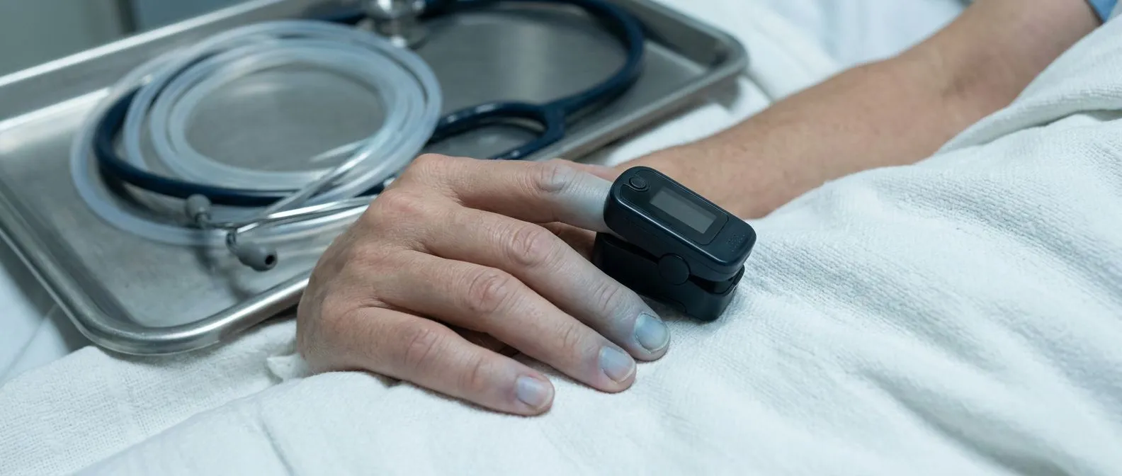
Seeing a blue tint on your own skin, or on the skin and lips of someone you love, is one of the most unsettling experiences a patient or family member can face. Cyanosis heart disease refers to a group of cardiac conditions in which blood circulating through the body carries too little oxygen, producing a visible bluish discoloration of the skin, lips, nail beds, or mucous membranes. The colour change is not a disease in itself — it is a sign that oxygen delivery to tissues has fallen below a critical threshold, typically when oxygen saturation in arterial blood drops below about 85 percent. Understanding what that sign means, and how quickly it must be addressed, is the first step toward making calm, informed decisions about care.
Normally, bright red oxygen-rich blood leaves the heart through the aorta and delivers oxygen to every organ. When that blood is oxygen-poor, it takes on a darker, purplish hue that shows through thin skin as blue. The mechanism behind the colour shift is straightforward: haemoglobin, the protein in red blood cells that carries oxygen, changes colour depending on how much oxygen is bound to it. If a structural heart problem causes poorly oxygenated blood to bypass the lungs and flow directly into the body's main circulation, that dark blood reaches the fingertips, lips, and toes — and the blue tone becomes visible. This mixing of unoxygenated blood into the systemic circulation is called a right-to-left shunt, and it is the mechanism behind most forms of cyanotic congenital heart disease.
It is important to distinguish a genuine cardiac or respiratory cause from something benign. Cold weather, tight jewellery, or even anxiety can briefly make fingertips look bluish without signalling a heart problem. But persistent or recurring central cyanosis — especially involving the lips, tongue, or face — should never be dismissed. According to Mayo Clinic, blue-coloured skin from a lack of oxygen is a symptom that warrants prompt medical evaluation, particularly when it appears suddenly or is accompanied by shortness of breath. The goal of any cardiac assessment is to determine whether the blue tint reflects a structural defect, a lung problem, or a circulatory issue that can be corrected.
Central Cyanosis vs Peripheral Cyanosis: Key Differences
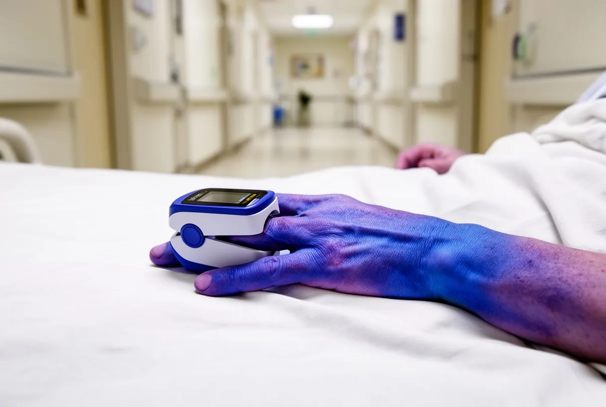
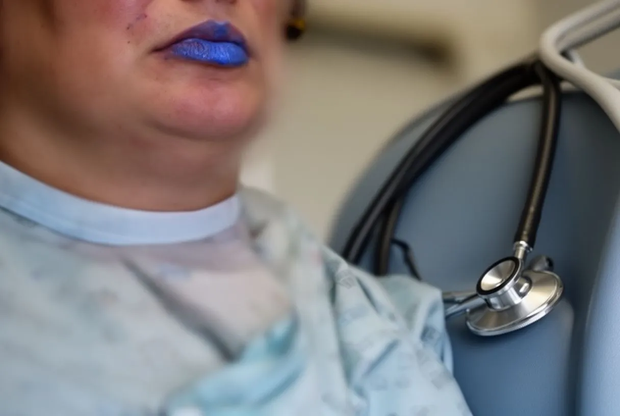
Not all blue skin is the same, and the location of the discolouration gives doctors important clues about its origin. Central cyanosis affects the core of the body — the lips, tongue, face, and the skin of the chest or trunk. It almost always indicates that arterial blood itself is carrying insufficient oxygen, which points to a problem in the heart or lungs. If the lips and tongue are blue but the hands and feet are warm, the issue is likely central: blood is leaving the heart without being properly oxygenated, either because it is bypassing the lungs through a shunt or because the lungs cannot transfer oxygen effectively. Central cyanosis does not improve with warming and usually signals a structural or functional problem that needs investigation.
Peripheral cyanosis, by contrast, affects the extremities — fingers, toes, earlobes — while the lips and tongue remain pink. It happens when blood flow to the limbs is reduced or slowed, allowing tissues to extract more oxygen than usual from otherwise normal blood. Cold exposure is the most common and least concerning cause. But peripheral cyanosis can also result from heart failure, where a weakened heart cannot maintain adequate circulation to the limbs, or from conditions that narrow the small vessels of the hands and feet. Warming the affected area will usually reverse peripheral cyanosis if the underlying circulation is adequate; if it does not, further evaluation is warranted.
The distinction matters because it changes the urgency and the type of testing required. Central cyanosis in a newborn, for instance, may prompt an immediate echocardiogram to look for a congenital defect such as transposition of the great arteries, where the two main vessels leaving the heart are connected the wrong way around. Peripheral cyanosis in an adult might lead to an assessment of cardiac output, peripheral vascular health, and medication review. In either case, the clinical team looks at the whole picture: oxygen saturation measured by pulse oximetry, a careful physical examination including auscultation — listening to the heart and lungs with a stethoscope — and a detailed history of when the colour change started, what makes it better or worse, and whether it is accompanied by other symptoms such as breathing difficulty or arm pain.
Common Cyanotic Heart Defects in Newborns and Children
When cyanosis appears in a newborn within hours or days of birth, it is often the first sign of a congenital heart defect — a structural problem that developed before birth. These defects are called cyanotic because they cause mixing of oxygen-poor and oxygen-rich blood, reducing the overall oxygen level reaching the body. Transposition of the great arteries is one of the most serious: the aorta, which should carry oxygen-rich blood to the body, arises from the right side of the heart, while the pulmonary artery, which should carry oxygen-poor blood to the lungs, arises from the left. Without prompt intervention, the body receives blood that has never been to the lungs for oxygen.
Tetralogy of Fallot is another well-known cyanotic defect, combining four abnormalities: a narrowed pulmonary valve, a hole in the wall between the two ventricles, an aorta that sits over the hole, and thickening of the right ventricle. Babies with Tetralogy of Fallot may appear pink at birth but develop cyanosis over weeks or months as the narrowing worsens. Some children experience "tet spells" — sudden episodes of deep cyanosis, rapid breathing, and irritability that occur when the narrowed pathway to the lungs restricts blood flow. Other cyanotic conditions include tricuspid atresia, where the valve between the right atrium and right ventricle fails to form properly, and total anomalous pulmonary venous connection, where oxygen-rich blood from the lungs returns to the wrong side of the heart.
Parents should know that these defects are rare and that most are treatable. The surgical options available today are well established, and many children go on to live full, active lives after repair. However, timing matters. Some defects require surgery in the first days of life; others are managed with a staged approach, performing an initial procedure in infancy and a more complete repair later. The decision about when and how to intervene depends on the specific anatomy, the child's size and overall health, and the oxygen saturation level. If your newborn has been diagnosed with a cyanotic defect, the most valuable step is to meet with a cardiac surgical team that can explain the anatomy in terms you understand and walk you through the planned approach.
What Causes Cyanosis in Adults With Heart Disease?
Cyanosis in adults is less commonly caused by a congenital defect from birth and more often linked to acquired heart or lung conditions. Advanced heart failure is one frequent cause: when the heart's pumping function declines significantly, circulation slows, oxygen extraction increases, and peripheral cyanosis can develop in the hands and feet. If heart failure is severe enough to cause fluid buildup in the lungs, oxygen exchange is impaired and central cyanosis may also appear. Adults with long-standing lung disease — particularly chronic obstructive pulmonary disease — can develop cyanosis because the lungs cannot move enough oxygen into the bloodstream, regardless of how well the heart is functioning.
Unrepaired or partially repaired congenital defects are another pathway. Some adults were born with heart defects that were managed palliatively in childhood, meaning they had procedures to improve blood flow but not a complete anatomical repair. Over time, these patients can develop cyanosis again if the palliative circulation becomes inadequate or if complications such as a leaky valve develop. This is sometimes seen in adults who had shunt procedures for conditions like Tetralogy of Fallot decades ago. The gradual return of a bluish tint in an adult with a history of childhood heart surgery should be evaluated promptly by a cardiologist familiar with grown-up congenital heart disease, because the cause may be correctable with a valve replacement, a revision of the original repair, or a catheter-based intervention.
Other adult causes include atrial flutter or other arrhythmias that severely reduce cardiac output, blood clots in the lungs that block oxygen exchange, and certain medications that reduce blood flow. Rarely, cyanosis results from a condition called Eisenmenger syndrome, in which a long-standing left-to-right shunt (such as an untreated hole in the heart) reverses direction over years of pressure buildup, eventually sending oxygen-poor blood into the systemic circulation. Eisenmenger syndrome is a serious complication of delayed treatment and is one of the strongest arguments for evaluating and repairing congenital defects early. The discussion about whether a combination of medical therapy and procedural intervention is appropriate depends on the individual's anatomy, functional capacity, and the expertise of the treating centre. For some patients, systemic review data on adjunctive therapies can inform the conversation; for instance, a systematic review on lipid-lowering therapies and cardiovascular risk reduction offers context on how medical management supports procedural outcomes Efficacy and safety of lipid-lowering therapies in combination with or without statin.
Signs and Symptoms That Accompany Blue Skin in Heart Conditions
Cyanosis rarely appears in isolation. The symptoms that accompany it tell the clinician a great deal about the underlying cause and the urgency of the situation. Respiratory distress — rapid breathing, flaring nostrils, or visible effort with each breath — suggests that the lungs are struggling to oxygenate the blood. This can happen with pneumonia, a pulmonary embolism, or severe heart failure with pulmonary oedema. When cyanosis is paired with laboured breathing, the situation is typically more urgent than cyanosis alone, and emergency evaluation is appropriate.
Clubbing of the fingers is a sign of long-standing cyanotic heart disease that develops slowly over months or years. The fingertips become bulbous and the nails curve downward, a change that reflects chronic low oxygen levels in the blood. Clubbing is not painful, but it is a visible marker that the body has been living with inadequate oxygenation for some time. In adults, clubbing may prompt a search for lung disease, a congenital heart defect, or even certain gastrointestinal conditions. In children with known cyanotic defects, clubbing is expected and does not by itself indicate an emergency — but it does confirm that the oxygen level has been chronically low.
Other accompanying symptoms include poor feeding in infants, low energy or exercise intolerance in older children and adults, and episodes of fainting or near-fainting. Some patients describe a sense of air hunger that does not resolve with rest. An atypical infarction — a heart attack that does not present with the classic crushing chest pain — can sometimes manifest as sudden cyanosis, confusion, or collapse, particularly in older adults or those with diabetes. The combination of blue skin and any sign of organ distress — altered consciousness, severe breathlessness, or inability to maintain blood pressure — requires immediate emergency care. The clinical team will work to stabilise oxygen levels while simultaneously searching for the root cause, and the speed of that response can be decisive in outcomes.
How Doctors Diagnose the Underlying Cause of Cyanosis
Diagnosis begins with a careful history and physical examination, followed by targeted testing. The doctor will ask when the cyanosis started, whether it is constant or intermittent, what triggers or relieves it, and whether there is a history of heart or lung disease in the patient or the family. Pulse oximetry — a small clip placed on the finger that measures oxygen saturation — provides an immediate numerical value. A reading below 92 percent at rest, or a significant drop with exertion, is a signal that further investigation is needed. In newborns, routine pulse oximetry screening in the first day of life has become a standard tool for detecting critical congenital heart defects before symptoms become severe.
The echocardiogram is the cornerstone of cardiac diagnosis. This ultrasound examination of the heart shows its structure in real time: the size of the chambers, the function of the valves, the presence of any holes in the walls between them, and the direction and speed of blood flow. If a structural defect is present, the echocardiogram usually identifies it. In complex cases, additional imaging such as a cardiac MRI or a CT angiogram may be used to build a three-dimensional picture of the anatomy before surgical planning. An arterial line — a thin tube placed in an artery — may be inserted in critically ill patients to provide continuous, accurate blood pressure monitoring and to allow repeated blood gas sampling, which measures oxygen and carbon dioxide levels directly from arterial blood rather than estimating them through the skin.
Other diagnostic tools include a chest X-ray to assess heart size and lung appearance, an electrocardiogram to evaluate the heart's electrical activity, and blood tests to check for anaemia, infection, or metabolic disturbance. If a lung cause is suspected, pulmonary function tests or a ventilation-perfusion scan may be ordered. For adults presenting with cyanosis and a question of coronary ischaemia without obstructive disease — a situation where the heart muscle is starved of oxygen despite open arteries — non-invasive functional testing is sometimes sufficient to guide management, as explored in the cardiology literature A debate: Isquemia sin enfermedad coronaria obstructiva. The goal of all of these tests is the same: to build a precise picture of why oxygen delivery is failing so that treatment can be targeted to the actual problem rather than to the symptom alone.
Treatment Options for Cyanotic Heart Disease
Treatment is dictated entirely by the underlying cause. There is no single therapy for cyanosis itself — the colour is a sign, not a condition. Oxygen therapy is the first step in nearly every case, delivered through a mask or nasal cannula to raise the amount of oxygen in the blood while the diagnostic workup proceeds. For some patients, especially those with lung disease, supplemental oxygen may be sufficient to correct the problem. For those with structural heart defects, however, oxygen alone will not resolve the cyanosis because the fundamental issue is blood bypassing the lungs entirely.
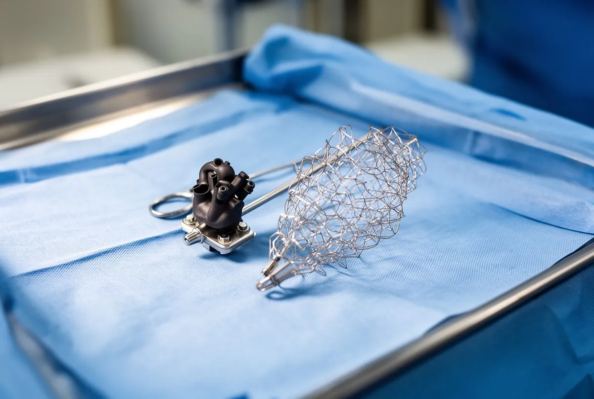
Surgical repair is the definitive treatment for most congenital cyanotic defects. The type of operation depends on the anatomy: a Tetralogy of Fallot repair involves closing the ventricular septal defect and widening the pulmonary outflow tract, sometimes using a patch or an arterial graft to enlarge the pathway. Transposition of the great arteries requires an arterial switch operation, in which the surgeon reconnects the aorta and pulmonary artery to their correct positions and reattaches the coronary arteries to the new aorta. Some defects are repaired in a single operation; others require a staged approach with an initial palliative procedure in infancy — sometimes using an autograft of the patient's own tissue — and a complete repair later in childhood. The decision about timing is individualised and depends on the defect, the child's weight, and the oxygen saturation trend.
For adults with acquired causes, treatment may involve heart failure medications, rhythm control for conditions like atrial flutter, valve repair or replacement, or catheter-based procedures. Some adults with congenital defects who had repairs decades ago develop complications such as a leaking pulmonary valve and may be candidates for a transcatheter pulmonary valve replacement, a less invasive approach than open surgery. The choice between surgical and catheter-based options depends on the individual's anatomy, overall health, and the expertise available. Key considerations for any patient and family weighing treatment options include:
- Identify the mechanism: Is the cyanosis central (heart/lung origin) or peripheral (circulation/blood flow)?
- Confirm the anatomy: Has an echocardiogram clearly shown the structural problem?
- Assess urgency: Does the defect need immediate repair, or is a staged approach safer?
- Review repair options: Open surgery, minimally invasive surgery, or catheter-based intervention — which is lowest risk for this patient's situation?
- Plan follow-up: What monitoring will be needed after the procedure, and for how long?
Heart Surgery for Cyanotic Defects: What to Expect in Recovery
Recovery from heart surgery for a cyanotic defect follows a recognisable path, though the timeline varies with the patient's age, the complexity of the operation, and any complications. Immediate postoperative care takes place in an intensive care unit, where the patient is closely monitored. An arterial cannula or central venous line may remain in place to allow continuous monitoring of blood pressure and access for blood tests. Mechanical ventilation supports breathing for a period ranging from a few hours to several days, depending on how quickly the patient's own lungs and heart stabilise. Pain is managed with medication, and the care team works to prevent complications such as infection, bleeding, or fluid buildup around the heart.
Mobilisation — getting the patient moving — begins sooner than most families expect. Once stable, patients are encouraged to sit up, stand, and take short walks with assistance within the first few days. Early movement reduces the risk of clots, helps re-expand the lungs, and speeds the return of normal digestion and sleep patterns. Children are often more resilient than adults in this phase, though they may be irritable or clingy as they recover. For adults, particularly those with long-standing cyanosis or multiple health issues, recovery can be slower and may require a structured cardiac rehabilitation programme lasting several weeks. The use of autologous blood — the patient's own blood, collected and returned during or after surgery — is one technique used to minimise the need for donor transfusion, especially in complex cases.
Long-term recovery involves regular follow-up with the cardiology team. After repair of a cyanotic defect, the oxygen saturation should rise to normal levels and the blue tint should resolve. If cyanosis persists or returns after surgery, further investigation is needed to look for residual shunts, valve problems, or other complications. Some repaired patients develop arrhythmias years later and may need monitoring or medication. The goal of follow-up is to catch any issue early, when it is most treatable. Families should be given a clear schedule of follow-up appointments, warning signs to watch for, and a direct contact for the surgical team. The relationship with the cardiac centre does not end at discharge — it continues for life, and open communication is essential to good outcomes.
Living With Cyanosis: Long-Term Management and Quality of Life
For patients who cannot undergo immediate or complete repair — whether because of anatomy, overall health, or personal circumstances — living with cyanosis becomes a management challenge that extends across daily life. Activity adaptation is central. Most patients with chronic cyanosis can be active, but they need to learn their own limits. Strenuous exercise may cause oxygen saturation to drop and symptoms to worsen, so a cardiologist should help define safe levels of activity. Children with unrepaired or partially repaired defects often modify their play, and schools may need to be informed about the condition. Adults may need to adjust their work duties or avoid high-altitude environments, where thinner air reduces the oxygen available even in healthy individuals.
Medical management focuses on optimising what the heart and lungs can do and preventing complications. Patients with chronic cyanosis are at increased risk of iron-deficiency anaemia because the body tries to produce more red blood cells to compensate for low oxygen, and the resulting high blood viscosity can paradoxically worsen circulation. Regular blood counts are important, and iron supplementation may be recommended — but only under careful supervision, because over-correction can increase the risk of clotting. Some patients benefit from aspirin or other blood-thinning strategies to reduce viscosity-related risk, but this must be individualised. Dental hygiene matters too: bacteria from the mouth can enter the bloodstream and infect the heart, so patients with cyanotic defects need antibiotic coverage before certain dental procedures.
Quality of life for patients living with cyanosis depends heavily on support, education, and access to specialist care. The psychological impact of a visible sign like blue skin should not be underestimated, especially for children and adolescents. Counselling, peer support groups, and clear communication with teachers and employers can help patients and families feel more in control. For those who eventually become candidates for late repair — and many adults with congenital heart disease do — the improvement in oxygen levels, energy, and appearance can be transformative. Practical strategies that support daily life with chronic cyanosis include:
- Monitor oxygen levels: Use a home pulse oximeter to track trends and recognise when to seek help.
- Stay hydrated: Adequate fluid intake helps keep blood from becoming too viscous.
- Plan for altitude: Avoid high-altitude travel or discuss supplemental oxygen with your cardiologist before flying.
- Maintain dental health: Regular dental care reduces infection risk; discuss prophylactic antibiotics with your team.
- Build a support network: Connect with congenital heart disease organisations for education and community.

When to Seek Immediate Medical Help for Blue Skin or Lips
Some episodes of cyanosis are managed at home with warming and rest. Others are emergencies. Sudden onset of blue lips or skin in a person who was previously well is always a reason to seek urgent medical attention, particularly if it is accompanied by chest pain, difficulty breathing, confusion, or loss of consciousness. In a newborn, cyanosis that does not resolve with warming, or that appears during feeding or crying, should be evaluated in an emergency setting without delay. The underlying cause could be a critical congenital defect, a lung infection, or a metabolic problem — all of which are treatable if identified quickly.
Progressive worsening of chronic cyanosis in a patient with a known heart condition also requires prompt evaluation. If oxygen saturation, which has been stable, begins to decline steadily, the cause may be a new complication: a leaking valve, an arrhythmia, a blood clot, or a worsening of heart function. Patients and families who have been given a pulse oximeter and a baseline saturation value should know the number that triggers a call to the cardiac team. A drop of more than five percentage points from baseline, especially if accompanied by new symptoms, is a reasonable threshold to seek advice.
In any situation where cyanosis is accompanied by signs of cardiac arrest — unresponsiveness, no breathing, or no pulse — call emergency services immediately. If an Automated External Defibrillator is available, it should be used without delay, as these devices are designed to guide even untrained users through the steps of resuscitation. For patients with known heart disease, a plan should be in place long before an emergency occurs: the cardiac team should provide written guidance on warning signs, emergency contacts, and the preferred hospital. Families who have rehearsed what to do report far less anxiety in a crisis. The message for anyone reading this and recognising a symptom in themselves or a loved one is simple: do not wait and do not self-diagnose. Cyanosis is the body's way of signalling that something is wrong with oxygen delivery, and the right response is to bring it to the attention of a qualified cardiac team who can determine the cause, explain it clearly, and guide you through the options. If you or a family member are experiencing blue skin, lips, or nail beds and have concerns about a heart condition, contact a cardiac specialist for a consultation to discuss your specific situation.