Endomyocardial Biopsy: Why It Is Done and What to Expect
Learn how an endomyocardial biopsy procedure works, why doctors perform it, and what recovery looks like.
When a cardiologist needs to understand exactly what is happening inside the muscle of your heart, imaging tests are not always enough. An endomyocardial biopsy offers a direct look at the cellular level, providing definitive answers when symptoms are confusing or when monitoring a transplanted organ. For many patients and families, the idea of a heart biopsy sounds daunting, but understanding the precise steps and reasons behind the test can transform anxiety into confidence. This guide walks you through every stage of the process, from preparation to recovery, ensuring you know exactly what to expect.
What is an endomyocardial biopsy and why is it performed?
Defining the procedure: An endomyocardial biopsy is a diagnostic test where a doctor removes a tiny sample of heart muscle tissue to examine it under a microscope. The heart is a complex pump, and sometimes blood tests or non-invasive images like echocardiograms cannot show specific cellular changes. By taking a small piece of the myocardium—the thick muscular layer of the heart wall—pathologists can identify inflammation, disease, or rejection. This procedure is usually done as part of a cardiac catheterization biopsy, where thin tubes are guided through your blood vessels directly into the heart chambers to retrieve the sample.
Unexplained heart muscle disease: One primary reason doctors perform this test is to diagnose cardiomyopathies, which are diseases of the heart muscle. If you have unexplained heart failure or arrhythmias like atrial flutter, the biopsy can reveal if a viral infection or a rare inflammatory condition is the root cause. As noted in research from NCBI, getting cellular evidence is crucial for guiding treatment plans rather than guessing based on symptoms alone. During auscultation, your doctor might hear unusual heart sounds that prompt this deeper investigation.
Systemic diseases affecting the heart: Sometimes, conditions that affect the whole body also target the heart. For example, high levels of apolipoprotein B can indicate severe cholesterol issues, but other systemic diseases can directly infiltrate the heart muscle. An endomyocardial biopsy helps detect these infiltrative diseases, such as amyloidosis or sarcoidosis. Knowing the exact disease process is critical, as it dictates whether you need specialized medical therapy or a different surgical intervention entirely.
Signs you need a heart tissue biopsy
Persistent unexplained symptoms: Patients do not usually request a heart biopsy test themselves; rather, it is recommended when specific clinical signs point to a deeper issue. If you experience new or worsening shortness of breath, profound fatigue, or swelling in your legs that does not respond to standard heart failure medications, your cardiologist might consider this test. If you have persistent arrhythmias, clinical trials like ARISTOTLE have shaped how we manage them medically, but when unexplained heart failure persists, looking directly at the tissue becomes the safest next step.
Rhythm disturbances and atypical presentations: Abnormal heart rhythms that appear suddenly and without an obvious cause are another trigger. An atypical infarction, or a heart attack that does not present with classic symptoms, can sometimes leave cellular changes that a biopsy might investigate if ongoing inflammation is suspected. If an electrocardiogram shows strange patterns or if you have persistent arm pain linked to cardiac stress without blocked arteries, your doctor needs to understand why. The biopsy helps differentiate between a one-time event and an ongoing disease process affecting the heart muscle.
Post-transplant monitoring protocols: The most common reason for an endomyocardial biopsy is routine surveillance after a heart transplant. Your immune system constantly surveys the new organ, and rejection can occur without any outward symptoms initially. Regular biopsies allow doctors to catch rejection early, adjusting immunosuppressive medications before the heart sustains permanent damage. This proactive monitoring is the standard of care, ensuring the longevity of the transplanted heart and the health of the patient.
How the endomyocardial biopsy procedure works
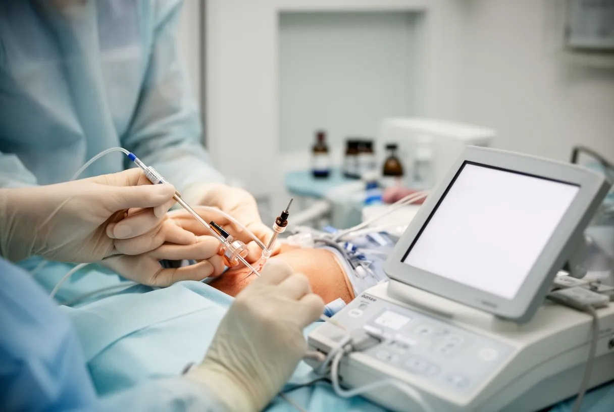
Accessing the heart safely: The biopsy is typically performed in a specialized hospital room called a catheterization lab. You will lie on an X-ray table, and the medical team will administer a local anesthetic to numb the skin. Unlike procedures requiring a large arterial cannula, the biopsy usually uses a venous approach, though an arterial line might be placed for continuous blood pressure monitoring. You may receive mild sedation to help you relax, but while complex life support systems use awake ECMO, for a routine biopsy, a gentle twilight sleep is standard.
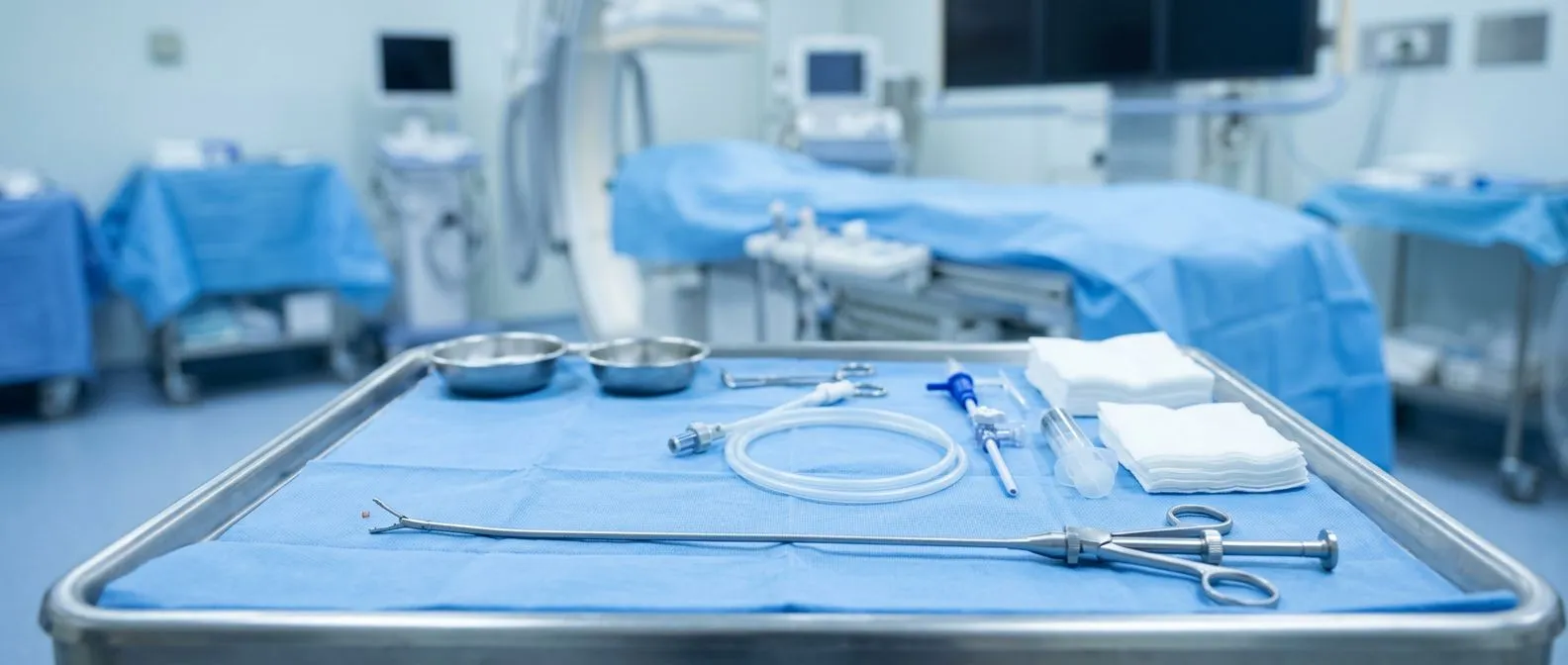
Guiding the bioptome: Through the catheter, the doctor advances a specialized instrument called a bioptome. This device has tiny jaws at its tip that can open and close. Using continuous X-ray imaging, or fluoroscopy, the doctor watches the bioptome travel through the superior vena cava and into the right atrium, one of the heart's upper chambers. From the atrium, the device is guided across the tricuspid valve into the right ventricle. The doctor presses the bioptome gently against the wall of the ventricle and takes tiny pinches of tissue, usually three to five samples.
Immediate post-procedure steps: Once the samples are collected, the bioptome and catheter are removed. Pressure is applied to the insertion site to prevent bleeding, and a small dressing is applied. The tissue samples are immediately placed in a preserving solution and sent to the pathology laboratory. The whole procedure usually takes less than an hour. Because the heart muscle lacks the same type of pain receptors as the skin, you generally will not feel the biopsy itself, though you might feel a brief flutter or pressure as the catheter moves.
Endomyocardial biopsy vs angiography: knowing the difference
Focusing on different structures: Patients often confuse a heart muscle biopsy with an angiogram because both involve catheters and both take place in a catheterization lab. However, they look at completely different parts of the heart. An angiography examines the coronary arteries, the blood vessels that supply oxygen to the heart muscle, including those branching off the ascending aorta. It looks for blockages caused by atheromatous plaque that might require a balloon angioplasty to open. An endomyocardial biopsy, on the other hand, examines the heart muscle tissue itself, looking for cellular disease, inflammation, or rejection.
Contrasting the techniques: During an angiogram, the catheter is directed to the opening of the arteries on the outside of the heart, and contrast dye is injected to highlight blockages. If a severe blockage is found, interventions like placing a stent, a balloon aortic valvuloplasty, or even evaluating for a balloon-expandable valve might be considered. In a biopsy, the catheter travels inside the heart chambers to reach the inner lining of the muscle. Sometimes, if a patient is undergoing an angiogram for suspected coronary disease and the doctor finds the arteries are clear, an endomyocardial biopsy might be performed to see if the muscle is diseased instead.
Why you might need both: It is not uncommon for a patient with complex cardiac symptoms to require both tests. For instance, a patient who has had a heart transplant might need an angiogram to check the coronary arteries for transplant vasculopathy, a form of chronic rejection, and an endomyocardial biopsy to check the muscle for acute rejection. While they serve different purposes, both provide vital, complementary information. Understanding which test your doctor is ordering helps you prepare mentally for the specific questions that test will answer.
Heart biopsy preparation: what to do beforehand
Medical history and medication review: Preparing for a myocardial biopsy doctor visit begins with a thorough review of your medications. Your cardiac team will give you specific instructions, especially regarding blood thinners. If you take aspirin or other anticoagulants, you may need to stop them a few days before the procedure to reduce the risk of bleeding at the puncture site. You should also inform your doctor about any allergies, and mention if you have ever had an arterial graft harvested, if you have stored autologous blood for emergencies, and what your complete medical history entails.
Fasting and logistical planning: You will typically be asked to fast for six to eight hours before the procedure, meaning no food or drinks, though sips of water for medications are usually allowed. Because you will receive sedation, you cannot drive yourself home afterward. You must arrange for a family member or friend to accompany you and stay with you overnight. Wearing comfortable, loose-fitting clothing is advisable. Leave valuables and jewelry at home, as you will need to change into a hospital gown for the procedure. Before the procedure, keep these key preparation points in mind:
- Medication list: Bring a complete list of all prescriptions and supplements.
- Hydration: Stay hydrated in the days leading up, but fast as instructed.
- Transportation: Arrange a responsible adult to drive you home safely.
Mental preparation and questions: It is completely normal to feel anxious before a heart procedure. Write down any questions you have beforehand so you do not forget them in the moment. Ask your doctor how long the procedure will take, who will be performing it, and what the immediate aftercare looks like. Knowing the steps and understanding that you will be awake but comfortable helps reduce pre-procedure stress. Your care team is there to ensure you feel safe and informed every step of the way.
What to expect during endomyocardial biopsy recovery
Immediate post-procedure care: Endomyocardial biopsy recovery is generally straightforward and much faster than recovery from open heart surgery. Immediately after the test, you will be moved to a recovery area where nurses will monitor your heart rhythm and blood pressure. You will need to lie flat for a few hours to prevent bleeding from the neck or groin site. The nurses will check the insertion site frequently. Once the doctor confirms there is no bleeding and your vital signs are stable, you will be allowed to sit up and eventually go home.
Managing site discomfort and limits: When you go home, you might experience mild soreness or a small bruise where the catheter was inserted. This is normal and should fade within a few days. You will be advised to avoid heavy lifting, strenuous exercise, or vigorous activity for at least 24 to 48 hours. If the site was in your neck, you may be asked to avoid turning your head sharply. Keeping the area clean and dry is essential to prevent infection. You can usually shower the next day, but you should not submerge the site in a bath until it is fully healed. Here are the standard recovery milestones to monitor:
- First 24 hours: Rest and avoid heavy lifting to prevent site bleeding.
- Day two: You can usually shower, keeping the puncture site clean.
- Follow-up: Return to the clinic to discuss your biopsy results.
When to resume normal activities: Most patients return to their normal daily routines within a day or two, though you should listen to your body and rest when needed. If you work a physically demanding job, you might need a few extra days off. Your cardiologist will schedule a follow-up appointment to discuss the results of the biopsy and to plan your ongoing care. Recovery from this procedure is a brief pause in your life, designed to gather critical information that will guide your long-term health strategy without requiring extensive downtime.
Endomyocardial biopsy risks and complications to watch for
Common minor risks: While an endomyocardial biopsy is generally safe, it is an invasive procedure and carries some risks. The most common complications are minor and related to the insertion site. You might experience mild bleeding, bruising, or soreness where the catheter was placed. Sometimes, a small hematoma or collection of blood forms under the skin, which usually resolves on its own. Infection at the puncture site is rare but possible, so keeping the area clean is vital. These minor endomyocardial biopsy complications are easily managed with basic wound care and rest.
Cardiac rhythm changes: Because the catheter touches the inside of the heart, it can temporarily irritate the heart's electrical system. This might cause transient arrhythmias, or irregular heartbeats, during the procedure. The medical team monitors your electrocardiogram continuously, and if an irregular rhythm occurs, they can treat it immediately. In very rare cases, the bioptome can puncture the heart wall, causing blood to leak into the sac surrounding the heart. This is a serious complication called cardiac tamponade, requiring immediate drainage. According to the Heart Disease and Stroke Statistics—2013 Update, continuous improvement in procedural techniques helps minimize these severe risks.
Recognizing warning signs at home: Once you are home, you need to watch for signs that warrant a call to your doctor. If you notice continuous bleeding from the site that does not stop with pressure, a growing lump, fever, or increasing pain, you should seek medical attention. If you feel sudden shortness of breath, chest pain, or fainting, these could be signs of a delayed complication and require an immediate emergency room visit. Having an automated external defibrillator nearby is not necessary for routine recovery, but being aware of severe symptoms ensures you act quickly if something feels wrong.
Biopsy for heart transplant rejection: monitoring your new heart
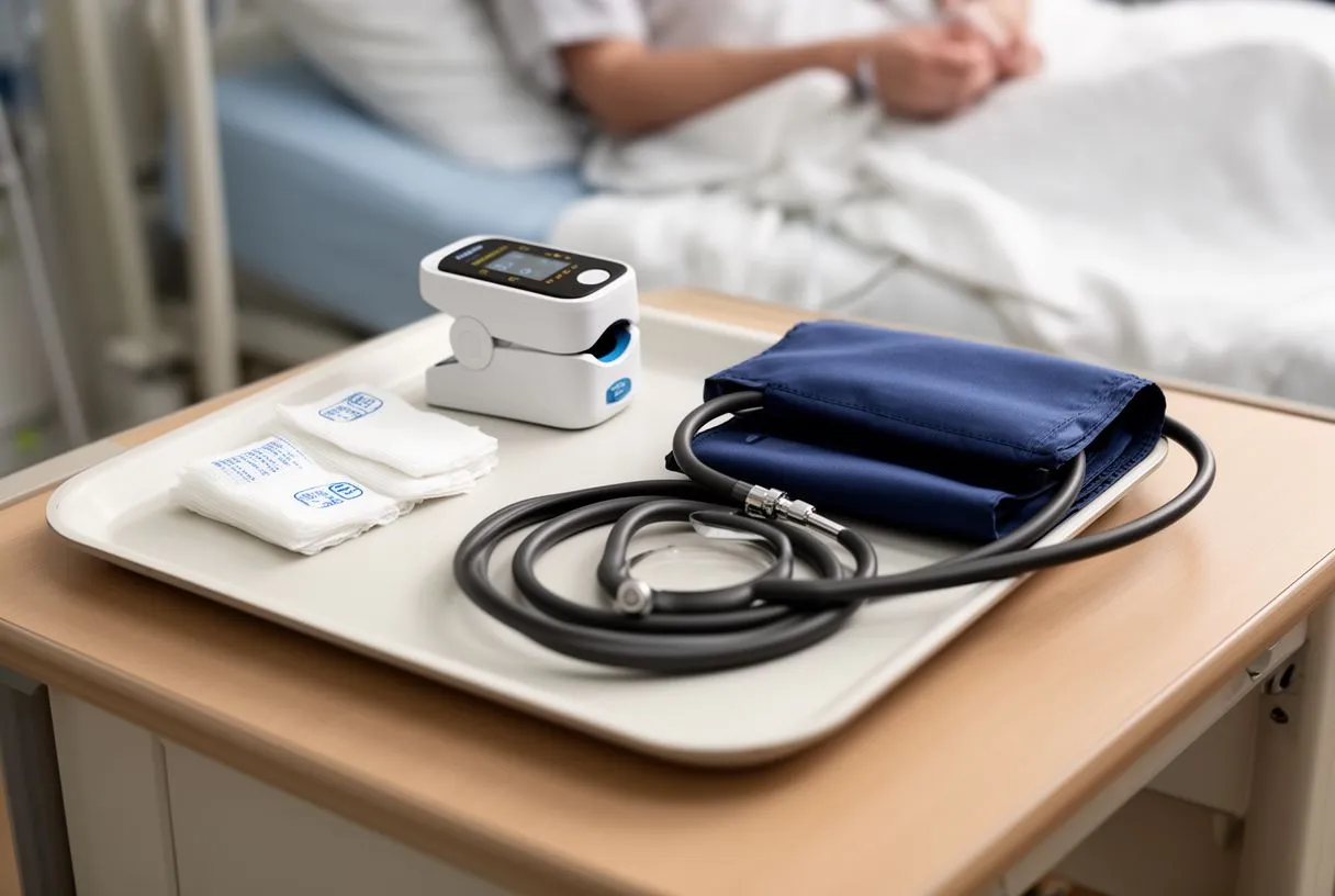
The role of surveillance biopsies: For heart transplant recipients, the endomyocardial biopsy is the gold standard for monitoring the new organ. Your immune system recognizes the transplanted heart as foreign and may try to attack it, a process called rejection. Often, rejection happens without any physical symptoms in its early stages. By performing routine biopsies—frequently in the first few months after surgery and gradually less often over time—doctors can detect cellular changes before they affect heart function. This allows for prompt adjustments to your immunosuppressant medications.
What rejection looks like under a microscope: When the pathologist examines the biopsied tissue, they look for specific immune cells invading the heart muscle. The results are graded based on the severity of these findings. A low-grade rejection might just require closer monitoring or a slight tweak in your medication dose. Higher grades might require a short course of stronger immunosuppressive therapy, sometimes given intravenously in the hospital. This tailored approach ensures that the heart is protected without over-suppressing the patient's immune system unnecessarily.
Psychological aspects of repeated biopsies: Undergoing repeated biopsies after a transplant can be emotionally taxing for patients and families. Each procedure brings a wave of anxiety while waiting for the results. It is important to acknowledge this stress and lean on your support system. Remember that these biopsies are a safety net, a proactive measure to ensure the long-term success of your transplant. As your body accepts the heart over time, the frequency of these tests decreases, allowing you to return to a more normal life.
Understanding your endomyocardial biopsy results
Waiting for the pathology report: It usually takes a few days to a week to receive endomyocardial biopsy results because the tissue must be carefully prepared, sliced, and stained in the laboratory. The pathologist examines the samples under a microscope to identify any abnormalities in the heart muscle cells. This waiting period can be stressful, but it is important not to rush the process, as an accurate diagnosis requires meticulous examination. Your cardiologist will schedule a follow-up appointment specifically to discuss these findings with you.
Interpreting the cellular findings: The results will explain exactly what the pathologist saw. For transplant patients, the report will include a rejection grade, ranging from mild to severe. For non-transplant patients, the report might identify specific diseases, such as myocarditis (inflammation of the heart), or infiltrative disorders where abnormal proteins or cells build up in the heart tissue. The doctor will explain what these medical terms mean in plain language, breaking down how these cellular changes relate to the symptoms you have been experiencing.
Next steps based on the diagnosis: The biopsy results are a tool to guide your treatment. If the biopsy shows inflammation, you might be prescribed anti-inflammatory medications or steroids. If it reveals an infiltrative disease, you might be referred to a specialist for targeted systemic therapies. Sometimes, the biopsy might show that the heart muscle is essentially normal, which can be reassuring and might direct your doctor to look for other causes of your symptoms, like an autograft issue or an unrelated nerve problem. The results provide a definitive roadmap for your cardiac care.
Who performs endomyocardial biopsy and finding cardiac care near you
The cardiac care team: A highly specialized team performs an endomyocardial biopsy. This includes an interventional cardiologist or a transplant cardiologist who physically performs the procedure, specialized nurses who monitor your vitals, and a pathologist who interprets the tissue. These teams work in advanced cardiac centers equipped with fluoroscopy and rapid laboratory processing. If you are looking for cardiac biopsy near me, it is essential to seek out centers that specialize in advanced heart failure or transplant cardiology, as they have the most experience with this specific technique.

International care and accessing treatment: For patients traveling for care, finding a center that coordinates complex diagnostics is vital. While some might explore options for a private heart biopsy Guadalajara offers highly trained specialists who manage these advanced procedures with international patients in mind. Research into complex cardiac interventions, such as the Transcatheter Aortic-Valve Implantation for Aortic Stenosis in Patients Who Cannot Undergo Surgery, highlights how specialized centers are pushing the boundaries of less invasive cardiac care. Patients should seek out teams that offer clear communication and comprehensive support for traveling families.
Making the decision to seek help: Deciding to undergo a heart biopsy is a significant step, but it is a step toward clarity and better health. If you or a loved one are experiencing unexplained cardiac symptoms or are navigating life after a heart transplant, you need a dedicated team by your side. At Cirugía de Corazón, Dr Alejandro Molina and his team provide compassionate, expert cardiac care in Guadalajara, Querétaro, and Hermosillo. We encourage you to discuss your specific situation with a qualified cardiac team rather than self-diagnosing, and to book a consultation to explore your options.
In summary, an endomyocardial biopsy is a highly specialized, generally safe procedure that provides critical answers when the heart muscle is in question. Whether it is used to diagnose a complex cardiomyopathy, investigate unexplained arrhythmias, or monitor the health of a transplanted heart, the insights gained from this tiny tissue sample are invaluable. By understanding the preparation, the procedure itself, and the recovery process, you can approach this test with confidence. If you are experiencing heart issues and need expert guidance, do not hesitate to seek professional evaluation to ensure your heart remains strong and healthy for years to come.