Finger Clubbing: A Sign of Heart and Lung Disease
Finger clubbing heart disease can reveal hidden cardiovascular or lung problems.
What is finger clubbing heart disease?
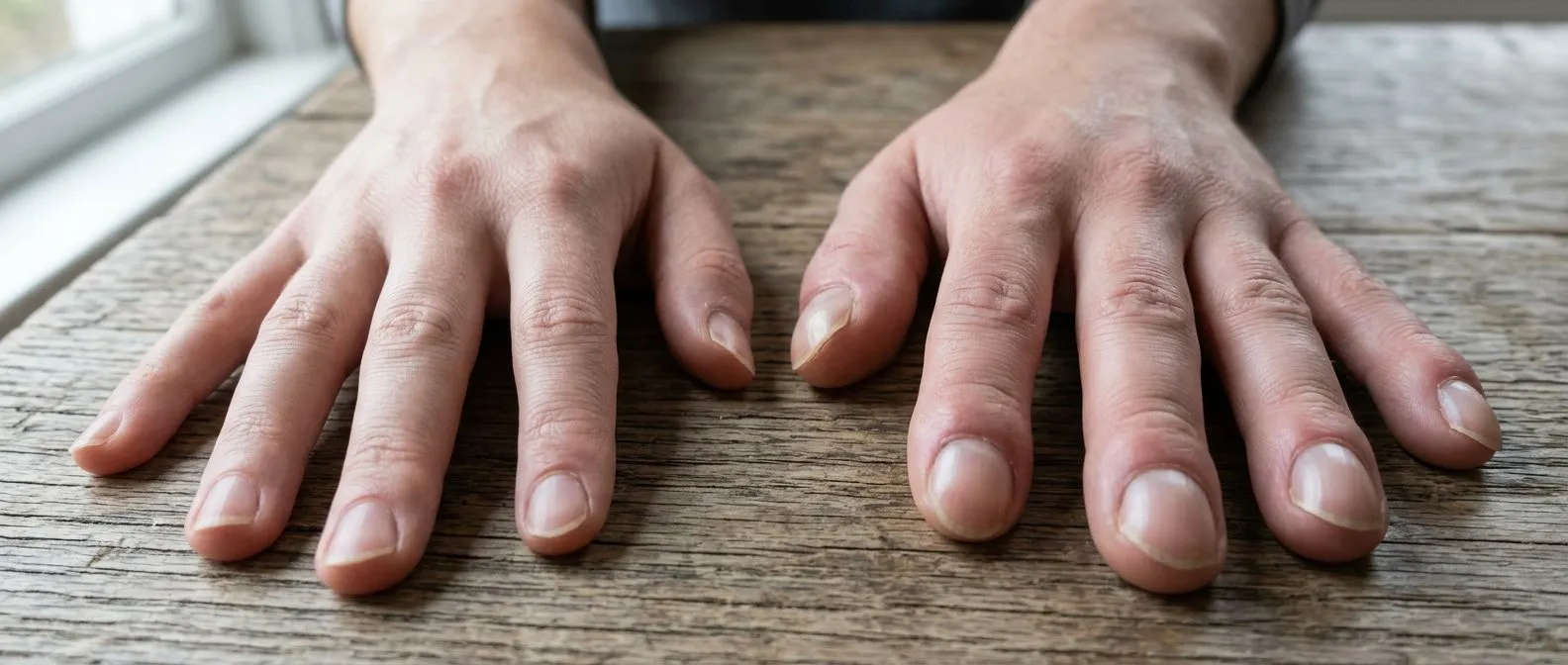
Noticing changes in the shape of your fingertips can be unsettling, especially when you learn that finger clubbing heart disease may be a warning sign of a deeper cardiovascular or respiratory issue. Finger clubbing refers to the soft tissue enlargement of the distal fingers, resulting in a bulbous, drumstick appearance. For patients and families navigating cardiac concerns, recognizing this physical change is an important step toward understanding what is happening inside the body. It is not a disease in itself, but rather a visible symptom of an underlying condition that warrants careful medical evaluation.
The anatomical mechanism: The exact biological process involves vasodilation of the tiny blood vessels in the fingertips, driven by altered blood flow. When the body repeatedly circulates oxygen-poor blood or experiences chronic inflammation, it triggers the release of growth factors that promote tissue swelling and the laying down of new bone matrix in the fingers. Conditions affecting the Ascending Aorta, for instance, can sometimes coexist with signs that manifest far from the chest, reminding us that the cardiovascular system is deeply interconnected.
From a patient perspective, these changes often happen so gradually that they go unnoticed until a family member points them out or a routine physical exam reveals them. The nails may feel spongy when pressed, and the normal angle between the nail bed and the finger fold begins to flatten. Understanding that your hands are providing a window into your cardiopulmonary health can help you approach this symptom with informed calm, rather than fear. If you notice these structural changes, it is simply a signal to seek a thorough cardiac assessment.
What causes finger clubbing in cardiac patients?
Finger clubbing is caused by a variety of conditions, but in the context of cardiac health, it is most strongly associated with cyanotic heart disease—conditions where blood bypasses the lungs and fails to pick up adequate oxygen. In a healthy heart, blood travels to the lungs to be oxygenated before pumping to the rest of the body. When structural defects allow blood to shunt from the right side of the heart directly to the left, the systemic circulation receives poorly oxygenated blood. This chronic hypoxia signals the body to develop the tissue changes seen in clubbing. A review of cardiovascular biomarkers cataloged by Europe PMC highlights how chronic hypoxia alters endothelial function throughout the peripheral vasculature.
Right-to-left shunting: The primary driver in cardiac patients is this right-to-left shunting. When blood is continuously directed away from the pulmonary circulation, the body attempts to compensate by increasing blood flow to peripheral tissues. Infective endocarditis, which is an infection of an Atheromatous Plaque or a heart valve, is another notable culprit that can trigger clubbing. The persistent presence of bacteria in the bloodstream leads to chronic immune system activation and vascular changes in the digits.
To determine if the heart is the source of the problem, your cardiologist will conduct a comprehensive physical exam, often starting by using Auscultation to listen for the heart murmurs that frequently accompany these structural defects. Routine blood work might also reveal elevated Apolipoprotein B levels or markers of infection, helping to clarify the underlying cause. Differentiating between a purely lung-driven cause and a cardiac one requires imaging, as the treatments and management pathways differ significantly based on the anatomical origin of the clubbing.
How to perform the Schamroth window test at home?
If you suspect that you or a loved one might be developing finger clubbing, there is a simple, non-invasive self-assessment you can perform to check for the loss of the normal nail bed angle. The Schamroth window test: This straightforward clinical maneuver involves placing the nails of your index fingers or ring fingers back-to-back, so that the dorsal surfaces of the terminal phalanges are touching. In a healthy hand, the angled profile of the nails will leave a small, clearly visible diamond-shaped window of light between the nail beds.
If finger clubbing is present, the angle of the nail bed is lost or flattened, and the nails become excessively curved. When you perform the Schamroth window test on clubbed fingers, the diamond-shaped window disappears entirely because the nails curve too closely around the fingertip. While this test is not a definitive diagnosis, the absence of the window is a reliable indicator that you should consult a medical professional. It provides a concrete, visual cue that often motivates patients to seek timely evaluation.

It is highly recommended to perform this test if you have a known family history of heart defects or if you experience unexplained Arm Pain alongside these digital changes. You should also pay attention to how your nails feel; a clubbed nail will often have a spongy, floating sensation when you press down on the nail bed. Remember to perform this assessment gently and document any noticeable changes to discuss with your cardiac team during your next appointment.
What are the physical signs of digital clubbing?
Recognizing the physical signs of digital clubbing requires looking at both the nails and the underlying tissue of the fingertips. The progression of finger clubbing is typically classified into stages, starting with the increased fluctuation and bogginess of the nail bed. As it advances, the angle between the nail and the skin fold (the Lovibond angle) straightens out and becomes greater than 180 degrees, giving the finger a bulbous appearance. The distal phalanx also appears broader and thicker, often taking on the characteristic "drumstick" or "watch-glass" shape that gives the condition its name.
Phalangeal depth ratio: Clinicians measure the diameter of the distal phalanx compared to the proximal joint to confirm clubbing. If the fingertip is wider than the joint just below it, clubbing is likely present. A study cataloged in PubMed 28512104 outlines the precise morphological criteria used to grade these changes, ensuring that physicians can objectively distinguish between early clubbing and natural variations in finger anatomy. For patients in a clinical setting, this might be measured while an Arterial Line is being placed for continuous blood pressure monitoring during a diagnostic workup.
Visually, the nails may also begin to curve downward and outward, enveloping the sides of the fingertips. The skin over the knuckles might appear shiny or stretched, and the overall contour of the hand changes significantly over time. These signs do not happen overnight; they are the result of chronic, sustained physiological changes. Observing these physical traits in yourself or a family member is a clear indication that a comprehensive cardiovascular and pulmonary assessment is necessary to rule out serious underlying conditions.
How does congenital heart disease cause finger clubbing?
Congenital heart defects are among the most common cardiac causes of finger clubbing, particularly those categorized as cyanotic congenital heart disease. Conditions such as Tetralogy of Fallot, transposition of the great arteries, or ventricular septal defects (when they lead to pulmonary hypertension) prevent blood from getting adequately oxygenated in the lungs. Because the defect is present from birth, patients with uncorrected or partially corrected cyanotic conditions often develop pronounced finger clubbing during childhood or early adolescence. The body adapts to the chronic lack of oxygen by increasing blood flow to the extremities, which ultimately changes the tissue structure.
Right Atrium dynamics: In these structural anomalies, deoxygenated blood returning to the right Atrium is shunted directly into the systemic circulation instead of traveling to the lungs. This mixing of oxygen-rich and oxygen-poor blood means the tissues are perpetually starved for oxygen, triggering the vascular changes that lead to clubbing. Patients with these conditions may also experience secondary complications, such as rhythm issues like Atrial Flutter, which can further complicate their cardiac management and exacerbate the feeling of breathlessness.

For families managing a child or adult with congenital heart disease, the presence of finger clubbing is often a baseline expectation rather than a sudden emergency. However, if the clubbing worsens rapidly or if the patient experiences a sudden increase in cyanosis (bluish tint to the lips or skin), it may indicate that the existing defect is progressing or that a new complication has arisen. Surgical correction of the underlying defect is the primary way to halt the progression of clubbing, though the physical changes in the fingers may not fully revert to normal if they have been present for many years.
What cardiovascular conditions lead to nail bed clubbing?
Beyond congenital defects, several acquired cardiovascular conditions can lead to the development of nail bed clubbing. Infective endocarditis: This infection of the heart valves or endocardium causes prolonged systemic inflammation, which can alter the peripheral vasculature and promote tissue growth in the fingers. Patients with endocarditis might also present with other signs like prolonged fever, heart murmurs, and small, painful nodules on the fingers known as Osler nodes, making prompt diagnosis critical. Atrial myxomas, which are rare, benign tumors in the heart, can also intermittently obstruct blood flow and cause clubbing due to transient hypoxia or inflammatory responses.
Research into structural heart interventions, such as the Clinical Outcomes of PIMSRA Treating for Hypertrophic Obstructive Cardiomyopathy: A Large-Sample Study, underscores the complexity of cardiovascular tissue changes. While hypertrophic obstructive cardiomyopathy itself is not a classic cause of clubbing, severe outflow tract obstructions and the subsequent heart failure can create a cascade of circulatory challenges. In extreme cardiac care scenarios, managing these profound circulatory deficits might require advanced support, such as supporting heart function with Awake ECMO, or ensuring an Automated External Defibrillator is nearby for sudden rhythm disturbances.
Additionally, any advanced heart failure that leads to profound, systemic hypoperfusion and chronic hypoxia can theoretically contribute to vascular changes in the periphery. During complex surgical interventions for these acquired conditions, the use of cell salvage techniques for the reinfusion of Autologous Blood helps maintain oxygen-carrying capacity, highlighting the critical link between healthy blood flow and tissue viability. Identifying which specific cardiovascular condition is driving the clubbing requires a detailed echocardiogram and careful clinical correlation to pinpoint the exact anatomical and functional failure.
What is hypertrophic osteoarthropathy and finger clubbing?
Hypertrophic osteoarthropathy (HOA) is a syndrome characterized by the triad of finger clubbing, periostosis (new bone formation on the long bones), and arthritis-like joint pain. Joint pain connection: While isolated finger clubbing is generally painless, HOA introduces a painful component that can significantly affect a patient's quality of life. The pain is typically localized to the wrists, ankles, knees, and elbows, and it often worsens with activity. This syndrome can be primary (hereditary) or secondary to an underlying disease, most commonly lung cancer or severe chronic pulmonary infections, but it can also stem from complex cardiovascular anomalies.
The pathophysiology of HOA involves abnormal production of growth factors and an exaggerated vascular response, leading not only to the soft tissue changes in the fingers but also to the inflammation of the outer layer of the long bones. When a patient presents with painful joints alongside finger clubbing, the diagnostic approach must be broadened. In some cardiac surgical contexts, the circulatory dynamics that lead to HOA can be as intricate as the vascular pathways managed when using an Arterial Graft to bypass a blockage, or the careful flow monitoring required when inserting an Arterial Cannula during cardiopulmonary bypass.
Treatment for the joint pain associated with HOA often focuses on managing the underlying heart or lung condition. In some cases, pain relievers or anti-inflammatory medications are prescribed to ease the musculoskeletal discomfort while the primary disease is being addressed. It is vital to differentiate HOA from other forms of arthritis, as the presence of finger clubbing changes the clinical picture entirely. A rheumatologist and cardiologist often work together to ensure that the vascular and inflammatory components of the syndrome are treated comprehensively.
How is finger clubbing diagnosed and evaluated?
Diagnosing finger clubbing and uncovering its root cause requires a meticulous, step-by-step medical evaluation. Comprehensive screening: Diagnosis is never based on finger shape alone; it requires a detailed patient history, a thorough physical examination, and targeted diagnostic testing. Your physician will look for the physical signs of clubbing, such as the loss of the Lovibond angle and the presence of the Schamroth window, while simultaneously listening to your heart and lungs. Sometimes, a patient might seek medical attention for an entirely different reason, such as an Atypical Infarction, and the physician will incidentally note the presence of clubbing, prompting a deeper investigation into their cardiopulmonary status.
- Physical examination: Checking the nail bed angle, assessing nail bed fluctuation, and using the Schamroth window test to confirm the presence of clubbing.
- Cardiac imaging: Echocardiograms and cardiac MRI to identify structural heart defects, valve infections, or congenital anomalies that might be causing right-to-left shunting.
- Laboratory tests: Blood work to check for infection markers, oxygen levels, and inflammatory indicators that might point to endocarditis or pulmonary disease.
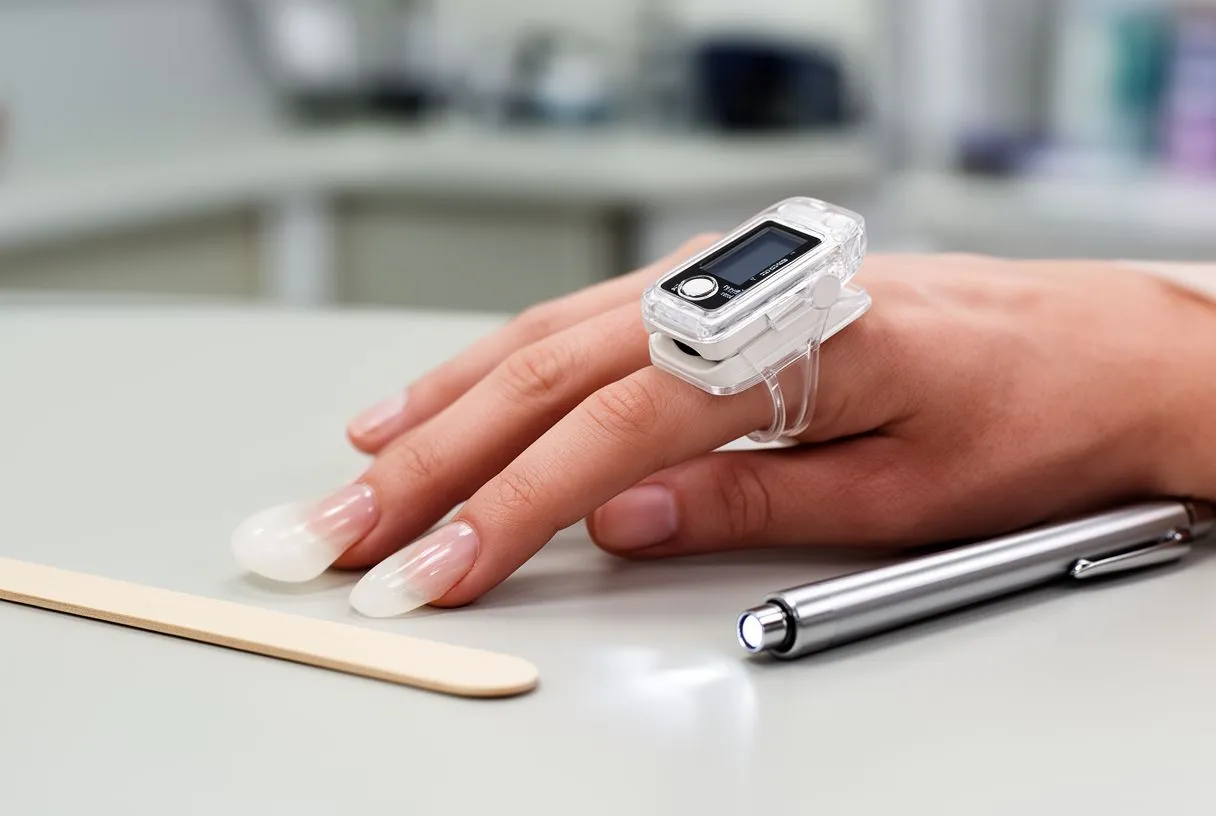
During the evaluation, if an obstruction in the coronary arteries is suspected as part of a broader circulatory issue, your doctor might discuss interventional options such as a Balloon Angioplasty procedure. However, it is crucial to understand that treating an isolated blockage will not reverse finger clubbing if the clubbing is driven by a separate, untreated structural heart defect. The evaluation must connect the dots between the symptom (clubbing) and the systemic cause to ensure that any proposed treatment plan addresses the true source of the physiological disruption.
Is finger clubbing reversible with treatment?
The reversibility of finger clubbing depends entirely on the ability to treat and resolve the underlying condition. Treating the root cause: Finger clubbing itself is not a disease that is treated in isolation; rather, it is a symptom that resolves or stabilizes when the primary cardiovascular or pulmonary issue is corrected. If a patient with a cyanotic congenital heart defect undergoes successful surgical repair to close the shunt, the chronic hypoxia is eliminated. Over time, the vascular and soft tissue changes in the fingers may slowly begin to recede, and the normal angle of the nail bed may partially or fully return, especially in younger patients whose tissues are more adaptable.
In cases where the clubbing is caused by an acquired condition, such as infective endocarditis, aggressive antibiotic therapy to clear the infection can halt the progression of clubbing. If the infection has severely damaged a heart valve, surgical replacement may be necessary. Modern cardiac interventions, such as a Balloon Aortic Valvuloplasty to open a narrowed valve, or a transcatheter aortic valve replacement (TAVR) using a Balloon-expandable Valve, restore healthy, oxygenated blood flow. When surgical repair requires replacing a vessel or valve, using your own tissue, known as an Autograft, can provide excellent long-term outcomes and reduce the need for lifelong blood thinners.
However, if finger clubbing has been present for many years, the structural changes to the bones and soft tissues may become permanent, and the fingertips may retain their bulbous shape even after the underlying condition is cured. The primary goal of treatment is always to address the life-threatening cardiovascular or pulmonary issue first. Reversing the physical appearance of the fingers is a secondary benefit, and patients should focus their expectations on improving their overall heart function, oxygenation, and longevity rather than purely cosmetic recovery.
When to seek help for heart disease finger symptoms?
If you notice progressive changes in the shape of your fingertips or the profile of your nails, it is a clear indication that you should seek medical advice. Monitoring changes: Because finger clubbing develops slowly, families should be vigilant if they have a history of heart disease. You should schedule a consultation if you observe the loss of the Schamroth window, increased sponginess in the nail beds, or a noticeable widening of the fingertips. It is especially important to seek prompt evaluation if these changes are accompanied by shortness of breath, chest pain, or a persistent cough, as these combined symptoms strongly suggest a serious cardiopulmonary condition.
- Progressive thickening: If your fingertips continue to enlarge or change shape over a period of weeks or months.
- Accompanying symptoms: Chest pain, severe shortness of breath, unexplained weight loss, or chronic fatigue alongside digital changes.
- Existing heart conditions: If you have a known heart defect or a history of heart disease and notice new changes in your nails or fingers.
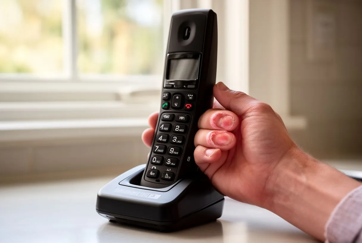
Patients managing cardiovascular risk factors might already be taking daily medications like Aspirin to prevent clots or participating in clinical research trials like the ARISTOTLE study for atrial fibrillation. For these patients, noticing finger clubbing is a sign to re-evaluate their current treatment plan and ensure their heart function remains optimized. Do not ignore these physical signs or wait for them to resolve on their own. We encourage you to discuss your specific situation with a qualified cardiac team to receive an accurate diagnosis. Booking a consultation with a specialist is the most proactive step you can take to protect your heart and overall health.