
High-Sensitivity Troponin: Detecting Heart Damage Early
A high-sensitivity troponin test detects heart damage hours earlier than standard tests.
The difference between a standard troponin test and a high-sensitivity troponin test could mean recognizing heart damage hours earlier — when treatment still has the power to prevent permanent injury. For patients and families navigating chest pain, unexplained fatigue, or planned cardiac procedures, understanding this biomarker brings clarity to moments of uncertainty. This guide explains how hs-troponin works, what your results mean, and why serial testing protocols have become the cornerstone of modern cardiac emergency care.
Testing for high-sensitivity troponin is often part of a comprehensive cardiac evaluation. Whether you arrive with acute symptoms or seek preventive screening, knowing how this test fits into your care helps you participate actively in decisions that protect your heart.
What Are High-Sensitivity Troponin Tests and Why Do They Matter
Immediate detection capability: A high-sensitivity troponin test measures proteins released when heart muscle cells suffer injury — even damage too slight for older tests to register. Troponin I and troponin T are structural proteins found almost exclusively in cardiac tissue. When the heart experiences ischemia (inadequate blood flow), trauma, or inflammation, these proteins leak into the bloodstream in detectable quantities.
Standard troponin assays, widely used through the early 2010s, required substantial myocardial damage before producing positive results. Patients with emerging acute coronary syndrome sometimes waited six to twelve hours for definitive answers. High-sensitivity platforms detect troponin concentrations ten to one hundred times lower, revealing injury within one to three hours of symptom onset. This acceleration transforms clinical decision-making in emergency departments worldwide.
The clinical significance extends beyond heart attack diagnosis. Elevated troponin signals myocardial strain in sepsis, pulmonary embolism, rapid Utility of exercise testing to assess athletes for post COVID-19 myocarditis, and other conditions where the heart faces systemic stress. Early identification guides resource allocation, risk stratification, and therapeutic intervention before irreversible damage accumulates.
Evolution from standard assays: The transition to high-sensitivity methodology represents more than technical refinement — it redefines what clinicians consider "normal." Earlier generations classified patients as "troponin-negative" or "troponin-positive" based on a single cutoff. Contemporary practice recognizes a continuous risk spectrum, where even minimally elevated values predict adverse cardiovascular events months later.
- Detects troponin at 10–100× lower concentrations than conventional assays
- Provides actionable results within 1–3 hours of chest pain onset
- Identifies rising or falling patterns through serial measurement
- Enables risk stratification even in patients without classic heart attack symptoms
For international patients traveling to Mexico for cardiac evaluation, high-sensitivity troponin testing offers particular value. A single blood sample, analyzed upon arrival, establishes baseline cardiac status and directs subsequent imaging or invasive procedures efficiently. This streamlines care coordination and reduces the need for extended diagnostic observation.
How High-Sensitivity Troponin Tests Detect Heart Muscle Injury
Biochemical mechanism explained: Troponin exists as a three-protein complex regulating muscle contraction. Cardiac-specific isoforms — troponin I and troponin T — differ subtly from skeletal muscle variants. Laboratory assays exploit these differences using monoclonal antibodies that bind exclusively to cardiac troponin, ignoring protein released from injured skeletal muscle.
When coronary artery plaque ruptures or an atheromatous plaque erodes, clot formation obstructs blood flow. Heart muscle downstream becomes ischemic, shifting from aerobic to anaerobic metabolism. Cell membranes destabilize; troponin leaks through intact membranes initially, then floods the circulation as cells necrose. High-sensitivity platforms capture this leakage during the earliest phase, when standard assays still read "undetectable."
The assay's sensitivity creates a methodological challenge: detecting tiny quantities risks false positives from analytical noise or minor, clinically irrelevant release. Manufacturers address this through rigorous quality control and by defining the 99th percentile of healthy reference populations as the diagnostic threshold. Values above this percentile — confirmed by serial testing — indicate genuine myocardial injury requiring clinical attention.
Clinical workflow integration: Emergency protocols now incorporate high-sensitivity troponin into accelerated diagnostic pathways. A patient presenting with chest pain receives initial testing, with repeat sampling at three and six hours. Rising values suggest acute coronary syndrome; stable or falling values may indicate chronic elevation or non-ischemic causes. This temporal pattern, more than any single measurement, drives therapeutic decisions.
Post-COVID cardiac evaluation illustrates this precision. Research in Nature and related publications documents persistent troponin elevation in recovery, reflecting ongoing myocardial inflammation. High-sensitivity detection identifies patients needing restricted activity, cardiac magnetic resonance imaging, or anti-inflammatory therapy before permanent dysfunction develops.
Troponin I vs Troponin T: Which Biomarker Your Doctor Uses
Structural differences: Troponin I and troponin T, while both cardiac-specific, occupy distinct positions within the contractile apparatus. Troponin I inhibits actin-myosin interaction in relaxed muscle; troponin T anchors the troponin complex to tropomyosin. These functional differences translate to distinct release kinetics after injury — knowledge that shapes test selection and interpretation.
Troponin I typically rises rapidly after myocardial infarction, peaks at 24–48 hours, and clears within 5–10 days. Troponin T demonstrates similar early kinetics but may remain elevated for 10–14 days, particularly with large infarcts. This prolonged elevation makes troponin T less useful for detecting recurrent injury within two weeks of an initial event. Conversely, troponin I's quicker clearance suits serial monitoring during hospitalization or early postoperative surveillance.
High-sensitivity assays exist for both markers, though regional preferences vary. North American institutions often favor high-sensitivity troponin T (hs-cTnT); European and Asian centers increasingly adopt high-sensitivity troponin I (hs-cTnI). Some centres select assays based on clinical context: troponin I for perioperative monitoring after coronary artery bypass grafting or valve replacement, troponin T for emergency department rule-out protocols.
Assay standardization challenges: Unlike cholesterol or glucose, troponin lacks universal calibration standards. A value of 14 ng/L on one manufacturer's hs-cTnI assay may not equate to 14 ng/L on another. This variability complicates comparison of results across institutions — relevant for medical tourists receiving initial care abroad and follow-up at home. Ask for assay-specific reference ranges and, when possible, repeat testing at the same laboratory.
The specificity of both markers for cardiac tissue remains their defining advantage. Skeletal muscle injury, even rhabdomyolysis, rarely elevates cardiac troponin I; cardiac troponin T shows marginally more skeletal cross-reactivity in some assays. For patients with concurrent trauma or vigorous exercise, troponin I offers slightly cleaner interpretation.
High-Sensitivity Troponin Normal Range and How to Read Your Results
Understanding your high-sensitivity troponin test results requires grasping three numerical concepts: the limit of detection, the 99th percentile upper reference limit (URL), and the "delta" or change between serial measurements. These concepts replace the simple "normal/abnormal" dichotomy of earlier testing eras.
The limit of detection represents the lowest concentration the assay can reliably distinguish from zero. For high-sensitivity platforms, this typically falls below 2–5 ng/L. Detectable values below the 99th percentile — often in the range of 5–14 ng/L for women and 6–22 ng/L for men — are not "abnormal" but indicate measurable baseline troponin in healthy individuals. Factors influencing baseline values include age, renal function, cardiovascular fitness, and genetic variation.
Gender-specific thresholds: The 99th percentile URL differs meaningfully between sexes. Women typically demonstrate lower baseline troponin concentrations; applying male thresholds to female patients historically underdiagnosed acute coronary syndrome in women. Contemporary guidelines mandate sex-specific cutoffs, improving diagnostic equity. A woman with hs-cTnI of 18 ng/L may exceed her 99th percentile while falling below the male threshold — a discrepancy with real clinical consequences.
| Population | Typical 99th Percentile hs-cTnI (ng/L) | Typical 99th Percentile hs-cTnT (ng/L) |
|---|---|---|
| Healthy women | 10–16 | 8–14 |
| Healthy men | 15–26 | 10–22 |
| Chronic kidney disease | Elevated baseline common | Elevated baseline common |
Values exceeding the 99th percentile warrant clinical correlation, not automatic alarm. Stable chronic elevations occur in heart failure, atrial fibrillation with rapid ventricular response, hypertensive heart disease, and renal impairment. The critical question becomes whether values are changing — rising or falling patterns indicate acute processes, while stable elevations suggest chronic myocardial strain. Your cardiologist interprets numbers within your complete clinical picture, never in isolation.
Serial Troponin Testing Protocol: Why One Test Is Never Enough
Dynamic biomarker principle: Cardiac troponin circulates as a concentration, not a binary switch. A single measurement captures one moment in time but cannot distinguish acute rising injury from stable chronic elevation. Serial troponin testing — repeated sampling over hours — reveals the trajectory that distinguishes emergency conditions from manageable chronicity.
Standard protocols vary by institutional risk stratification. For low-risk chest pain patients, European Society of Cardiology guidelines endorse a 0/1-hour or 0/2-hour algorithm: testing at presentation and again at one or two hours. A negative result with absolute change below validated thresholds permits safe discharge without coronary angiography. Higher-risk patients undergo 0/3-hour or 0/6-hour protocols with concurrent electrocardiographic monitoring.
The mathematical concept of "delta" — absolute concentration change between measurements — has transformed emergency practice. Smaller deltas exclude acute coronary syndrome with high negative predictive value; larger deltas mandate invasive evaluation regardless of absolute values. This shift from threshold-based to change-based decision-making reduces unnecessary catheterizations while maintaining safety.
Postoperative applications: After coronary artery bypass grafting, aerial line monitoring and serial troponin testing track myocardial protection during the vulnerable perioperative period. Expected troponin rises reflect surgical manipulation; excessive elevation prompts evaluation for graft dysfunction or perioperative infarction. The same principles apply after transcatheter aortic valve replacement (TAVR), where procedural myocardial injury must be distinguished from clinically significant coronary obstruction.
Patients undergoing cardiac surgery may follow a structured serial testing plan set before discharge. Clear instructions on follow-up timing — typically 48 hours, one week, and one month postoperatively — ensure continuity of care regardless of where subsequent monitoring occurs.
Troponin Elevation Without Heart Attack: Other Causes Doctors Watch For
Non-ischemic myocardial injury: Elevated troponin does not equate to myocardial infarction. Clinicians increasingly recognize diverse conditions causing troponin release without coronary artery obstruction. Understanding these alternatives prevents unnecessary interventions and directs appropriate therapy.
Sepsis and critical illness exemplify this pattern. Bacterial toxins, inflammatory cytokines, and catecholamine surges increase myocardial oxygen demand while impairing microvascular perfusion. Troponin rises proportionally to illness severity, predicting mortality independent of cardiac history. Treatment targets the underlying infection and hemodynamic support; coronary angiography rarely benefits unless concurrent acute coronary syndrome is suspected.
Atrial arrhythmias — particularly atrial flutter with rapid ventricular response — elevate troponin through demand ischemia. The atrium's sustained rapid contraction stresses the ventricle, especially with pre-existing coronary disease or hypertrophy. Rate control and rhythm restoration typically resolve troponin elevation without anticoagulation or revascularization.
- Myocarditis: Viral or autoimmune inflammation directly injures myocytes; troponin may markedly exceed myocardial infarction ranges
- Pulmonary embolism: Right ventricular strain from sudden pressure overload releases troponin proportionally to clot burden
- Renal failure: Reduced clearance elevates baseline troponin; superimposed acute injury may be subtle to detect
- Extreme exertion: Marathon runners and elite athletes demonstrate post-event troponin elevation that typically resolves within 48–72 hours
Post-COVID cardiac considerations: The pandemic illuminated troponin elevation in contexts previously less appreciated. Case reports in Outcome of survivors of COVID-19 in the intermediate phase of recovery: A case report documents persistent cardiac symptoms with biomarker abnormalities months after acute infection. High-sensitivity testing identifies this population for specialized cardiac magnetic resonance evaluation and guided rehabilitation, preventing dangerous return-to-activity decisions.
Accurate interpretation requires clinical context — history, examination, electrocardiogram, and imaging alongside troponin values. The test is extraordinarily sensitive but deliberately nonspecific; its power lies in prompting appropriate investigation, not providing standalone diagnoses.
Troponin Monitoring After Heart Surgery: Tracking Recovery Safely
Perioperative myocardial injury: Every cardiac surgical intervention — from coronary bypass to ascending aorta replacement to valve procedures — involves some degree of planned myocardial manipulation. The heart-lung machine, aortic cross-clamping, cardioplegic arrest, and direct surgical handling all release measurable troponin. Distinguishing expected postoperative release from clinically significant injury is a central monitoring challenge.
Troponin monitoring begins in the intensive care unit with arterial line access enabling frequent sampling. Peak values typically occur 6–12 hours postoperatively, declining over subsequent days. Excessive elevation — variously defined as >5×, >10×, or >20× the 99th percentile depending on surgery type — triggers evaluation for graft occlusion, procedural infarction, or inadequate myocardial protection. Echocardiographic assessment and possible coronary angiography follow.
Valve surgery specifics: Aortic valve replacement, whether surgical or transcatheter, poses unique troponin considerations. Balloon aortic valvuloplasty and TAVR involve direct compression of the left ventricular outflow tract and coronary ostia proximity. Transcatheter approaches generally produce lower troponin peaks than open surgery, reflecting reduced myocardial incision. However, procedural complications — coronary obstruction, annular rupture, or ventricular perforation — cause dramatic troponin surges requiring immediate intervention.
Serial testing continues through hospital discharge and early follow-up. Rising troponin after an initial decline suggests new ischemic events, prosthetic dysfunction, or pericardial complications. We provide patients and families with explicit warning signs: recurrent chest pain, dyspnea disproportionate to activity level, syncope, or palpitations with hemodynamic compromise. These symptoms, especially with new electrocardiographic changes, mandate urgent re-evaluation regardless of scheduled follow-up timing.
Rapid Troponin Test in the Emergency Room: Speed vs Accuracy
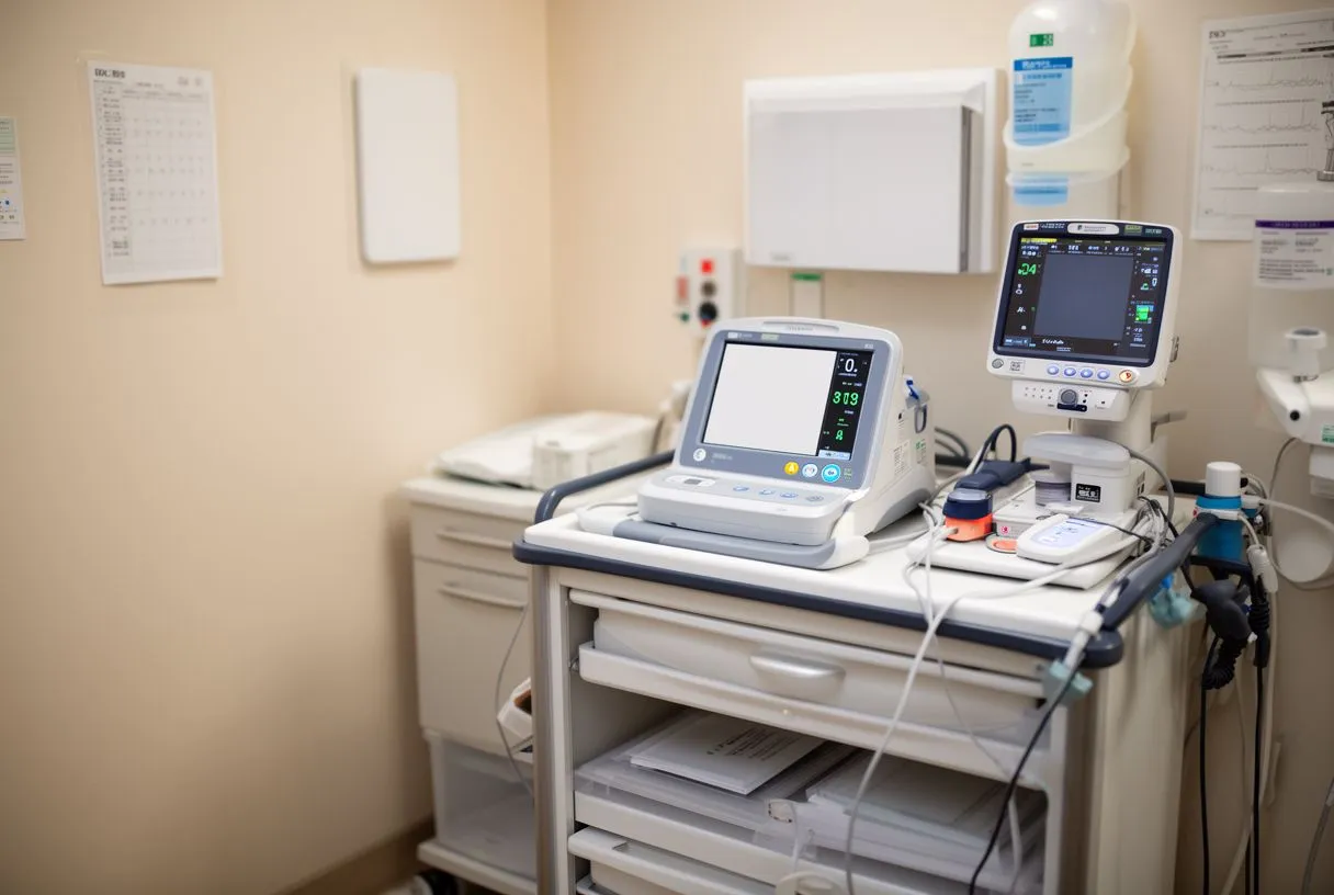
Emergency department triage: The first hours of acute coronary syndrome determine myocardial salvage and survival. Rapid troponin testing — point-of-care platforms returning results in 15–20 minutes —accelerates initial risk stratification, though with trade-offs against centralized laboratory high-sensitivity assays.
Point-of-care devices sacrifice sensitivity for speed. Their analytical limits of detection exceed laboratory platforms tenfold or more; early presenter with sub-threshold injury may receive falsely reassuring results. Contemporary practice reserves point-of-care testing for initial triage of patients with classic symptoms plus unambiguous electrocardiographic changes, while routing others for immediate laboratory high-sensitivity analysis.
The concept of "troponin-only" pathways has gained traction where access to immediate cardiology consultation is limited. Negative high-sensitivity troponin at 0 and 3 hours, combined with normal electrocardiogram and validated risk score, permits discharge with outpatient follow-up. This approach, studied extensively in low-prevalence settings, requires strict adherence to timing and interpretive criteria; applicability varies by regional heart attack incidence and healthcare resource availability.
Limitations and pitfalls: Rapid testing fails when clinical urgency overrides analytical certainty. A patient with crushing chest pain, diaphoresis, and anterior ST-elevation requires emergent catheterization without awaiting troponin results. Conversely, exclusively relying on rapid testing in atypical presentations — particularly women, elderly patients, and those with diabetes — risks missed diagnoses where symptoms manifest as dyspnea, nausea, or altered mental status rather than classic chest pain.
For patients evaluating cardiac services internationally, understanding local testing infrastructure matters. Facilities with 24-hour high-sensitivity laboratory availability offer different diagnostic reliability than those dependent on daytime batch processing or point-of-care alternatives. We transparently discuss our capabilities and limitations during pre-operative consultation.
Best High-Sensitivity Troponin Approach for Preventive Heart Screening
Risk stratification beyond symptoms: Can high-sensitivity troponin testing benefit individuals without acute symptoms? Emerging evidence supports selective screening in intermediate-risk populations — those with cardiovascular risk factors but no established disease. Detectable troponin below the 99th percentile, previously considered "normal," predicts future heart failure and death in population studies.
This "troponin-first" preventive paradigm remains investigational for universal screening but increasingly guides targeted evaluation. Patients with diabetes, chronic kidney disease, strong family history, or inflammatory conditions may benefit from baseline high-sensitivity testing. Identifying subclinical myocardial strain prompts intensified risk factor modification — blood pressure control, statin therapy, glycemic management, and lifestyle intervention — before symptomatic disease develops.
Integration with other biomarkers: Troponin alone offers incomplete cardiovascular risk assessment. Complementary markers include B-type natriuretic peptide (BNP) for hemodynamic stress, high-sensitivity C-reactive protein for inflammation, and lipoprotein(a) for genetic lipid risk. The combination of elevated troponin plus elevated BNP particularly predicts adverse outcomes, identifying patients warranting echocardiographic evaluation and specialist referral even when asymptomatic.
Preoperative cardiac risk assessment before non-cardiac surgery represents another preventive application. Revised cardiac risk indices incorporate troponin monitoring into postoperative surveillance for high-risk procedures. Detecting myocardial injury after non-cardiac surgery (MINS) — often asymptomatic and electrocardiographically silent — enables initiation of cardioprotective medications that reduce mortality.
Screening approaches should be personalized, considering individual risk profiles, prior testing, and patient preferences rather than applying uniform protocols.
When to Seek High-Sensitivity Troponin Testing: Symptoms and Risk Factors
Acute presentation indications: Seek emergency evaluation for chest pressure, pain, or discomfort lasting more than 20 minutes, especially if accompanied by radiation to the arm, jaw, or back, diaphoresis, nausea, or dyspnea. These symptoms warrant immediate high-sensitivity troponin testing regardless of age or apparent health. Women and patients with diabetes particularly may experience atypical presentations — unexplained fatigue, indigestion-like symptoms, or breathlessness without chest pain — that nonetheless represent acute coronary syndrome.
Risk factor thresholds for elective evaluation: Consider scheduled cardiac assessment including high-sensitivity troponin if you carry multiple cardiovascular risk factors: hypertension requiring multiple medications, diabetes with end-organ complications, chronic kidney disease (estimated glomerular filtration rate <60 mL/min), established peripheral or carotid arterial disease, or premature family history (first-degree relative with heart attack before age 55 for men, 65 for women). Smoking history, even remote, adds incremental risk.
Postoperative patients following cardiac intervention should maintain scheduled troponin monitoring even when feeling well. The asymptomatic period after balloon angioplasty, stent placement, or bypass surgery can mask subclinical graft dysfunction or restenosis. Scheduled testing catches these before symptomatic decompensation.
For families considering cardiac surgery in Mexico, we encourage pre-travel consultation with your local physician to establish baseline biomarkers and document current functional status. Bring prior echocardiograms, angiographic reports, and medication lists. This preparation enables our team to focus on decision-making rather than redundant data collection, accelerating your path to definitive treatment.
High-sensitivity troponin testing has transformed cardiac diagnosis from a delayed, threshold-based discipline to a dynamic, pattern-recognition art. The test's extraordinary sensitivity demands equally sophisticated interpretation — accounting for baseline variation, serial changes, clinical context, and individual patient factors. Whether you face acute symptoms, navigate recovery from heart surgery, or seek preventive understanding of your cardiovascular risk, troponin biomarkers provide objective insight into your heart's cellular health. At Cirugía de Corazón, we integrate this technology with experienced clinical judgment to guide decisions that protect your most vital organ. Discuss your personal situation with our qualified cardiac team to determine whether high-sensitivity troponin testing deserves a place in your care — and how we can support your heart health journey from initial evaluation through long-term follow-up.