Ischaemia Testing: Finding Blocked Arteries
Get ischaemia testing heart specialists in Guadalajara use to find blocked arteries early.
Ischaemia testing for the heart is a set of examinations that show whether the heart muscle is receiving enough oxygen-rich blood, especially when it is made to work harder. If you have chest pain, breathlessness or unusual fatigue, your doctor may recommend one of these tests to find out whether an artery is narrowed or blocked. The idea is simple: a healthy heart can increase its blood supply when you exercise, but a heart with a narrowed coronary artery may fall short, and the right test can reveal that shortfall before it becomes a heart attack. This guide explains each type of test, what to expect, and what the results mean for your decisions ahead.
What Is Ischaemia Testing for the Heart?
Why testing matters. The heart is a muscle, and like any muscle it needs a constant supply of blood. The Coronary Arteries are the small vessels that feed it, and when one of them narrows from cholesterol deposits, the muscle downstream may not get enough oxygen during exertion. That temporary shortage is called ischaemia. Ischaemia testing for the heart is the family of examinations designed to detect it, ideally before permanent damage occurs.
What the tests actually measure. These tests answer one central question: does the heart muscle receive adequate blood flow when its demand rises? Some tests watch the electrical activity of the heart, others watch the motion of the muscle itself, and still others produce images of blood flow through the muscle. No single test suits every patient, which is why doctors choose based on your symptoms, your age, and other conditions you may have.
In our practice, every case is reviewed by the multidisciplinary team against international practice guidelines before any recommendation is made, and the patient receives a clear proposal with the alternatives, so the choice of test is never one-size-fits-all.
How Is Myocardial Ischaemia Detected?
Stress as the trigger. Most ischaemia only appears when the heart is pushed to work harder, which is why the heart is "stressed" during testing, either by exercise on a treadmill or with medication that mimics exercise for patients who cannot walk comfortably. The heart's response under this stress is what reveals a narrowed artery that may look normal at rest.
Signs the tests look for. Each test detects ischaemia through a different signal. Together they form a graduated pathway from the simplest to the most detailed:
- Electrocardiogram (ECG) changes: sticky patches on the chest record the heart's electrical signals; certain changes under stress suggest the muscle is short of oxygen.
- Imaging of the muscle: ultrasound, nuclear scans or MRI show whether parts of the heart wall move poorly or receive less blood under stress.
- Direct measurement: a cardiac catheterization, where a thin tube is threaded into the Coronary Arteries, can show the exact site and severity of a blockage.
Most patients begin with the simplest test and move to more detailed imaging only if the results are unclear or concerning. Myocardial ischaemia is well described in the peer-reviewed literature, and an overview of symptoms, causes and diagnosis can be found through the National Institutes of Health.
What Are the Causes and Symptoms of Ischaemic Heart Disease?
Where the disease comes from. Ischaemic Heart Disease is the condition in which the Coronary Arteries become narrowed by fatty deposits called plaque, a process that develops over years. Risk factors include high blood pressure, high cholesterol, smoking, diabetes, obesity and a family history of early heart disease. Over time, a narrowed artery limits blood flow, or a plaque can rupture and cause a sudden blockage, which is what happens during a heart attack.
How it usually feels. Symptoms vary widely, and some patients, especially women, older adults and people with diabetes, have little or no typical pain. The most common warning signs include:
- Pressure or tightness in the chest: often described as squeezing or heaviness, typically triggered by exertion and relieved by rest.
- Breathlessness: finding that stairs or hills that used to be easy now leave you unusually short of air.
- .Fatigue and weakness:
What Is Subendocardial Ischaemia?
Understanding the layers of the heart wall. The heart muscle has an inner layer and an outer layer. The inner layer, called the subendocardium, is the most vulnerable to reduced blood flow because it is the furthest from the surface vessels and endures the greatest pressure as the heart squeezes. Subendocardial ischaemia therefore means the deepest layer of the heart muscle is short of oxygen, and it is often seen on an ECG as a subtle depression of the ST segment during stress.
What it implies for you. Subendocardial ischaemia is common in situations where the demand on the heart exceeds supply across a broad area, such as severe narrowing in one or more Coronary Arteries, severe anaemia, or very high blood pressure. It is a finding your cardiologist will interpret together with your symptoms and the rest of your testing; on its own, the word describes a pattern, not a prognosis.
When we review results like these in consultation, we take the time to explain what the studies actually show, in plain words, and to answer every question before any decision is made.
What Does Inferior Ischaemia Mean?
Location matters on the ECG. The heart can be divided into regions on an electrocardiogram, and "inferior" refers to the underside of the heart, the part that sits on the diaphragm. Inferior ischaemia means the pattern of oxygen shortage appears in that particular region. This part of the heart is supplied mainly by the Right Coronary Artery, so an inferior pattern points your cardiologist to look more closely at that vessel.
Why the region matters. Knowing which region of the heart is affected helps guide which artery is likely responsible, which in turn shapes the choice of further testing and treatment. Sometimes an inferior pattern appears on a routine ECG as an incidental finding in a person with no symptoms, and in that situation the cardiologist will judge whether further testing is genuinely needed or whether the finding can simply be monitored.
Exercise Tolerance Test: How the Heart Stress Test Works
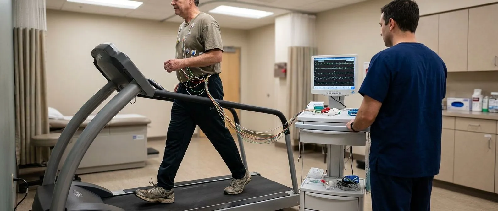
What to expect during the test. The exercise tolerance test, sometimes called a treadmill stress test or exercise ECG, is the most established form of ischaemia testing for the heart. Electrodes are placed on your chest and you walk on a treadmill that gradually becomes faster and steeper, roughly every three minutes, while your heart rhythm, blood pressure and symptoms are monitored. The goal is to reach a target level of exertion so the heart is genuinely stressed.
Who it suits and who it does not. This test is simple, widely available and inexpensive, which makes it a sensible first step for many patients. However, it gives less precise information about location and severity than imaging-based tests, and it is not suitable for people who cannot walk safely, have severe arthritis, or have certain ECG abnormalities at rest that make interpretation difficult. For those patients, a pharmacological stress test, using medication that simulates exercise, or an imaging-based stress test is usually chosen instead.
Nuclear Stress Test and Cardiac Perfusion Imaging
How the scan works. A nuclear stress test, also called myocardial perfusion imaging, uses a tiny amount of a radioactive tracer injected into a vein. A special camera then takes pictures of the heart, both at rest and under stress, showing exactly which areas of the heart muscle receive good blood flow and which do not. This technique, known as cardiac perfusion imaging, offers precise information about the location and extent of any ischaemia.
What the images tell your doctor. A nuclear scan can show a narrowed region clearly and can also distinguish between muscle that is temporarily short of blood and muscle that has already been permanently scarred by a previous heart attack. That distinction matters greatly, because muscle that is still alive and merely underfed may recover its function if blood flow is restored, whereas scarred muscle will not. This is a key piece of information when weighing whether an intervention is worthwhile.
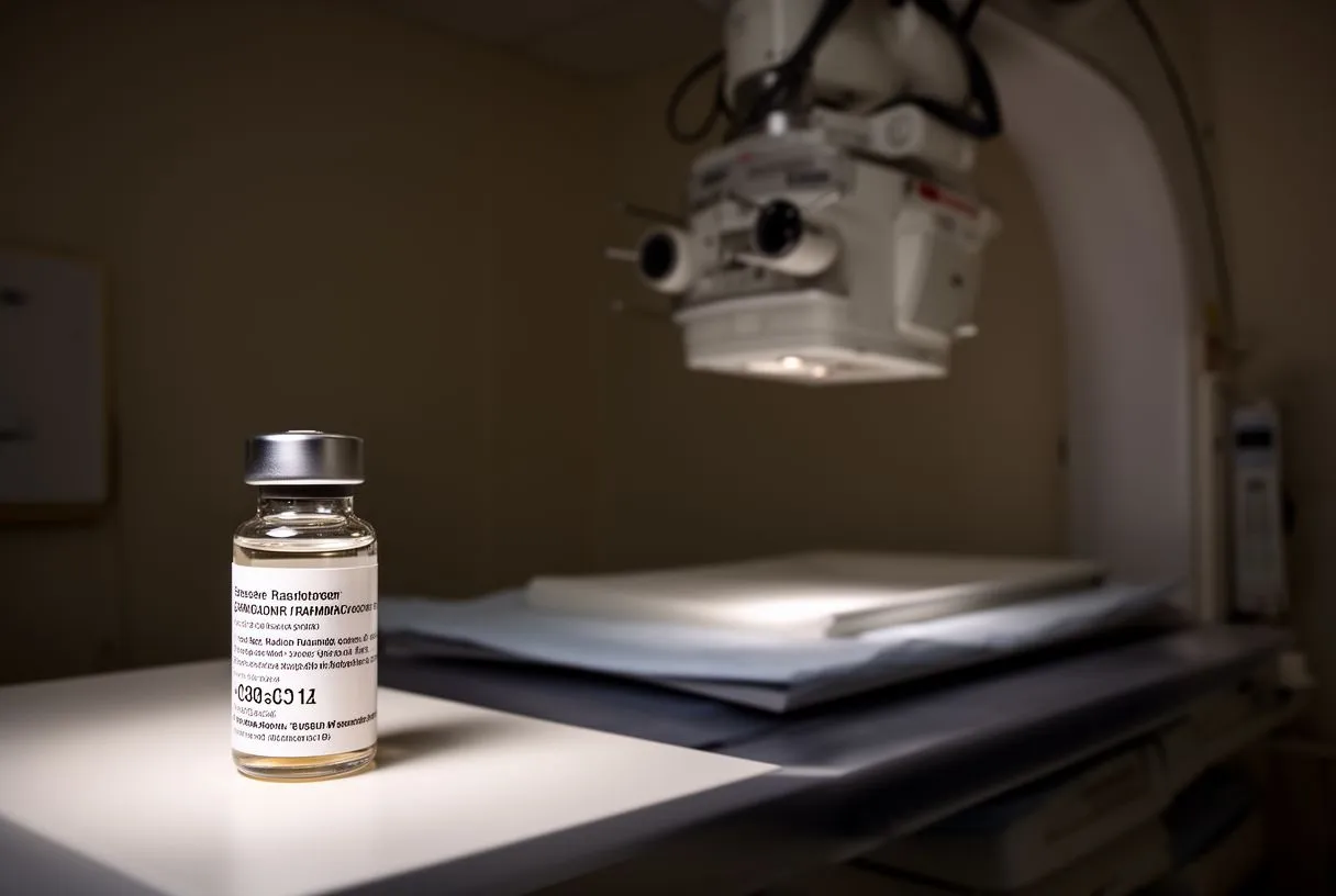
Safety and radiation. The radiation dose from a nuclear stress test is modest and comparable to other medical imaging; the tracer used is cleared from the body within a day or so. The test is not suitable during pregnancy, and patients with certain kidney problems will have their doctor adjust the approach, sometimes choosing an alternative test altogether.
Stress Echocardiogram and Cardiac MRI Options
Stress echocardiogram explained. An echocardiogram is an ultrasound scan of the heart, using the same kind of sound waves that produce images of a baby before birth. In a stress echocardiogram, ultrasound images of the heart are taken before and immediately after the heart is stressed, either by exercise or with medication. Ischaemia shows up as a segment of the heart wall that was moving normally at rest but moves sluggishly under stress, which is a highly informative finding.
Cardiac MRI as a detailed option. Cardiac MRI offers very detailed images of the heart muscle without radiation, and stress MRI or perfusion MRI can show blood flow differences with great clarity. It is particularly useful when other tests have been inconclusive, or when the question involves scar tissue, inflammation, or cardiomyopathy alongside ischaemia.
Choosing between imaging tests. Your doctor will consider availability, your body habitus, kidney function, the presence of a pacemaker, and the exact question to be answered when recommending one imaging test over another. In our multidisciplinary team, this choice is discussed case by case, and we recommend the less invasive route only where the evidence supports it for that particular patient.
When Ischaemia Testing Leads to Coronary Artery Disease Treatment
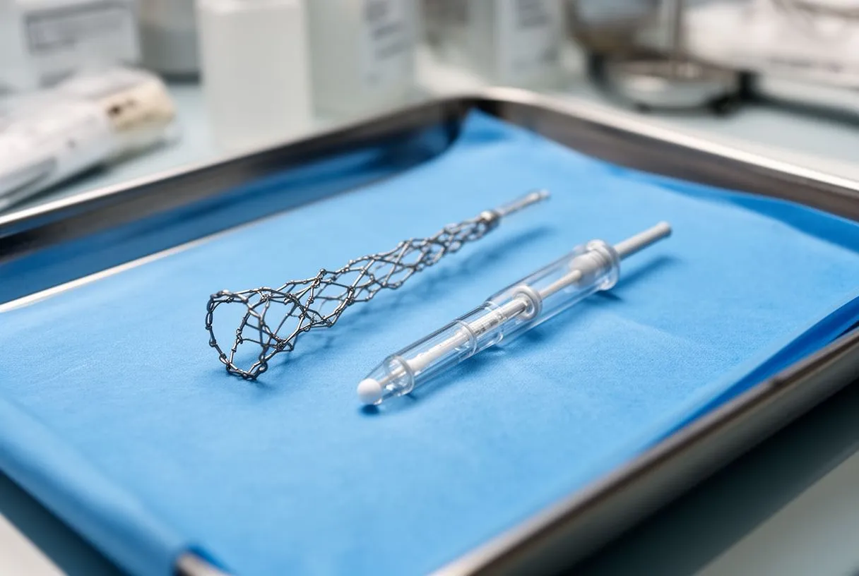
From test to treatment plan. When ischaemia testing confirms significant narrowing, the next step depends on how severe the blockage is and which artery is involved. A critical stenosis of the Left Main Coronary Artery, for example, is treated differently from a smaller blockage in a side branch. Treatment options range from medication to control symptoms and slow plaque growth, to Percutaneous Coronary Intervention, where a stent is placed through a catheter in the wrist or groin, to Coronary Artery Bypass Grafting, open surgery that uses a healthy vessel, often the Left Internal Mammary Artery, to route blood around the blockage.
How your team decides. Decisions are rarely driven by a single result. The team considers the anatomy, how much muscle is at risk, how severe your symptoms are, and how well medication alone is controlling them. In our practice, every such recommendation is reviewed by the whole multidisciplinary team, led by Dr Alejandro Molina, so that the patient hears a clear proposal with the alternatives and always makes the final decision. We also encourage second opinions and treat seeking one as a normal part of deciding about heart surgery.
Ischaemia testing before other surgery. Sometimes ischaemia testing is needed for reasons beyond chest pain. Patients with Valvular Heart Disease who are being assessed for surgery, or those facing other major operations, may undergo testing to confirm that the coronary arteries are not an additional hidden problem that should be addressed at the same time.
Choosing the Right Heart Ischaemia Test for You
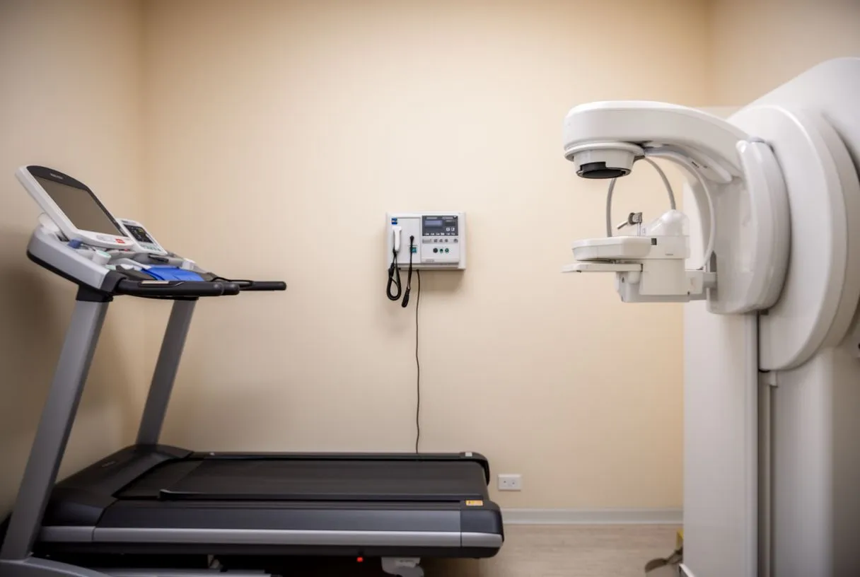
Start with the right question. The best test is not the most advanced one, but the one that answers your specific question with the least risk and cost. If you have chest pain during exertion and can walk comfortably, an exercise tolerance test may be the right first step. If you cannot exercise, if your resting ECG makes interpretation difficult, or if your doctor needs to know precisely which artery is involved and how much muscle is at risk, an imaging-based test such as a nuclear stress test, stress echocardiogram, or a Coronary Calcium Score and CT angiography pathway will usually give clearer answers.
Practical considerations for international patients. For patients travelling from abroad, the sequence matters just as much as the test itself. Arriving with your previous ECGs, reports and images saves time and avoids repeating studies. The first contact with our team is a free video consultation: we get to know you, talk through your condition, and say which studies are still needed so that no unnecessary ones are ordered, and a face-to-face visit at our consulting rooms in Guadalajara, Querétaro or Hermosillo follows when you decide to go ahead.
What happens after the results. A normal test is reassuring but does not mean the risk of Coronary Artery Disease disappears forever; it means your current risk is low enough that invasive testing is not warranted today. An abnormal test always needs interpretation in the context of your symptoms, and sometimes further testing or treatment follows. Dr Molina trained in cardiothoracic surgery at the Universidad de Guadalajara, in cardiac, minimally invasive, robotic and structural surgery at the University of Montreal, and in structural cardiology and TAVR at CHU Bordeaux, so whatever pathway the results suggest, the options are reviewed with the full picture in front of the whole team before anything is proposed. If you have chest pain, breathlessness or unexplained fatigue, the most valuable next step is to discuss your situation with a qualified cardiac team rather than trying to diagnose yourself; a consultation is the place where the right test, and the right plan, get chosen for you.