


Low Ejection Fraction: What It Means and How It Is Treated
Get low ejection fraction treatment from heart specialists at Cirugía de Corazón.
Understanding heart function percentage can feel overwhelming when you are first faced with a cardiology report. If you or a loved one has been told that the heart is not pumping as strongly as it should, you likely have many questions about what this means for your daily life and future health. Seeking low ejection fraction treatment is a critical step toward stabilizing heart function and improving your overall quality of life. At Cirugía de Corazón, our approach focuses on clear, compassionate explanations combined with advanced cardiac care tailored to your specific anatomy and lifestyle. We bridge the gap between complex medical terminology and your personal healthcare journey, ensuring you understand every treatment option and recovery milestone.
What Is Low Ejection Fraction and What Does the Percentage Mean?
Defining ejection fraction in everyday terms: Ejection fraction (EF) is a measurement of the percentage of blood leaving your heart each time it squeezes. When the left ventricle, the main pumping chamber, contracts, it pushes blood out to your body. A normal heart function percentage usually sits between 50% and 70%. When that number falls below 40%, it indicates that the heart muscle is weakened and not pumping efficiently. This condition is often referred to as systolic dysfunction, meaning the squeezing phase of the heartbeat is compromised. Understanding this baseline measurement is the first step in building a low ejection fraction treatment plan, as it guides the medical team in determining how aggressively the heart needs support.
The difference between left and right ventricular function: While most discussions of EF focus on the left ventricle, the right ventricle and the top chambers, known as the atrium, also play vital roles in circulation. The left side is most commonly measured because it is responsible for pushing oxygen-rich blood to the entire body. However, if the right side weakens, blood backs up into the body's veins, causing swelling in the legs and abdomen. Measuring the specific percentage of blood ejected from the left ventricle remains the standard diagnostic metric because it reflects the heart's ability to meet the body's metabolic demands.
Why a lower percentage does not mean the heart stops working: A low EF does not mean your heart has stopped beating or is about to stop. It simply means the heart is working harder to achieve the same result, or it is achieving a lower output with each beat. Many people live for years with an EF of 30% to 40% by carefully managing their underlying conditions and adhering to prescribed therapies. The heart adapts to its reduced capacity by beating faster or dilating, but these compensatory mechanisms eventually strain the muscle. Therefore, identifying a reduced percentage early allows your cardiac team to intervene before the heart undergoes irreversible structural changes.
How Low EF Causes Heart Pumping Problems and Cardiac Output Reduction
How a weak squeeze reduces blood flow to the body: When the heart's squeezing power diminishes, the volume of blood delivered to your organs drops, a condition known as cardiac output reduction. Your body relies on a constant supply of oxygenated blood to function correctly. If the heart cannot pump enough blood forward, you may feel fatigued, weak, or short of breath because your muscles and lungs are not receiving the oxygen they need. The body attempts to compensate by holding onto fluid and constricting blood vessels, which ironically increases the workload on the already struggling heart. This vicious cycle is what makes low ejection fraction treatment so essential; it aims to break this cycle and reduce the strain on the cardiac muscle.
Why fluid backs up into the lungs and tissues: As the left ventricle fails to pump blood forward effectively, blood backs up into the left atrium and eventually the lungs. This backward flow causes fluid to leak into the air sacs, leading to a condition often described as congestive heart failure. You might experience a persistent cough, difficulty breathing when lying flat, or waking up gasping for air. Similarly, if the right side of the heart struggles to pump blood into the lungs, fluid builds up in the lower extremities, causing swelling in the ankles and legs. Managing this fluid overload is a primary goal of early heart failure ejection fraction therapies.
The connection between arrhythmias and heart failure: A weakened heart muscle is highly susceptible to electrical disturbances. The structural changes in a failing heart can stretch the muscle fibers, disrupting the electrical pathways that keep the heartbeat regular. According to research indexed by the ncbi.nlm.nih.gov, patients with reduced EF are at a significantly higher risk for dangerous arrhythmias, including ventricular tachycardia. These irregular rhythms can further reduce cardiac output and, in severe cases, lead to sudden cardiac arrest. Because the pumping capacity is already compromised, even a minor electrical misfire can have a profound impact on how you feel physically on a day-to-day basis.
Common Low EF Causes Including Heart Attacks and Atypical Infarction
How heart attacks damage the pumping muscle: The most frequent cause of a weakened heart muscle is a heart attack, or myocardial infarction. When a coronary artery becomes blocked, usually by an atheromatous plaque, the heart muscle downstream is starved of oxygen and dies. Scar tissue forms in place of healthy muscle, and because scar tissue does not contract, the overall pumping ability of the heart drops permanently. The extent of the damage directly correlates with the resulting ejection fraction. A massive heart attack can drop EF below 40% immediately, requiring urgent low ejection fraction treatment to stabilize the patient. Sometimes, patients experience an atypical infarction without classic crushing chest pain, delaying diagnosis and leading to unexpected muscle damage.
The role of cholesterol and genetics in plaque formation: The buildup of atheromatous plaque is driven by several factors, including high levels of Apolipoprotein B, a protein that carries harmful cholesterol in the blood. Genetics also play a crucial role; some families carry traits that predispose them to early coronary artery disease. As noted in the Heart Disease and Stroke Statistics—2013 Update, identifying these risk factors early is vital for preventing the kind of progressive arterial disease that ultimately weakens the heart. Recognizing these underlying causes allows cardiologists to treat the root of the problem rather than just the symptoms.
- Coronary Artery Disease: Blockages that limit blood flow, leading to muscle death and reduced EF.
- Cardiomyopathy: Genetic or viral infections that directly weaken the heart muscle tissue.
- Valvular Heart Disease: Leaky or tight valves forcing the heart to work too hard over time.
Other structural issues that weaken the heart: Beyond heart attacks, conditions like cardiomyopathy—a disease of the heart muscle itself—can cause EF to drop. Viral infections, severe high blood pressure, and leaky heart valves force the heart to pump against higher resistance or pump the same blood twice. Over years, this constant overwork exhausts the muscle, leading to a reduced squeeze. Identifying the specific low EF causes is a cornerstone of effective care, as treating a blocked artery requires a vastly different approach than treating a viral cardiomyopathy.

Recognizing the Symptoms of Systolic Dysfunction and HFrEF Management
Identifying early shortness of breath and fatigue: The symptoms of a reduced heart function percentage often develop slowly, making them easy to dismiss as normal aging or lack of fitness. You might notice you become unusually winded walking up a flight of stairs or carrying groceries. This fatigue happens because your muscles are receiving less oxygenated blood. Sometimes, heart disease presents with referred pain; you might experience jaw or arm pain rather than chest discomfort. Early recognition of these signs is critical for successful HFrEF management. If you notice a sudden decrease in your exercise tolerance, it is essential to consult a cardiologist before the symptoms progress to severe shortness of breath at rest.
Recognizing the signs of fluid retention: When the heart fails to keep up with the body's demands, fluid begins to accumulate. You might notice your shoes fitting tighter by the end of the day, or you may experience sudden weight gain over just a few days. This swelling, known as edema, occurs because blood backs up into the venous system. In some cases, patients might take Aspirin for suspected chest discomfort, but if the underlying issue is heart failure, fluid retention will persist. Monitoring your daily weight is a simple, effective way to catch fluid overload early and adjust your low ejection fraction treatment before a hospitalization is necessary.
When symptoms require urgent medical attention: Certain symptoms indicate that your heart function is critically compromised and requires immediate evaluation. If you experience sudden, severe shortness of breath that wakes you from sleep, a persistent cough producing pink frothy sputum, or an inability to lie flat without gasping for air, you need emergency care. These signs suggest acute fluid buildup in the lungs. At our clinics in Guadalajara, Querétaro, and Hermosillo, we educate patients on these warning signs so they can seek timely interventions, preventing a downward spiral that could lead to a life-threatening cardiac event.
How Doctors Diagnose Ejection Fraction Below 40 Using Auscultation and Imaging
What happens during a physical exam: The diagnostic journey often begins with a thorough physical examination. Your cardiologist will perform auscultation, carefully listening to your heart and lungs with a stethoscope. They are listening for specific sounds, such as a gallop rhythm or a murmur, which suggest the heart is struggling to pump effectively or that a valve is leaking. They will also check your neck veins for swelling and examine your legs for fluid retention. While a physical exam provides vital clues, it cannot definitively measure your heart function percentage. It simply points the medical team toward the need for more advanced imaging to confirm if your ejection fraction is below 40.
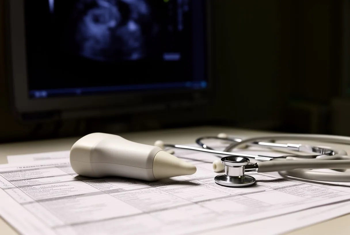
The role of echocardiograms in measuring EF: An echocardiogram is the gold standard for diagnosing low EF. This painless ultrasound test uses sound waves to create detailed moving images of your heart. It allows the cardiologist to see exactly how the left ventricle contracts and measures the precise percentage of blood pumped out with each beat. It also reveals the size of the heart chambers and the function of the valves. Understanding your exact EF measurement is the foundation of your low ejection fraction treatment plan, as it dictates whether you need simple medication adjustments or if you require evaluation for advanced structural interventions.
Additional tests to uncover the underlying cause: Once a low EF is confirmed, further testing is necessary to determine why the heart weakened. A coronary angiogram can reveal if blocked arteries are depriving the muscle of blood. Blood tests can check for elevated cholesterol, thyroid dysfunction, or markers of heart strain. Sometimes, a cardiac MRI is ordered to look for inflammation or scar tissue deep within the muscle walls. By combining these diagnostics, your cardiac team can pinpoint the exact low EF causes and tailor a comprehensive recovery strategy aimed at stabilizing and improving your heart health.
Low Ejection Fraction Treatment with Ejection Fraction Medication
How beta-blockers protect the heart muscle: Medication is the cornerstone of low ejection fraction treatment. Beta-blockers are often prescribed to slow the heart rate and reduce the force of the heart's contractions. While it might seem counterintuitive to slow a weak heart, reducing the workload allows the muscle to rest and recover. Over time, this specific ejection fraction medication has been shown to actually improve the pumping function and help remodel the heart back toward a healthier shape. By blocking the body's stress hormones, these drugs prevent the heart from overworking itself into further failure.
Using ACE inhibitors to reduce workload: Another critical class of drugs includes ACE inhibitors or ARBs. These medications widen the blood vessels, lowering the blood pressure the heart must pump against. By reducing this resistance, the heart can eject blood more easily, improving cardiac output reduction symptoms. For patients who also experience irregular heartbeats like atrial flutter, these medications help stabilize the heart's rhythm and geometry. Proper medication adherence is essential; skipping doses can lead to a rapid decline in heart function and a worsening of symptoms.
Diuretics for symptom relief and fluid management: To manage the fluid backup that often accompanies a low EF, doctors prescribe diuretics, commonly known as water pills. These medications help the kidneys flush out excess sodium and water, relieving the swelling in the legs and the congestion in the lungs. While diuretics do not directly improve the heart's pumping percentage, they drastically improve quality of life and prevent hospitalizations. They are often adjusted based on your daily weight and symptoms, working in harmony with other medications to provide comprehensive heart strengthening therapy.
Advanced Heart Strengthening Therapy and Device Options
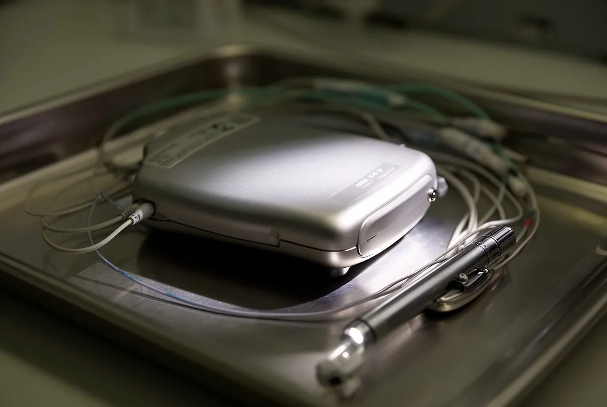
When medications are not enough: When a patient's ejection fraction remains below 35% despite optimal medication, advanced therapies must be considered. The weakened heart muscle is prone to dangerous, rapid heartbeats that can cause sudden cardiac arrest. To mitigate this risk, cardiologists may recommend an implantable cardioverter-defibrillator, a device similar to an Automated External Defibrillator but placed internally. This device constantly monitors the heart rhythm and delivers a life-saving shock if a lethal arrhythmia occurs. Deciding to implant a device is a significant step in HFrEF management and requires a detailed discussion about the risks and benefits for your specific situation.
- ICD Implantation: Protects against sudden cardiac arrest by monitoring and correcting lethal arrhythmias.
- Cardiac Resynchronization Therapy: Uses a specialized pacemaker to coordinate the left and right ventricles, improving the efficiency of each heartbeat.
How specialized pacemakers synchronize the heartbeat: In many cases of heart failure, the left and right sides of the ventricle do not beat in perfect synchrony, reducing the efficiency of the squeeze. Cardiac Resynchronization Therapy involves placing a pacemaker with a special lead on the left ventricle to coordinate the timing of the beats. This advanced heart strengthening therapy can significantly improve EF and reduce symptoms of heart failure. By ensuring the muscle contracts in a unified, organized manner, the heart maximizes the blood pumped to the body with each beat.
Monitoring device function and adjusting settings: After a device is implanted, it requires regular monitoring, often done remotely from the comfort of your home. The data is transmitted to the clinic, allowing the cardiac team to check battery life, lead integrity, and any recorded arrhythmias. This continuous oversight is a vital component of long-term low ejection fraction treatment. It ensures the device is functioning perfectly and allows doctors to adjust your medications based on the real-time data collected by the pacemaker or defibrillator.
When Surgery Becomes Necessary for Low EF Causes
Coronary bypass surgery for blocked arteries: If your low EF is caused by severe coronary artery disease, surgery may be necessary to restore blood flow to the starving heart muscle. Coronary Artery Bypass Grafting uses a healthy Arterial Graft, often taken from your chest or arm, to bypass the blockages. Surgical teams may use techniques to minimize blood loss, including the use of Autologous Blood recovered during the procedure. By restoring blood flow, the heart muscle that was merely hibernating can recover its function, leading to significant EF improvement. This surgery addresses the root low EF causes directly, offering a chance for the heart to heal.
Valve repair and replacement options: Sometimes, a low ejection fraction is driven by a severely leaky or tight heart valve. The heart exhausts itself trying to pump against a tight valve or pumps blood backward through a leaky one. Surgical repair or replacement of the valve can dramatically reduce the workload on the heart. For patients with a narrowed aortic valve, a procedure called Balloon Aortic Valvuloplasty can temporarily stretch the valve, though it is often a bridge to a more permanent solution. Fixing the valve allows the ventricle to pump more efficiently, stabilizing the heart function percentage over time.
Transcatheter options for high-risk patients: When patients are too frail for open-heart surgery, transcatheter approaches offer a lifeline. For severe aortic stenosis, transcatheter aortic valve replacement (TAVR) allows cardiologists to insert a Balloon-expandable Valve through a small catheter, usually in the leg, threading it up to the heart. This valve is deployed inside the diseased one, immediately improving blood flow. As detailed in a study on Transcatheter Aortic-Valve Implantation for Aortic Stenosis in Patients Who Cannot Undergo Surgery, this approach provides a viable option for those who previously had no surgical alternatives, significantly improving their quality of life and heart function.
Monitoring EF Improvement and Adjusting Congestive Heart Fraction Care
Tracking heart function with regular echocardiograms: Low ejection fraction treatment is not a one-time event; it requires ongoing monitoring. Regular echocardiograms are scheduled every few months to track the heart function percentage. These follow-up imaging tests allow your cardiologist to see if the medications or surgical interventions are working. If the EF is rising, the treatment plan is continued. If the percentage remains stagnant or drops, the medical team must reassess the situation, looking for new blockages, worsening valve disease, or arrhythmias that may be sabotaging the recovery process.
The role of inpatient monitoring during acute episodes: If your symptoms suddenly worsen, hospitalization may be required to stabilize your condition safely. In the intensive care unit, an Arterial Line is often placed in the wrist to provide constant, beat-to-beat blood pressure readings. This continuous data is crucial for adjusting powerful intravenous medications. An Arterial Cannula might also be utilized if mechanical support is needed to rest the heart. During these acute episodes of congestive heart fraction exacerbation, precise monitoring ensures that fluid is safely removed and blood pressure is kept at an optimal level to protect the heart muscle.
Adjusting medications based on symptoms and tests: As your body changes and your heart adapts, your medications will need adjustments. Your kidney function, blood pressure, and heart rate dictate how much of each drug you can tolerate. Fine-tuning your ejection fraction medication is a delicate balance managed by your cardiologist. You play a vital role in this process by tracking your weight, reporting new symptoms, and adhering strictly to your dosing schedule. This collaborative approach is what ultimately drives sustained EF improvement and keeps you out of the hospital.

Long-Term Outlook and Quality of Life with Systolic Dysfunction Treatment
Setting realistic expectations for heart recovery: The goal of treating a weak heart is not always to return the heart function percentage to normal. For many patients, the realistic goal of systolic dysfunction treatment is to stabilize the condition, prevent further decline, and dramatically improve the quality of life. With proper medical management, many individuals with an EF of 30% return to their daily activities, travel, and enjoy time with their families without debilitating symptoms. Recovery depends on the underlying cause, your adherence to the treatment plan, and the structural state of your heart when treatment began.
Advanced support for severe heart failure: In the most severe cases where the heart does not respond to medication and devices, advanced mechanical support may be discussed. Ventricular assist devices can take over the pumping function of the left ventricle, acting as a bridge to a heart transplant or as a permanent therapy. In specialized scenarios, Awake ECMO might be utilized to support both heart and lung function without requiring full sedation, allowing the patient to remain conscious and interact with their medical team. These highly advanced interventions are typically coordinated at major cardiac centers with specialized surgical teams.
Partnering with your cardiac team for the future: Managing a low ejection fraction is a lifelong journey that requires a strong partnership between you and your medical team. At Cirugía de Corazón, we are committed to walking this path with you, offering expert guidance, advanced surgical options, and compassionate care across our locations in Guadalajara, Querétaro, and Hermosillo. Do not self-diagnose or delay seeking care. If you are experiencing symptoms of heart failure or have been told your heart function is reduced, we encourage you to discuss your specific situation with a qualified cardiac team and book a consultation to explore your personalized low ejection fraction treatment options.






