

Radiation Dose in Heart Imaging: How Much Is Safe
Radiation dose heart imaging explained—learn safe exposure levels in millisieverts, how cardiac CT and nuclear tests compare, and ways to minimize your risk.
What Is Radiation Dose in Heart Imaging?
Understanding the basics: Radiation dose heart imaging refers to the small amount of ionizing radiation a patient receives during certain cardiac diagnostic tests. When your cardiologist orders a scan to evaluate blood vessels, heart muscle, or valve function, some of these tests use X-rays or radioactive tracers to create detailed images. The dose is measured in millisieverts (mSv), a unit that quantifies biological effect on human tissue. To put this in perspective, everyone on Earth receives about 3 mSv of natural background radiation each year from the sun, rocks, and soil — so many cardiac scans involve a dose comparable to a few months of simply living on the planet.
For patients facing decisions about their cardiac care, understanding radiation dose matters because it helps you weigh the real diagnostic benefit against a small but measurable risk. Modern cardiology has made tremendous progress in lowering the radiation delivered during imaging. Dose-reduction protocols, newer scanner technology, and alternative non-radiation options like cardiac MRI have shifted the landscape significantly. The principle every cardiac team follows is ALARA — "As Low As Reasonably Achievable" — meaning your exposure should be the minimum needed to get a clinically useful answer. Research compiled through resources like Europe PMC consistently shows that informed dose management reduces cumulative exposure without sacrificing diagnostic accuracy.
If your doctor has recommended a test involving radiation, it is reasonable and important to ask about the expected dose. A good cardiac team will explain why the scan is necessary, what alternative options exist, and how they plan to keep your exposure as low as possible. The goal is never to avoid imaging that could save your life, but to ensure each test is justified, optimized, and performed with the safest available technique.
How Cardiac Imaging Radiation Exposure Works
The physics, simplified: During a cardiac CT scan, a rotating X-ray tube fires a thin beam of radiation through your chest while detectors on the opposite side capture the attenuated signal. A computer then reconstructs these measurements into cross-sectional slices of your heart and coronary arteries. The dose you receive depends on several variables: the scanner type, the area scanned, your body size, the scanning speed, and whether dose-modulation software is used. A coronary CT angiogram, for example, images a smaller volume than a full chest CT, but it requires high spatial resolution to visualize vessels as narrow as 1-2 millimeters.
Why body size changes the dose: Larger patients require more radiation to produce images of acceptable quality because thicker tissue absorbs more of the X-ray beam before it reaches the detector. This is not a flaw in the system — it is physics. Modern scanners use automatic exposure control, which adjusts the radiation output in real time based on the thickness and density of the tissue being imaged. This means a slim patient receives a lower dose than a larger patient for the same scan, even though both are imaged on the same machine. Your cardiac team accounts for this when planning your study.
In nuclear cardiology, the mechanism is different. A small amount of a radioactive tracer — typically technetium-99m or thallium-201 — is injected into a vein, usually in the arm. The tracer travels through your bloodstream and concentrates in heart muscle in proportion to blood flow. A gamma camera then detects the radiation emitted from inside your body. The dose here comes from the tracer itself, which decays over several hours and is excreted through urine. The radiation you receive is internal, and the total exposure depends on the tracer type, the administered activity (measured in megabecquerels), and how quickly your kidneys clear it.
Types of Heart Imaging and Their Radiation Levels
Coronary CT angiography: This test visualizes the coronary arteries noninvasively and is one of the most common cardiac imaging studies involving radiation. A typical effective dose ranges from 1 to 5 mSv on modern scanners, though older technology or protocols without dose optimization can deliver 8-15 mSv. High-pitch dual-source CT scanners have pushed some coronary angiograms below 1 mSv — comparable to a few months of natural background radiation. The dose depends on whether your heart rate is low enough for a single-heartbeat acquisition or whether multiple beats must be averaged.
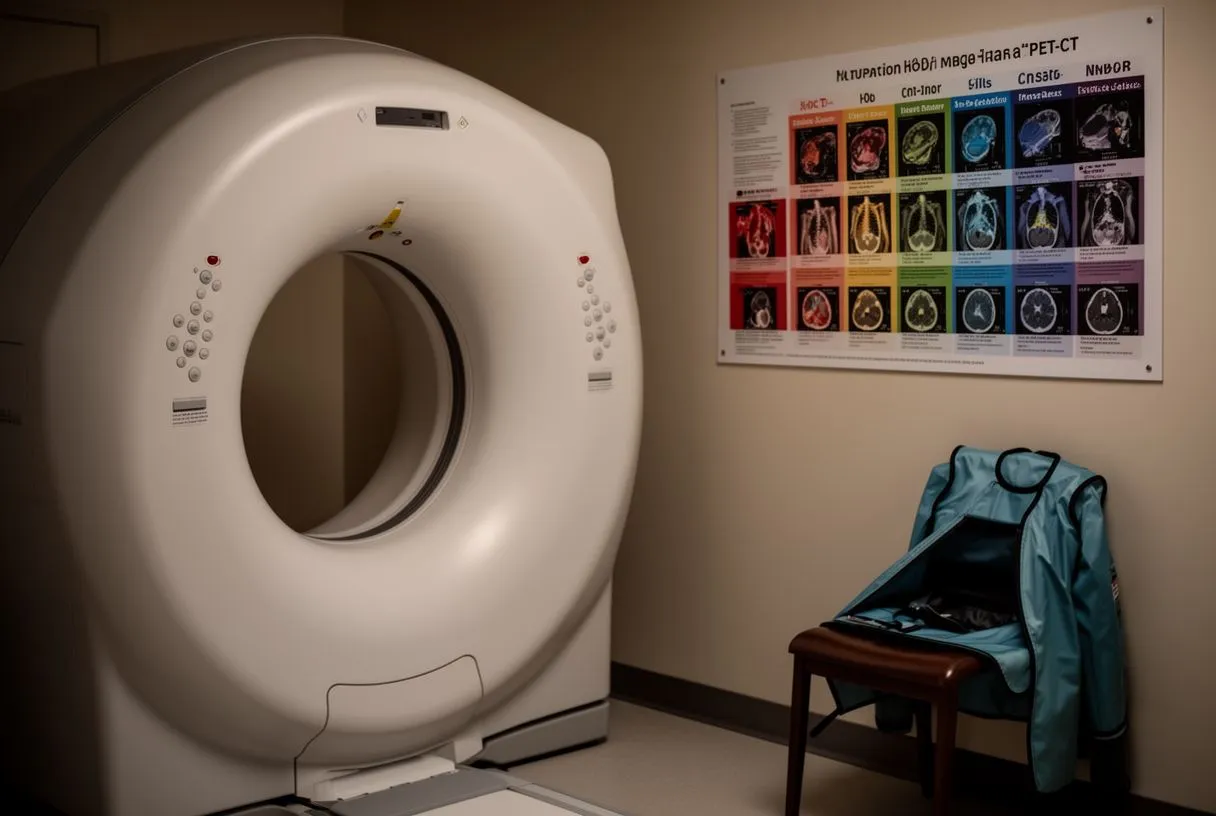
Myocardial perfusion imaging and cardiac catheterization: Nuclear stress tests using technetium-99m tracers typically deliver 10-15 mSv for a single-day rest-stress protocol. If a thallium-based protocol is used, the dose may be slightly lower. Diagnostic cardiac catheterization, which involves fluoroscopy and cineangiography to directly visualize coronary arteries, delivers an average of 5-10 mSv, though complex interventional cases — such as those involving an atheromatous plaque in the ascending aorta or revascularization with a balloon angioplasty — can reach higher doses. Each of these tests has a specific clinical purpose, and the radiation is a trade-off for information that cannot be obtained another way.
When comparing options, the key question is not simply which test has the lowest dose, but which test answers the clinical question with the least radiation. Your cardiologist balances diagnostic yield, radiation exposure, contrast agent requirements, and your individual risk factors before recommending a specific study. Understanding these trade-offs helps you participate meaningfully in that decision.
What Is a Safe Radiation Dose for Heart Scans?
The concept of "safe" in radiation dose heart imaging is not a single number but a reasoned judgment. Regulatory bodies and scientific societies generally agree that doses below 100 mSv per year do not produce clinically detectable increases in cancer risk. Most cardiac imaging studies fall well below this threshold — a coronary CT at 3 mSv, for instance, is roughly equivalent to one year of natural background radiation. However, the principle of minimizing exposure still applies because radiation effects are cumulative over a lifetime. A comparative analysis of treatment approaches for myocardial infarction patients, as documented by the Journal of Rawalpindi Medical College, reinforces the importance of standardized guideline adherence when cardiac imaging and interventions are involved.
How to think about risk: The theoretical risk from a single cardiac CT scan is small — estimated at roughly one additional cancer case per several thousand scans, and even lower for younger patients imaged with modern dose-reduction techniques. For comparison, the baseline lifetime risk of developing cancer in the general population is approximately 40%. A 3 mSv scan adds a fraction of a percent to this baseline. For patients with chest pain, a family history of coronary disease, or symptoms suggesting atypical infarction, the clinical benefit of identifying a treatable coronary blockage far outweighs this small theoretical risk.
That said, safety also means avoiding unnecessary tests. If you have had a recent coronary CT or nuclear study at another facility, your current cardiac team should be informed. Duplicate imaging multiplies radiation exposure without adding clinical value. In some cases, your previous images can be reviewed and used for current decision-making, sparing you a repeat scan. Always ask whether the information from a prior study could be sufficient before proceeding with new imaging.
Cardiac CT Radiation Dose: What Patients Should Know
What has changed in recent years: Cardiac CT has undergone one of the most dramatic dose-reduction transformations in all of medical imaging. A decade ago, a standard coronary CT angiogram routinely delivered 10-20 mSv. Today, with prospective triggering, high-pitch helical acquisition, and advanced iterative reconstruction algorithms, many scans are performed at 1-3 mSv. Some centers achieve sub-mSv imaging in appropriately selected patients with low, regular heart rates. This represents a reduction of over 80% without compromising the ability to detect clinically significant coronary artery disease.
Practical preparation tips: If you are scheduled for a coronary CT, your heart rate matters enormously. Scanner technology that images the heart in a single heartbeat requires a heart rate typically below 60-65 beats per minute. If your resting heart rate is higher, your cardiologist may prescribe a beta-blocker to lower it temporarily. Patients with atrial flutter or other rhythm irregularities may not be ideal candidates for the lowest-dose protocols, and your team will select an alternative approach. Holding your breath for 5-10 seconds during the scan is also critical — motion artifacts degrade image quality and can necessitate a repeat acquisition, doubling your dose.
It is also worth understanding that not every cardiac CT is the same. A coronary calcium score, which screens for calcified plaque in the coronary arteries, uses far less radiation — typically 1-3 mSv — and does not require contrast injection. A full coronary CT angiogram, which visualizes both calcified and non-calcified plaque and assesses the degree of narrowing, requires intravenous contrast and may involve a slightly higher dose. Your cardiac team will determine which study is appropriate for your clinical scenario.
Nuclear Cardiology Radiation Dose Explained
What nuclear imaging involves: Myocardial perfusion imaging is a nuclear cardiology technique that evaluates blood flow to the heart muscle at rest and under stress. A radioactive tracer is injected into a vein, and a gamma camera (SPECT) or PET scanner detects the tracer's distribution within the myocardium. Areas of reduced tracer uptake indicate diminished blood flow, often due to a coronary artery narrowed by atheromatous plaque. The radiation dose comes from the tracer itself, not from an external X-ray beam, and the total exposure depends on the isotope used, the injected activity, and whether imaging is performed at rest only, stress only, or both.
Typical doses you can expect: A standard single-day rest-stress SPECT study using technetium-99m sestamibi or tetrofosmin typically delivers 10-12 mSv. Two-day protocols may use slightly lower injected activities per injection but achieve a similar total dose. PET myocardial perfusion imaging, which uses tracers such as rubidium-82 or nitrogen-13 ammonia, can be performed at lower doses — often 2-5 mSv — because PET cameras are more sensitive and the tracers have shorter half-lives. Emerging research on the use of stem cells in the treatment of myocardial infarction, as reviewed by the Journal of Applied Biological Sciences, highlights how advanced imaging strategies are being integrated into post-infarction care pathways.
One way to reduce dose in nuclear cardiology is the use of stress-only protocols. If your stress images are normal and show homogeneous tracer uptake throughout the left ventricle, the rest study may be omitted entirely, cutting your radiation exposure in half. Additionally, solid-state cadmium-zinc-telluride (CZT) gamma cameras, which are more efficient than traditional sodium-iodide detectors, can achieve diagnostic-quality images at significantly lower injected activities. Ask your cardiac team whether a stress-first or stress-only protocol is appropriate for your situation.
Heart Imaging Safety Guidelines and Dose Limits
Who sets the rules: Several international and national bodies publish guidance on radiation dose heart imaging safety. The International Commission on Radiological Protection (ICRP) provides the foundational framework for dose limits and the principle of justification — every exposure must have a net benefit. Professional societies such as the American College of Cardiology, the American Heart Association, and the European Association of Cardiovascular Imaging have published appropriateness criteria that help clinicians select the right test for the right patient. These guidelines emphasize that no imaging study involving ionizing radiation should be performed if an alternative non-radiation modality — such as echocardiography or cardiac MRI — can answer the clinical question equivalently.
Dose-tracking and registries: Many institutions now participate in dose registries that benchmark their radiation exposure against national averages. The goal is to identify outliers and drive systematic dose reduction. If your cardiac center participates in such a registry, it signals a commitment to dose optimization and quality improvement. You can ask your team whether they track and report their imaging doses, and how their levels compare to national benchmarks. This transparency is a positive indicator of a safety-conscious practice.
In the catheterization laboratory, where fluoroscopy is used for coronary angiography and interventions such as balloon aortic valvuloplasty or valve procedures, real-time dose monitoring is standard. The interventional cardiologist sees a running display of cumulative air kerma and dose-area product throughout the procedure. These metrics guide decisions about imaging time, angulation, and frame rate. For complex procedures involving an arterial graft assessment or placement of a balloon-expandable valve, the team actively balances the need for visualization against the dose accumulated over the course of the intervention.
Comparing Radiation Exposure Across Cardiac Tests
When patients ask which cardiac test involves the most radiation, the answer depends on the specific protocol and the clinical indication. Below is a summary of typical effective doses for common cardiac imaging studies. These values are approximate and vary by institution, equipment, and patient factors.
- Coronary calcium score: ~1-3 mSv, no contrast, screening test for calcified plaque burden
- Coronary CT angiogram: ~1-5 mSv on modern scanners, requires IV contrast, visualizes coronary anatomy
- SPECT myocardial perfusion (rest-stress): ~10-15 mSv, assesses blood flow to heart muscle
- PET myocardial perfusion: ~2-5 mSv, higher sensitivity, lower tracer doses
- Diagnostic coronary angiography (cath): ~5-10 mSv, invasive, gold standard for coronary anatomy
Putting these numbers in context: A coronary calcium score at 2 mSv is equivalent to about 8 months of natural background radiation. A SPECT perfusion study at 12 mSv is roughly equivalent to 4 years of background radiation. These comparisons help patients and families calibrate the magnitude of exposure. None of these doses approach levels where acute radiation effects occur — those thresholds are thousands of times higher. The concern is long-term, cumulative, theoretical cancer risk, which is why minimizing unnecessary exposure remains the standard of care.
Cardiac MRI: The No-Radiation Alternative
How cardiac MRI works: Cardiac magnetic resonance imaging uses a powerful magnetic field and radiofrequency pulses — not ionizing radiation — to generate detailed images of the heart. It excels at evaluating heart muscle function, valve anatomy, congenital defects, and tissue characterization, including scar detection after a heart attack. For many clinical questions, cardiac MRI can replace or complement radiation-based studies. For example, if the clinical question is whether heart muscle has been damaged by a prior infarction or an inflammatory process, late gadolinium enhancement imaging during MRI provides this information without any radiation exposure.
When MRI is the right choice: Cardiac MRI is particularly valuable for younger patients, pregnant patients (when imaging is unavoidable and MRI is deemed appropriate), and patients who have already accumulated significant radiation exposure from prior tests. It is also the gold standard for assessing right ventricular function, pericardial disease, and complex congenital anatomy. However, cardiac MRI cannot visualize coronary arteries with the same resolution as CT or invasive angiography, so it is not a substitute when the clinical question specifically requires coronary artery anatomy.
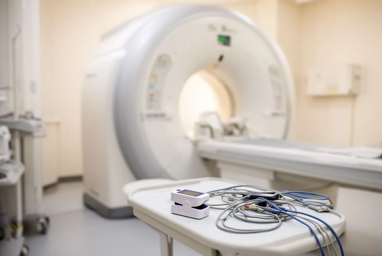
Practical limitations exist. MRI scanners have narrower bores than CT scanners, which can be challenging for claustrophobic or larger patients. The scan takes longer — typically 30-60 minutes versus a few minutes for a coronary CT — and requires breath-holding at intervals. Patients with certain implanted devices, such as older pacemakers or defibrillators, may not be eligible, though modern MRI-conditional devices are increasingly available. Your cardiac team will assess whether cardiac MRI is feasible and appropriate for your situation.
How to Minimize Radiation Risk in Cardiac Imaging
Questions to ask before any cardiac scan: The single most effective way to reduce radiation exposure is to avoid tests that are not necessary. Before agreeing to any cardiac imaging study, consider asking your cardiologist a few key questions that can help ensure the test is justified and optimized for your situation.
- What clinical question will this test answer? If the answer is vague or the test is being done "routinely," that may warrant further discussion.
- Is there a non-radiation alternative? Echocardiography or cardiac MRI may answer the question without ionizing radiation.
- What is the expected radiation dose? Ask for a specific number in mSv, and compare it to the background radiation benchmark.
- Can a prior study be used instead? If you have had imaging at another facility, ask whether those results can be obtained and reviewed.
Beyond your role as a patient, the imaging team has specific responsibilities. They should use dose-modulation software, employ the lowest tube voltage that produces diagnostic images, limit the scanned field to the cardiac region, and use iterative reconstruction to reduce noise without increasing dose. For nuclear studies, they should consider stress-first protocols and CZT camera technology when available. For catheterization, they should use low frame rates, minimize fluoroscopy time, and use collimation to limit the irradiated field. These measures collectively reduce dose without compromising diagnostic quality.
In summary, radiation dose heart imaging is a manageable risk when approached thoughtfully. Modern technology, standardized guidelines, and active dose-tracking have made cardiac imaging safer than ever. The key takeaways are simple: choose the right test for the right reason, prefer non-radiation alternatives when they provide equivalent information, and ask your cardiac team about dose optimization. If you are facing a cardiac imaging decision and want to discuss your options with a qualified team, we encourage you to book a consultation. A careful, personalized assessment of your situation is the best way to ensure that any imaging you undergo is both necessary and as safe as possible.






