

TEVAR: Stent Graft Repair of the Thoracic Aorta
TEVAR thoracic aorta repair from Guadalajara heart specialists: minimally invasive stent graft procedure with faster recovery.
What is TEVAR Thoracic Aorta Repair and How Does It Work
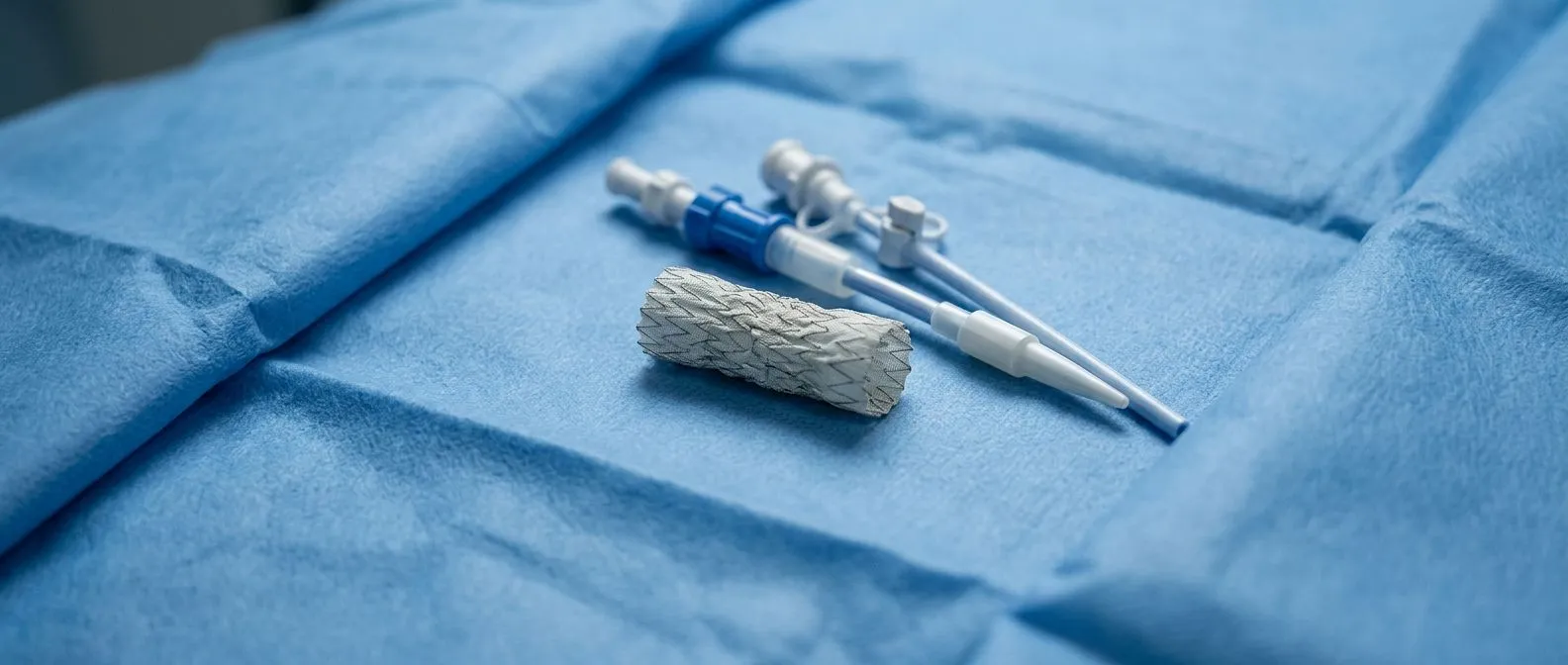
TEVAR, or Thoracic Endovascular Aortic Repair, is a minimally invasive medical procedure used to treat life-threatening conditions of the descending aorta, the major artery carrying blood from the heart down through the chest. For many patients facing an aortic aneurysm or dissection, the thought of open-chest surgery is daunting. TEVAR offers a crucial alternative, utilizing a fabric-covered metal mesh tube called a stent graft to reinforce the damaged vessel wall from the inside. This approach avoids large chest incisions, drastically changing the patient experience and recovery trajectory. This guide explains TEVAR thoracic aorta repair from candidacy to recovery.
How TEVAR supports the damaged aorta: The procedure works by threading a catheter through small incisions in the groin, advancing it up to the thoracic aorta under live X-ray guidance. Once the catheter reaches the diseased segment, the compressed stent graft is deployed, expanding to fit snugly against the aortic walls. This creates a new, stable pathway for blood flow, effectively sealing off the aneurysm or the tear in a dissection, preventing rupture.
Understanding the anatomy helps clarify why TEVAR is so effective. The aorta exits the heart, ascends, arches, and then descends through the chest. While conditions of the ascending aorta often require open surgery, pathologies in the descending segment are frequently excellent candidates for this endovascular approach. By excluding the weakened wall from blood pressure, TEVAR acts as an internal shield, stabilizing the vessel and preventing catastrophic rupture, according to comprehensive overviews from institutions like the Mayo Clinic.
TEVAR vs Open Surgery for Thoracic Aortic Aneurysm
When facing a thoracic aortic aneurysm, the choice between TEVAR and open surgical repair depends heavily on your anatomy, overall health, and the specific location of the disease. Open surgery involves a large thoracotomy, where the chest is opened to manually replace the diseased aortic segment with a synthetic arterial graft. This traditional approach is highly effective but carries significant trauma to the body, requiring the use of an arterial cannula and an arterial line for cardiopulmonary bypass support during the operation.
Comparing recovery and physical impact: Open surgery typically requires extensive hospitalization, often involving days in the intensive care unit, and a prolonged recovery period at home. TEVAR, by contrast, is performed through small groin incisions, avoiding the need to open the chest. While autologous blood is often reserved for open procedures due to anticipated higher bleeding, TEVAR patients experience significantly less blood loss, fewer transfusions, and shorter ICU stays.
Clinical guidelines emphasize that high-volume centers yield better outcomes for complex cardiovascular interventions. Research indicating that procedural volume significantly impacts patient survival, such as the Clinical Outcomes in Relation to Total Hospital Surgical and Transcatheter Aortic Valve Replacement Volumes study, reinforces the importance of seeking experienced teams for aortic repairs. While that specific study focuses on valve replacements, the principle of volume-based expertise applies directly to complex aortic stent grafting, ensuring your team is prepared for any anatomical challenge.
Who is a Candidate for TEVAR Procedure
Determining candidacy for a TEVAR procedure requires detailed imaging, typically a CT scan, to evaluate the precise dimensions of your aorta. Not every patient is suited for endovascular repair. The anatomy of the aorta must have healthy segments of the vessel above and below the aneurysm or tear where the stent graft can securely anchor and form a seal. If the blood vessels in the groin are too narrow or tortuous, accessing the aorta might be difficult.
Navigating complex anatomy and comorbidities: Patients with severe calcification or atheromatous plaque in the access vessels may face challenges, as navigating large catheters through diseased femoral arteries carries a risk of dislodging debris. Furthermore, if the aneurysm is located very close to the left subclavian artery, covering that branch might be necessary, requiring a preemptive bypass to ensure blood flow to the arm and brain is maintained.
Sizing the stent graft accurately is critical to prevent endoleaks, which are instances of blood leaking around the graft. This anatomical precision is paramount, much like how device selection dictates outcomes in transcatheter valve procedures. Insights from resources like the Self-expanding or balloon-expandable TAVR in patients with a small aortic annulus: A review highlight how matching a metallic device to specific vascular dimensions prevents complications, a principle that perfectly mirrors the careful sizing required for a successful TEVAR deployment.
TEVAR Recovery Time and Hospital Stay
One of the most significant advantages of TEVAR is the remarkably short recovery time compared to open surgery. Because the chest wall is not opened, patients experience far less postoperative pain and respiratory compromise. Most patients spend only a day or two in the intensive care unit for monitoring, followed by a few days in a regular hospital room before discharge. Total hospital stay is typically under a week.
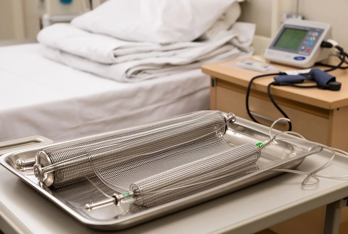
What to expect during the first weeks at home: While the incisions in the groin are small, you will still need to avoid heavy lifting and strenuous activity for several weeks to allow the access sites to heal and the stent graft to settle. You might experience mild groin discomfort or bruising, which is normal. While severe respiratory failure might necessitate techniques like Awake ECMO in rare cases, most TEVAR patients breathe easily on their own shortly after the procedure and are encouraged to begin gentle walking to promote circulation.
Regular follow-up imaging is essential to ensure the stent graft remains in place and continues to function correctly. You will typically undergo a CT scan or ultrasound before discharge, again at one month, six months, and annually thereafter. This lifelong surveillance ensures that no endoleaks develop and that the repaired segment of the descending aorta remains stable, giving you and your cardiac team peace of mind.
Potential TEVAR Complications and Risks
Although TEVAR is minimally invasive, it is still a major cardiovascular procedure and carries inherent risks. The most common complications are vascular injuries at the access site in the groin, which can cause bleeding or the formation of a pseudoaneurysm. Rarely, patients may experience arm pain if the left subclavian artery is covered by the graft without a prior bypass, though this is usually anticipated and managed preemptively by your surgical team.
Neurological and cardiac risks explained: Because the stent graft is placed in the chest near the spinal cord's blood supply, there is a small risk of spinal cord ischemia, which can lead to paralysis. To mitigate this, blood pressure is carefully managed, and sometimes a drain is placed in the spine to relieve pressure. Patients are also monitored for cardiac arrhythmias, such as atrial flutter, and atypical infarction, though these risks are generally lower than in open chest surgeries.
Another specific risk is an endoleak, where blood continues to flow into the aneurysm sac outside the stent graft. If an endoleak is detected on follow-up imaging, it may require a secondary procedure to fix, sometimes utilizing balloon angioplasty to expand the graft further or placing an additional overlapping stent. Understanding these risks candidly helps patients and families prepare for the realities of the procedure and the necessity of lifelong follow-up.
Aortic Dissection TEVAR: Repairing the Tear
An aortic dissection occurs when the inner layer of the aorta tears, allowing blood to surge through the split, creating a false channel for blood flow. This is a life-threatening emergency. In the descending aorta, TEVAR is often the treatment of choice for complicated dissections, particularly when blood supply to vital organs is compromised or the aorta is at risk of rupture.
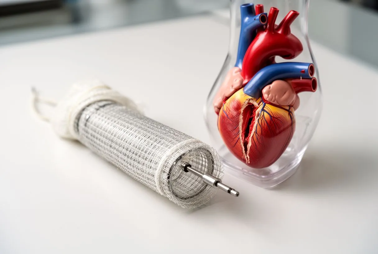
The mechanism of dissection repair: The goal of TEVAR in a dissection is to cover the primary tear in the inner wall. By deploying the stent graft over this entry point, blood flow is redirected back into the true lumen, the original, healthy pathway of the aorta. This depressurizes the false lumen, encouraging it to clot and shrink over time, while restoring proper blood flow to the branches that feed the kidneys, intestines, and legs.
- Primary Tear Coverage: The stent graft blocks the entry tear to stop blood from entering the false channel.
- True Lumen Expansion: Restoring flow to the true lumen improves circulation to critical organs.
- False Lumen Thrombosis: Depressurization encourages clotting in the false lumen, promoting aortic remodeling.
Even after a successful TEVAR for dissection, the aorta remains a fragile structure. Patients must maintain strict blood pressure control to prevent further tearing. The underlying connective tissue weakness that led to the dissection remains, meaning that close monitoring of the entire aorta is critical, as other segments may dilate over time and require future intervention.
Stent Graft Placement: Step-by-Step Process
The stent graft placement process begins with precise preoperative planning. Your surgeon will map the exact length and diameter of the diseased aorta to select the appropriate device. In the operating room, you will receive general or regional anesthesia. The surgeon makes small incisions in the groin to access the femoral arteries, the main highways to the legs, through which the graft will be delivered.
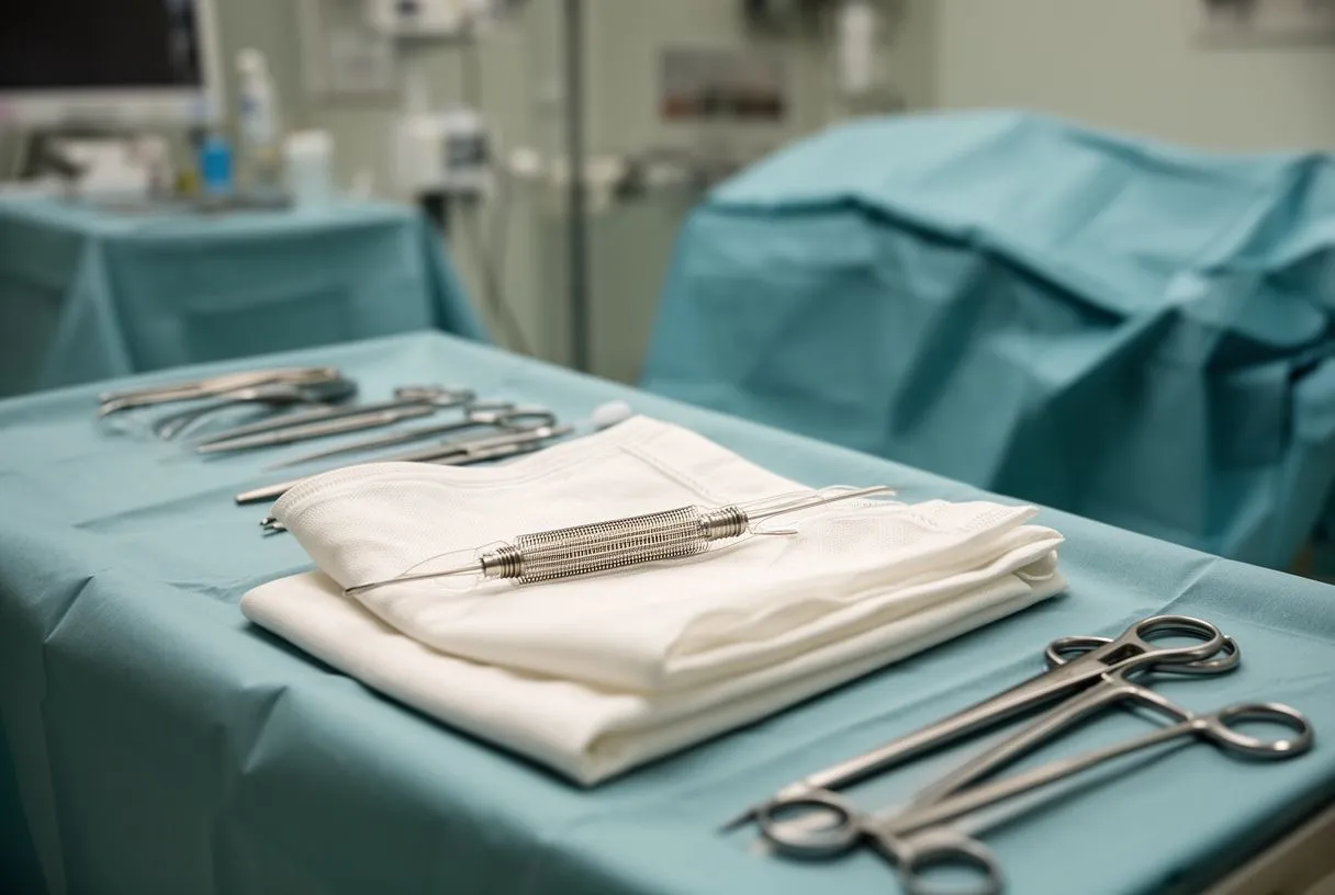
Device delivery and deployment: A guidewire is advanced up to the aortic arch, and the catheter containing the compressed stent graft is slid over the wire. Using real-time fluoroscopy (X-ray imaging), the surgeon positions the graft exactly across the aneurysm or tear. The sheath is withdrawn, allowing the stent to spring open. Unlike a balloon-expandable valve used in heart valve replacements, most aortic stent grafts are self-expanding, relying on their own radial force to anchor against the vessel walls.
Once deployed, the surgeon uses a balloon catheter to gently press the ends of the graft against the healthy aortic tissue, ensuring a tight seal. Unlike a balloon aortic valvuloplasty used to crack calcified heart valves, the balloon used here is not meant to stretch tissue, but simply to mold the stent graft securely. An angiogram is then performed by injecting contrast dye to confirm that blood is flowing through the new graft and that the aneurysm sac is excluded before removing the catheters and closing the incisions.
TEVAR Hospital Guadalajara: What to Expect
For patients traveling to Mexico for cardiac care, seeking TEVAR in Guadalajara offers access to advanced cardiovascular technology paired with highly specialized surgical teams. From initial teleconsultations to postoperative care, the focus is on continuity and clarity.
Preparing for your international cardiac journey: Patients are typically asked to provide recent CT scans and medical records for review. Upon arrival, you will undergo a comprehensive physical examination, and the surgical plan will be finalized based on local imaging protocols. The team evaluates various risk factors, including lipid profiles like apolipoprotein B levels, which indicate the burden of atherosclerosis and help stratify cardiovascular risk prior to the intervention.
- Comprehensive Evaluation: Review of imaging, history, and cardiovascular risk factors upon arrival.
- Coordinated Care: Dedicated coordinators assist with travel, translation, and hospital logistics.
- Expert Auscultation: Physical exams and careful monitoring ensure no hidden murmurs or pulse deficits are missed.
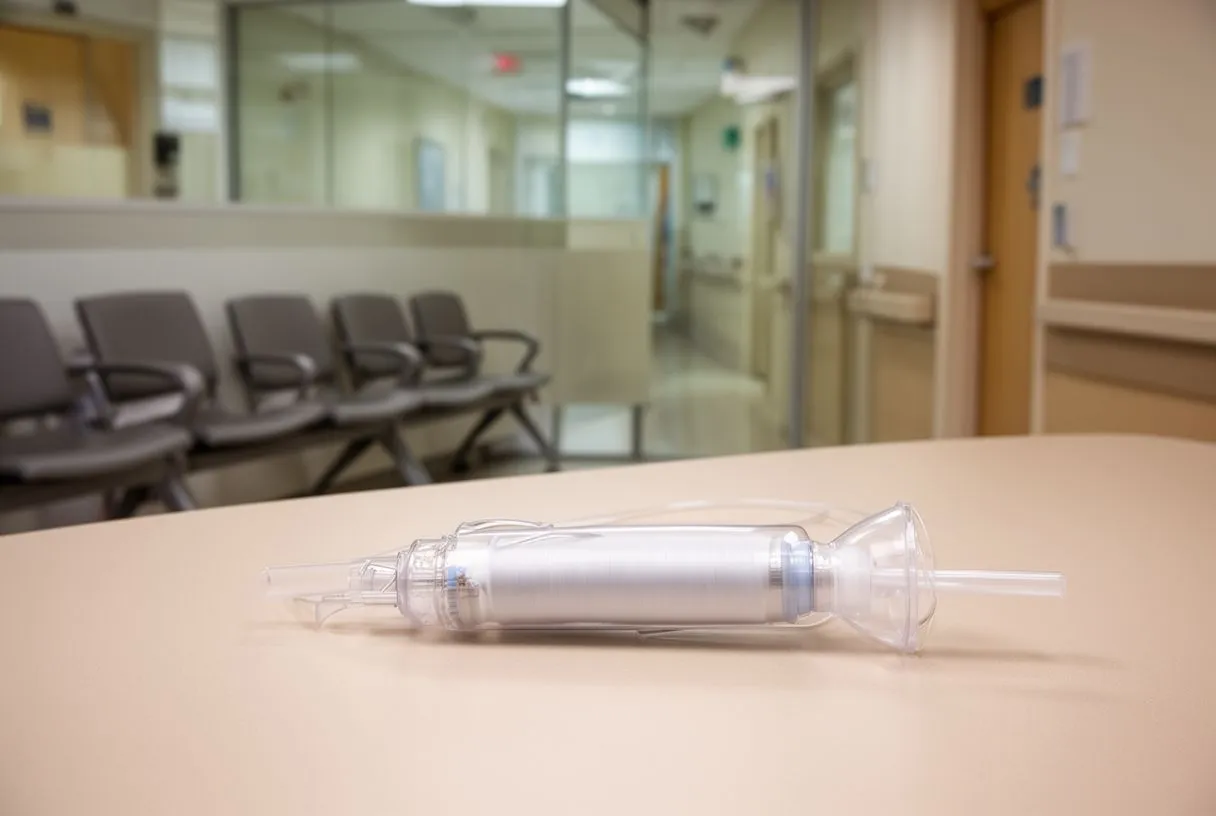
During your hospital stay, the nursing and medical staff are trained to manage the specific needs of aortic patients. Pain management, blood pressure control, and early mobility are prioritized. Arrhythmias or other atrial issues that arise after the procedure are monitored and treated during the hospital stay.
Understanding the cost of a thoracic stent graft in Mexico involves several variables, including the specific type of device used, the length of the hospital stay, and the need for any adjunctive procedures. The stent graft itself is a precision-engineered piece of medical equipment, which represents a significant portion of the total cost.
Navigating insurance and self-pay options: Many international patients choose to self-pay for their procedures in Mexico, finding the out-of-pocket costs highly competitive. It is essential to request an itemized quote that covers surgeon fees, anesthesia, hospital days, ICU stays, and the prosthesis. If you have international health insurance, it is advisable to check whether your policy covers treatments abroad and what pre-authorization is required.
Patients with coexisting conditions, such as atrial fibrillation, may need to manage medications like aspirin or blood thinners according to protocols derived from studies like the ARISTOTLE trial prior to surgery. Managing these comorbidities carefully is crucial, as they can impact both the complexity of the procedure and the overall cost.
Thoracic stent graft cost in Mexico and insurance
The cost of a thoracic stent graft in Mexico depends on several variables, including the specific type of device used, the length of the hospital stay and the need for complementary procedures. The stent graft itself is a precision-engineered piece of medical equipment that accounts for a significant share of the total cost.
Managing insurance and self-pay options: Many international patients choose to pay for their procedures themselves in Mexico, since the direct costs are very competitive. It is essential to request an itemised estimate that includes the surgeon's fees, anaesthesia, hospital days, the ICU stay and the prosthesis. If you have international health insurance, we recommend checking whether your policy covers treatment abroad and what prior authorisation you need.
If you have concomitant conditions such as atrial fibrillation, you may need to adjust medicines such as aspirin or anticoagulants before surgery according to protocols derived from studies such as the ARISTOTLE trial. Managing these comorbidities carefully is essential, since they can affect both the complexity of the procedure and the total cost.
Long-Term Outlook After Minimally Invasive Aorta Surgery
The long-term outlook after TEVAR is generally excellent, provided patients adhere to their follow-up schedules and medical regimens. The stent graft effectively takes the pressure off the weakened aortic wall, preventing rupture. However, the graft is a permanent implant, and the underlying vascular disease that caused the aneurysm or dissection remains, requiring ongoing management of blood pressure, cholesterol, and lifestyle factors.
Life after TEVAR and maintaining vascular health: Most patients return to their normal daily activities without feeling the presence of the stent. You will need to take medications to control your blood pressure and protect your remaining aorta. While serious emergencies are rare after successful repair, knowing the signs of aortic distress is vital. Although an automated external defibrillator is used for sudden cardiac arrest, recognizing chest or back pain early and seeking immediate medical evaluation is the best defense against graft complications.
In conclusion, TEVAR represents a profound advancement in the treatment of thoracic aortic disease, offering a less invasive, effective alternative to open surgery. By reinforcing the aorta from the inside, it stabilizes life-threatening conditions while preserving quality of life. If you or a loved one has been diagnosed with a thoracic aneurysm or dissection, we encourage you to discuss your specific anatomy and risk profile with a qualified cardiac team. Booking a consultation is the first step toward understanding if TEVAR is the right path for you.






