


The Thoracic Cavity: How Surgeons Reach the Heart
Thoracic cavity heart surgery explained: learn how surgeons safely access the heart through the chest wall.
When facing thoracic cavity heart surgery, understanding the anatomical journey your surgical team takes to reach your heart can ease much of the anxiety surrounding the procedure. The thoracic cavity serves as the body's primary protective chamber for vital organs, and navigating it requires precision, experience, and a deep understanding of cardiovascular anatomy. This guide explains the surgical approaches used to access the heart, what patients physically experience during recovery, and how modern techniques aim to provide the lowest risk for your specific situation. Whether you are exploring options in Mexico or seeking a second opinion, understanding these mechanisms empowers you to make informed decisions alongside your cardiac care team.
What is the thoracic cavity and why is it important for heart surgery?
The thoracic cavity is the enclosed space within the chest, bounded by the rib cage, the diaphragm below, and the upper chest structures above. It houses the heart, the lungs, the esophagus, and major blood vessels like the aorta. For cardiac surgeons, this cavity is not just a location; it is a complex anatomical landscape that must be navigated carefully to avoid damaging surrounding structures. The heart itself sits centrally in the mediastinum, wrapped in the pericardial sac, which provides an additional layer of protection but must be opened to access the heart muscle and valves.
Anatomical landmarks guide the surgical approach. Surgeons use specific landmarks within the thoracic cavity to plan their entry. The ascending aorta, the first section of the body's largest artery as it leaves the heart, is a critical structure that must be isolated safely. The right atrium, one of the upper chambers of the heart, is often the site where intravenous lines are connected during cardiopulmonary bypass. Understanding these structures is essential because an incision that is optimal for one valve might offer poor visibility for another.
Clinical thresholds dictate the need for intervention. Surgeons do not enter the thoracic cavity lightly; the decision is driven by strict clinical guidelines. According to guidelines published by ahajournals.org, surgical intervention is often recommended when an aortic aneurysm reaches a diameter greater than 5.5 centimeters, as the risk of rupture increases significantly. Similarly, severe blockages in the coronary arteries or failing heart valves prompt the need to access the thoracic cavity. These thresholds ensure that the risks of surgery are outweighed by the risks of the underlying disease.
How do surgeons reach the heart during open heart surgery access?
Traditional open heart surgery access is most commonly achieved through a median sternotomy. This involves making a vertical incision down the center of the chest, over the sternum (breastbone), and carefully dividing the bone to spread the rib cage apart. This approach provides the widest possible view of the heart and surrounding structures within the thoracic cavity. It allows surgeons the necessary space to perform complex procedures, such as multiple coronary artery bypasses or extensive valve replacements, with direct manual access to the heart.
Cardiopulmonary bypass takes over the work of the heart. Once the thoracic cavity is open, the surgical team must stop the heart to operate on it safely. To do this, the patient is placed on cardiopulmonary bypass. An arterial cannula is inserted into the heart or major vessels to drain blood away, where it is oxygenated and returned to the body. An arterial line is continuously monitored to ensure blood pressure remains stable throughout the procedure. Because the patient's blood is circulating outside the body, maintaining precise fluid management is critical, often utilizing the patient's own autologous blood to minimize the need for donor transfusions.
The sternum is secured for long-term healing. After the cardiac repair is complete, the heart is restarted, and the bypass machine is gradually withdrawn. The most critical part of the closure involves wiring the sternum back together. Stainless steel wires are threaded through the bone to hold it firmly in place, allowing it to heal over the next several months. Patients are given strict precautions against heavy lifting or pushing to ensure this bone heals properly, as the structural integrity of the thoracic cavity relies on this reunion.
What are the types of minimally invasive heart access?
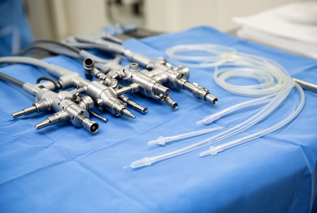
Minimally invasive heart access aims to provide the same surgical repair but through smaller incisions, avoiding the full division of the sternum. The most common alternative to a sternotomy is a thoracotomy, where the surgeon makes an incision between the ribs on the side of the chest. Specialized instruments and cameras are inserted through these intercostal spaces. This approach significantly reduces the trauma to the thoracic cavity and the chest wall, leading to less postoperative pain and a shorter hospital stay for appropriate candidates.
Smaller incisions are matched to specific procedures. Minimally invasive approaches are highly tailored to the specific cardiac issue. For instance, certain valve repairs can be achieved through a mini-thoracotomy, allowing the surgeon to reach the mitral valve from the right side of the chest. In contrast, some catheter-based procedures, such as balloon angioplasty to open narrowed arteries or balloon aortic valvuloplasty to stretch a stenotic valve, do not require opening the thoracic cavity at all, as they are accessed through blood vessels in the groin or neck.
Transcatheter options expand access for high-risk patients. For patients whose age or frailty makes traditional open heart surgery access too risky, transcatheter approaches offer a vital alternative. Transcatheter aortic valve replacement (TAVR) involves delivering a compressed, balloon-expandable valve through a catheter, usually via the femoral artery in the leg. The valve is positioned within the diseased native valve and expanded, immediately restoring blood flow. This method completely bypasses the need to enter the thoracic cavity surgically, representing a significant shift in how structural heart disease is managed with the lowest risk for your situation.
Is a thoracotomy cardiac surgery right for your valve procedure?
Deciding if a thoracotomy cardiac surgery is the right choice depends on the specific valve involved, the patient's anatomy, and the surgeon's expertise. A thoracotomy approach is frequently used for mitral valve repairs because of the direct line of sight it provides to the right side of the heart. However, accessing the aortic valve through the ribs can be more challenging due to its central location. Your surgical team will conduct thorough imaging to determine if the anatomical layout of your thoracic cavity permits a safe and effective minimally invasive repair.
Preoperative assessment minimizes intraoperative surprises. Before recommending a specific incision type, the cardiac team performs a comprehensive evaluation. This includes detailed echocardiograms to visualize valve function and computed tomography scans to map the exact dimensions of the thoracic cavity. During physical exams, careful auscultation—listening to the heart with a stethoscope—helps identify murmurs that suggest valve dysfunction. Surgeons must also consider the patient's history; an atypical infarction or prior chest surgeries might alter the surgical plan, as scar tissue can make navigating the thoracic cavity more hazardous.
Patient factors heavily influence the surgical plan. The decision between a full sternotomy and a thoracotomy is never one-size-fits-all. Here are some key factors your surgical team considers:
- Valve location: Mitral valve surgery is often more straightforward via a right thoracotomy, while aortic valve surgery might require a partial sternotomy.
- Lung health: Patients with reduced lung capacity might face higher risks with a thoracotomy, as it requires deflating one lung to access the heart.
- Surgical complexity: Complex repairs requiring extensive visualization or multiple procedures are often managed more safely with an open approach.
How does robotic heart surgery thoracic entry work?
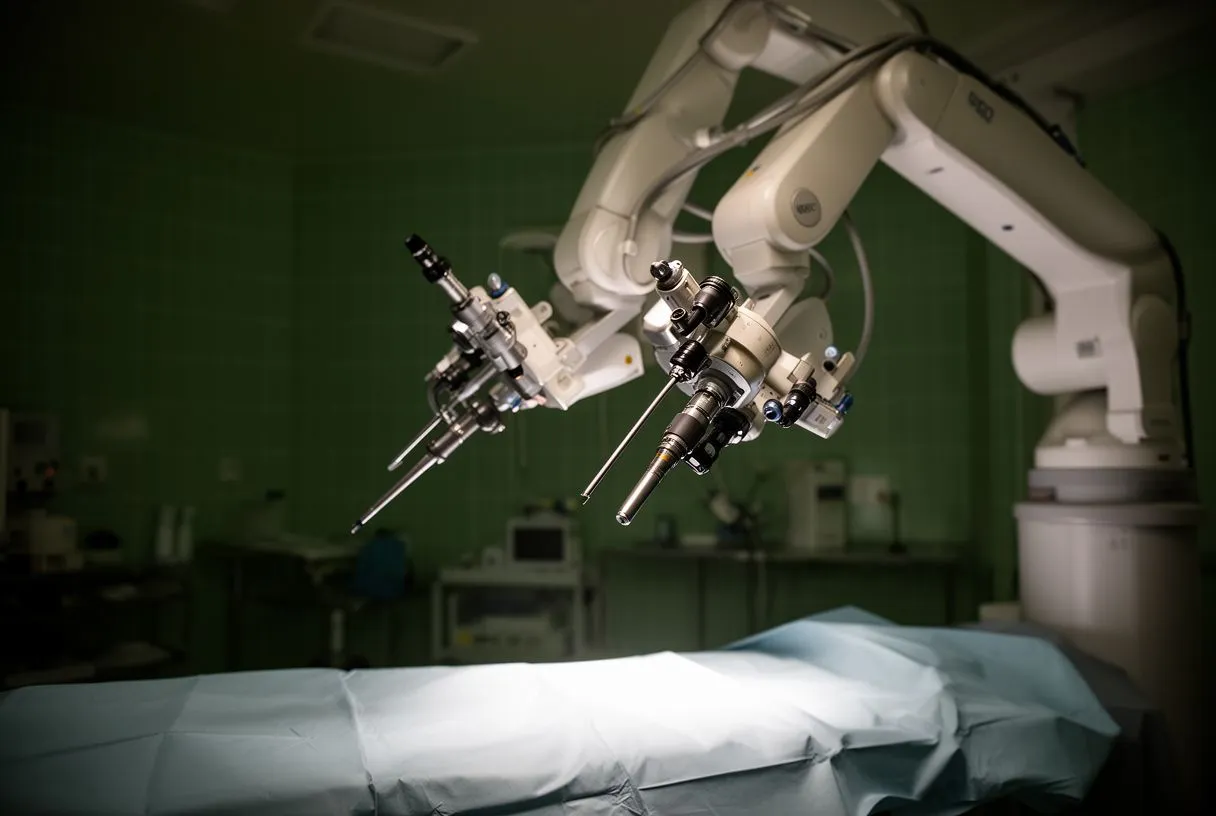
Robotic heart surgery thoracic entry represents the most advanced evolution of minimally invasive cardiac care. Instead of opening the chest, the surgeon makes several tiny incisions—often the width of a pencil—between the ribs. A high-definition 3D camera and robotic instruments equipped with tiny wrists are inserted into the thoracic cavity. The surgeon sits at a console a few feet away, translating their hand movements into precise, scaled motions inside the heart, eliminating the natural tremor of the human hand.
Enhanced precision treats complex blockages. The magnified, high-definition view allows surgeons to perform intricate repairs on tiny vessels. When treating severe atheromatous plaque buildup—the accumulation of cholesterol and fats in the artery walls—robotic assistance can help meticulously clear the blockages or attach bypass grafts with extreme accuracy. Understanding a patient's overall cardiovascular risk profile, including markers like apolipoprotein B (a protein linked to cholesterol particles), helps the team decide if robotic precision is necessary for complex plaque removal or grafting.
Robotic techniques demand specialized expertise. While the technology is remarkable, it requires extensive training and is not available at every medical center. The steep learning curve means these procedures are typically reserved for specialized centers with dedicated robotic surgical teams. For patients, the benefit lies in the preservation of the thoracic cavity's structural integrity, as keeping the sternum intact drastically reduces recovery time and the risk of deep bone infections.
What happens to the chest wall during coronary bypass incision type?
When treating coronary artery disease, the coronary bypass incision type is typically a median sternotomy, though less invasive options are emerging. During a traditional coronary artery bypass grafting (CABG) procedure, the chest is opened to access the heart. The surgeon harvests a healthy blood vessel, usually from the chest wall or the leg, to bypass the blockage in the coronary artery. An arterial graft, such as the left internal mammary artery, is highly favored because it remains open and durable for many years compared to vein grafts.
The bypass restores critical blood supply. Once the thoracic cavity is accessed, the surgeon attaches the harvested graft above and below the blockage in the coronary artery. This creates a new pathway for oxygen-rich blood to reach the heart muscle. Because manipulating the ascending aorta can dislodge debris, careful surgical technique is vital. After the grafts are secured, the patient is typically placed on long-term medication, including aspirin, to prevent clots from forming in the newly placed grafts and to protect the long-term health of the heart.
Alternative grafts minimize secondary trauma. In some cases, surgeons may use an autograft—a vessel taken from the patient's own body—rather than a synthetic material, as natural vessels have a lower risk of rejection and clotting. The choice of graft and the specific coronary bypass incision type depend on the number and location of the blockages. For patients with isolated blockages, minimally invasive direct coronary artery bypass (MIDCAB) might be an option, avoiding the heart-lung machine and minimizing disruption to the thoracic cavity.
Are there pediatric heart surgery access differences?
Yes, pediatric heart surgery access requires highly specialized approaches because a child's thoracic cavity and cardiovascular anatomy are vastly different from an adult's. The structures are much smaller, and congenital defects often involve complex anatomical rearrangements. Despite these differences, the primary access point for most pediatric open-heart procedures remains a median sternotomy. The surgeon must be exceptionally gentle, as the rib cage is more flexible, and the margins for error when placing an arterial cannula for bypass are minuscule.
Congenital defects require precise anatomical correction. Pediatric surgeons correct a wide array of congenital heart defects, ranging from holes in the heart's walls to abnormal connections between the atrium and ventricles. Some children present with abnormal heart rhythms, such as atrial flutter, which might require surgical ablation in addition to structural repair. The surgical strategy is meticulously planned based on fetal echocardiograms and postnatal imaging to ensure that the single entry into the thoracic cavity allows for comprehensive repair.
Critical support systems are tailored for infants. Because the pediatric heart is small and sensitive, the bypass circuit must be miniaturized to reduce the amount of foreign blood surfaces the child is exposed to. In extreme cases of cardiac failure before or after surgery, children might require advanced life support. Awake ECMO (Extracorporeal Membrane Oxygenation) can be used to support the heart and lungs while the child remains conscious, a delicate balance in the pediatric intensive care unit. Should a dangerous arrhythmia occur, the rapid use of an automated external defibrillator (AED) is part of the emergency response protocols safeguarding these young patients.

How does thoracic cavity anatomy surgery affect recovery?
Recovery from thoracic cavity anatomy surgery is a gradual process dictated largely by the type of incision made. If a sternotomy was performed, the bone requires about six to eight weeks to heal fully, and sometimes longer. Patients must avoid heavy lifting, pushing, or pulling during this time to prevent the wires from loosening. The muscles and tissues of the chest wall also undergo trauma, leading to general fatigue and discomfort that slowly subsides over the first few weeks.
Postoperative pain requires active management. Pain management is a crucial aspect of cardiac recovery, not just for comfort, but to allow patients to breathe deeply and cough effectively. Many patients experience referred arm pain or sharp twinges across the chest wall as the nerves heal. This pain is typically managed with medication and gentle stretching. Refusing to cough due to pain can lead to fluid buildup in the lungs, a common complication after any thoracic cavity surgery. Patients are taught to hold a pillow tightly against their chest when coughing to stabilize the sternum and reduce discomfort.
Optimizing lung health is critical for healing. Because the lungs share the thoracic cavity with the heart, protecting respiratory function is vital. For patients with pre-existing lung conditions, optimizing their condition before surgery is a priority. Understanding the Global Strategy for the Diagnosis, Management, and Prevention of Chronic Obstructive Pulmonary Disease helps surgical teams tailor respiratory therapy, ensuring that lung capacity is maximized to handle the stress of intubation and the recovery phase. Pulmonary rehabilitation and incentive spirometry are standard tools used to keep the lungs clear and expanded.
What are the risks of chest cavity heart procedure approaches?
Every entry into the chest cavity carries inherent risks, and understanding these is part of giving informed consent. Bleeding is a primary concern, as the thoracic cavity contains the body's largest blood vessels. Infection is another risk, particularly deep sternal wound infections following a sternotomy, which can prolong recovery significantly. Surgeons use strict sterile techniques and prophylactic antibiotics to minimize this risk, but it cannot be eliminated entirely. The approach chosen—sternotomy, thoracotomy, or robotic—alters the specific risk profile.
Evidence-based frameworks guide risk assessment. Clinical teams evaluate the risks of different surgical paths using standardized frameworks. The GRADE: an emerging consensus on rating quality of evidence and strength of recommendations approach ensures that the benefits of a chosen surgical method are weighed against the potential complications using the best available clinical data. Tools like the EuroSCORE II are also utilized to calculate a patient's specific mortality risk based on age, gender, and comorbidities, helping the team choose the approach with the lowest risk for your situation.
Specific complications dictate postoperative care. The nature of the risks varies by procedure. Here are key risks associated with chest cavity heart procedures:
- Bleeding: Requires careful monitoring of chest tube drainage and sometimes a return to the operating room for exploration.
- Infection: Deep infections of the sternum are rare but severe, requiring aggressive antibiotic courses and sometimes reconstructive surgery.
- Arrhythmias: Irritation of the heart tissue can cause irregular heartbeats, often managed with medication or temporary pacemakers.

When should you discuss adult cardiac surgery approach with your team?
You should discuss your adult cardiac surgery approach the moment your cardiologist recommends surgical intervention. Do not wait until the night before the operation. Early discussions allow you time to understand the proposed plan, seek a second opinion if desired, and mentally prepare for the recovery. If you have conditions like advanced atheromatous plaque or symptoms such as atypical infarction that prompted the need for surgery, understanding how your specific disease dictates the surgical entry point is empowering.
Medication management requires careful planning. If you are already taking blood thinners—such as those studied in the ARISTOTLE trial, which compared different anticoagulants for atrial fibrillation—your surgical team must coordinate the timing of your surgery. Some medications need to be stopped days in advance to prevent excessive bleeding, while others might be bridged with shorter-acting alternatives. Having an open conversation about your current medication regimen ensures a smooth transition into the operating room and minimizes the risk of complications related to blood clotting.
Choosing the right team for your specific needs. Ultimately, the decision of how to access the thoracic cavity is a partnership between you and your surgeon. Dr Alejandro Molina and the team at Cirugía de Corazón, serving Guadalajara, Querétaro, and Hermosillo, are dedicated to explaining these complex procedures in plain, reassuring language. We encourage you to book a consultation to discuss your specific cardiac condition, review your imaging, and determine the surgical approach that offers the lowest risk and the best long-term outcome for your unique anatomy. Do not self-diagnose; let a qualified cardiac team guide your next steps.






