

TMVR or TEER: Transcatheter Mitral Options Compared
TMVR vs TEER — compare transcatheter mitral valve repair and replacement side by side.
When the mitral valve starts leaking badly enough to cause heart failure symptoms, patients face a fork in the road: repair the valve through a catheter or replace it with a new one. That decision is often framed as tmvr vs teer, and it is one of the most consequential choices in modern structural heart care. This guide walks you through both options in plain language — how each is done, who is a candidate, what recovery looks like, and what the published evidence actually says — so you can arrive at a consultation knowing the right questions to ask.
What Is Transcatheter Mitral Valve Repair and Replacement?
Two families of treatment. Transcatheter mitral valve options fall into two broad categories. The first is repair: instead of removing the valve, the procedure modifies the patient's own tissue so it leaks less. The second is replacement, known as TMVR — transcatheter mitral valve replacement — in which a new artificial valve is delivered through a catheter and positioned inside the failing one. Both belong to the world of percutaneous mitral valve procedures, meaning the heart is reached through a blood vessel rather than through an open chest.
Why "transcatheter" matters. A transcatheter procedure avoids opening the chest and stopping the heart, which is what traditional mitral valve surgery requires. For older patients, people with previous chest surgery, or those with lung disease or frailty, avoiding an open operation can change the risk profile dramatically. That is why transcatheter mitral valve replacement vs repair has become such an important discussion at heart teams around the world.
Which valve is the hardest to replace? The mitral valve is widely regarded as the most challenging heart valve to replace with a catheter. Unlike the aortic valve, which sits in a relatively rigid ring of calcium, the mitral valve sits in a saddle-shaped, flexible, moving structure — the annulus — close to two papillary muscles and the outflow tract of the left ventricle. Every TMVR device must work with that anatomy rather than against it. In our practice, every candidate for a transcatheter mitral valve procedure is reviewed by the full multidisciplinary team against international practice guidelines before any recommendation is made, and the alternatives are laid out clearly so the final decision stays with the patient.
How Do TMVR vs TEER Procedures Work?
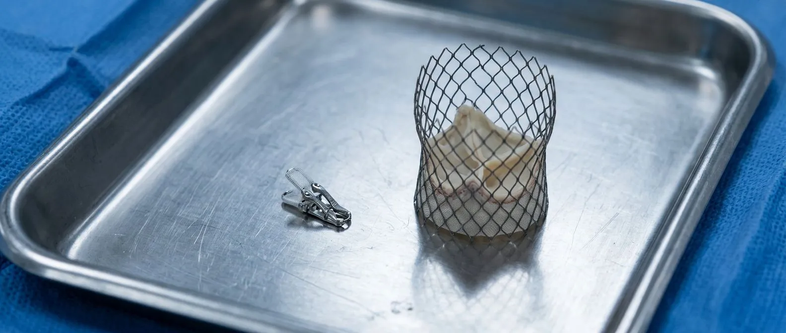
The clip mechanism. An edge-to-edge repair device is a small metal clip delivered through a vein in the groin, advanced to the heart, and steered from the left atrium down onto the middle of the two mitral valve leaflets. It clips them together at the point where they fail to meet, converting one leaky orifice into two smaller, better-closing ones. This approach is known as Transcatheter Edge-to-edge Repair, or TEER, and it is the most commonly performed transcatheter mitral procedure worldwide.
The TMVR mechanism. TMVR, by contrast, delivers a complete replacement valve — usually a balloon-expandable or self-expanding valve built for the mitral position — inside the patient's own leaky valve. The diseased leaflets are pushed aside or used as an anchor, and the new valve takes over all the work of preventing backflow. There is nothing left to repair; the old valve tissue stays in place but no longer carries the load.
The access difference. Both procedures typically begin in the catheterization lab. The clip enters through the femoral vein and crosses the wall between the right and left atria with a small puncture. TMVR can follow the same venous route or, in some designs, enter through a small incision near the ribs at the heart's apex, with the catheter guided directly into the left ventricle. We take the time in consultation to show patients on the anatomy exactly where each route goes and what each device does, in plain words, before any decision is discussed.

What Are the Symptoms of a Leaky Mitral Valve?
Early signs. A leaky mitral valve — mitral regurgitation, in medical language — often begins quietly. The most common early symptoms are unusual breathlessness on stairs or hills, a drop in exercise tolerance, and tiredness that rest does not fully fix. Some people notice a fluttering or pounding sensation, especially lying on the left side, because the left atrium enlarges as it absorbs the extra volume.
Later signs. As the leak worsens and the heart strains, symptoms shift to classic heart failure patterns: breathlessness lying flat, waking at night gasping, swelling of the ankles and legs, rapid weight gain from fluid, and sometimes a dry cough. An irregular heartbeat — atrial fibrillation — is a frequent companion of a long-standing leak, and it may be the first sign something is wrong.
When to act. Severe mitral regurgitation does not usually improve on its own, and guideline thresholds recommend intervention once symptoms appear or once the heart's pumping function begins to decline. Because the symptoms can creep up so gradually that patients normalize them, anyone with unexplained breathlessness should ask their doctor about an echocardiogram rather than wait.
Who Qualifies for TEER? Candidacy Explained
The anatomy that fits. The single biggest factor in TEER procedure candidacy is the shape of the leak. The clip works best when the leak originates at the center of the valve and the leaflet tissue is adequate and not badly calcified. Regurgitation caused by a prolapsing leaflet (degenerative disease) and regurgitation caused by a poorly closing, dilated heart (functional disease) can both be suitable, but the leaflet geometry must be measured carefully.
The patient profile. Edge-to-edge repair was designed for patients who are too high-risk for open mitral surgery — typically older adults, those with weakened heart function, or people with other serious conditions. The landmark COAPT trial, performed with the original clip device, established its role in patients with severe functional mitral regurgitation whose hearts are dilated and who remain symptomatic on medication. The EVEREST II trial, earlier, proved the concept in a broader group.
What disqualifies it. Heavy calcification of the leaflets or annulus, extreme tethering of the leaflets down into the ventricle, a very wide or very eccentric leak jet, and certain coexisting valve problems can make the clip impractical or unlikely to reduce the leak enough. When a patient comes to us for an evaluation, we review the echocardiogram measurements against these criteria ourselves and are candid when the anatomy will not support a good clip result, because a half-effective repair is not a fair trade for the procedure.
- Best fit: central leak, adequate pliable leaflets, high surgical risk.
- Poor fit: heavily calcified valve, extreme leaflet tethering, very eccentric jet.
- Gray zone: multiple leak jets or barlow-type prolapse — needs expert imaging review.
TMVR vs TEER: Which One Fits Your Anatomy?
Repair preserves what works. When deciding between tmvr vs teer, the core question is whether the patient's own valve tissue can be made to work again. If the leaflets are healthy but misaligned, a clip — or a surgical Mitral Valve Repair — preserves the native valve, avoids the need for lifelong anticoagulation in many cases, and keeps future options open.
Replacement solves what repair cannot. When the valve is destroyed — heavily calcified mitral annular calcification, severe stenosis, or leaflets too diseased for any repair — a replacement is the only realistic transcatheter route. TMVR also fits patients who had a previous surgical repair or ring annuloplasty that failed, a situation where the existing ring actually helps anchor the new valve.
The role of imaging. This is where the decision is truly made. Modern echocardiography and CT scanning map the annulus size, leaflet lengths, calcium distribution, and the distance to the left ventricular outflow tract. A new TMVR valve that sits too low can obstruct outflow; one that sits too high can dislodge. In our consultations we explain what each study actually shows — annulus measurements, calcium score, outflow clearance — so patients understand why we are recommending one route over the other, and we welcome a second opinion as a normal part of the process.
Edge-to-Edge Repair vs Valve Replacement: What Outcomes Show
What the evidence says for repair. The published data on edge to edge repair vs valve replacement come from very different levels of maturity. Edge-to-edge repair has a decade-plus track record: the COAPT trial, which used the original clip device, showed durable reductions in heart failure hospitalization and improved survival at two years in the right patients, and a recent meta-analysis of two edge-to-edge repair systems — the original clip and a newer design — found consistently high procedural success and meaningful symptom improvement across both platforms (Journal of Cardiothoracic Surgery).
What the evidence says for TMVR. TMVR is newer and the evidence base is thinner. In patients with severe mitral annular calcification — a hardened, calcified ring that makes both surgery and clipping nearly impossible — a study of transcatheter mitral valve replacement reported encouraging one-year outcomes for a dedicated TMVR system, with the caveat that complications such as valve-in-valve issues and access complications remain relevant considerations (1-Year Outcomes of Transcatheter Mitral Valve Replacement in Patients With Severe Mitral Annular Calcification). The message is not that one always beats the other; it is that each has a defined territory where it performs best.
Trade-offs in teer vs tmvr outcomes. Repair keeps the native valve and typically avoids mandatory anticoagulation; replacement provides a definitive seal in valves that cannot be repaired but introduces risks specific to placing a large prosthesis in a moving position. Because our team performs conventional, minimally invasive, robotic and transcatheter procedures, we recommend the less invasive route only where the evidence supports it for that specific patient — never as a default.
TMVR Valve Replacement Recovery: What to Expect
The first days. Both procedures usually require one to a few nights in hospital. TMVR valve replacement recovery is often slightly longer than a clip procedure, particularly when an apical incision near the ribs is used, since that small wound needs a few extra days to settle. Most patients stand and walk the day after the procedure.
The first weeks. Expect fatigue for two to four weeks — the body has processed a significant procedure even though the chest was never opened. Breathlessness improves progressively as the heart stops being overloaded by the leak; many patients report noticeable improvement in exercise tolerance within weeks to a few months. Anticoagulation or antiplatelet therapy is prescribed according to protocol, and follow-up echocardiograms verify that the valve or clip is seated and functioning.
What helps recovery. Attending cardiac rehabilitation, taking medications as directed, controlling blood pressure, and reporting new symptoms early all improve long-term results. Recovery is best seen as a monitored process with structured follow-up points, rather than a single hospital discharge.
Transcatheter Heart Valve Comparison: TMVR, TAVR and the Tricuspid
TAVR: the mature sibling. In any transcatheter heart valve comparison, TAVR — transcatheter aortic valve replacement — is the benchmark. The aortic valve sits in a rigid calcified ring that makes an excellent landing zone, which is why TAVR is now routine for high-risk aortic stenosis and is expanding to lower-risk patients. Transcatheter Aortic Valve Implantation is the same procedure known by another acronym, but we use TAVR in line with common usage.
TMVR: the frontier. TMVR applies similar valve technology to a far more difficult position, as explained above. Devices continue to evolve specifically for the mitral annulus geometry, and clinical experience is growing year by year, but it remains a procedure for carefully selected patients, often with complex valve anatomy such as severe annular calcification.
The tricuspid valve. The same catheter principles have moved to the right side of the heart. Transcatheter Tricuspid Intervention — mostly edge-to-edge clipping of the tricuspid valve — is now an established option for severe tricuspid regurgitation in patients who cannot undergo surgery, and it shares much of its technique with mitral TEER. Dr Molina trained in structural cardiology and TAVR at CHU Bordeaux.
TMVR vs Surgical Mitral Valve Replacement
What surgery still does better. Open surgery remains the gold standard for durability in younger, lower-risk patients. Surgical Mitral Valve Repair by an experienced team can last for decades, and surgical replacement gives the surgeon direct vision to address every part of the valve and annulus. Minimally invasive mitral surgery — a small right-chest incision without opening the breastbone — offers much of surgery's durability with a faster recovery than a full sternotomy.
What transcatheter offers. TMVR vs surgical mitral valve replacement is essentially a risk-versus-durability trade. Transcatheter treatment avoids cardiopulmonary bypass and a chest incision, which matters enormously for elderly and frail patients; surgery offers proven long-term durability and the ability to correct multiple problems in one operation.
Who should not be pushed one way. A healthy 55-year-old with a repairable valve should almost always be offered surgical or robotic repair. An 84-year-old with severe annular calcification and lung disease may have transcatheter treatment as the only reasonable option. Dr Molina performed the first robotic mitral valve repair in the Ángeles Health System and the first robotic heart surgery in Guadalajara, and we bring that full range of options to the table rather than steering every patient toward one technique.
Second Opinions and Choosing Your Care
Understanding cost drivers. Transcatheter mitral valve repair cost and TMVR cost depend on the device used, the hospital, the length of stay, and the imaging workup required beforehand. Ask for an itemized estimate covering the valve or clip device, hospital stay, imaging, and follow-up.
Insurance and logistics. Confirm coverage in writing, ask about travel timing — plan to stay near the hospital for the first follow-up — and make sure your records, especially the echocardiogram and CT, travel with you in digital form.
How to choose a team. Ask how many transcatheter mitral procedures the team performs, whether a full heart team discusses your case, and what the alternatives are. A trustworthy team explains options, respects your decision, and never rushes you. The first contact with our team is a free video consultation: we get to know you, talk through your condition, and tell you which studies are genuinely still needed — so no unnecessary tests are ordered — and a face-to-face visit at our consulting rooms in Guadalajara, Querétaro or Hermosillo follows only if you decide to move forward.
Where to learn about your valve. Reliable background reading on the mitral valve itself, its anatomy and diseases — including Mitral Valve Prolapse, Mitral Stenosis, and regurgitation — helps you arrive at a consultation with informed questions. Sources such as Stanford Health Care provide dependable overviews of transcatheter valve therapies for patients.
In summary, the choice between tmvr vs teer is not a contest with a universal winner — it is an anatomy-and-patient decision. A clip repairs a valve that can still be repaired; a transcatheter replacement replaces a valve that cannot. The honest path forward is a complete imaging workup, a heart team discussion against international guidelines, a clear presentation of every alternative including surgery, and a decision you make with full understanding. If you are living with a leaky mitral valve, do not self-diagnose your options — bring your studies to a qualified cardiac team, ask every question you have, and book a consultation to find out which route truly fits your heart.






