Transoesophageal Echocardiogram: Preparation and What to Expect
Transoesophageal echocardiogram preparation guide: fasting rules, medication steps, and what happens during TEE.
What Is a Transoesophageal Echocardiogram and Why Is It Ordered?
A transoesophageal echocardiogram — often shortened to TEE — is a specialised heart imaging test that gives your cardiologist a remarkably detailed view of your heart's structures and valves. Unlike a standard chest ultrasound, which is performed by pressing a probe against the skin of your chest, a transoesophageal echocardiogram uses a thin, flexible probe that is gently guided down your oesophagus (the swallowing tube that runs from your mouth to your stomach). Because the oesophagus sits directly behind the heart, the ultrasound images produced are far clearer than those from a surface scan, with virtually no interference from ribs, lungs, or body tissue.
Why your doctor may order this test instead of a standard echo: When a routine transthoracic echocardiogram (the kind done through the chest wall) does not provide enough detail, your cardiac team may need the higher-resolution images that only TEE can deliver. This is especially common when assessing the mitral valve, looking for small blood clots in the left atrium, or evaluating infections on the heart valves (a condition called endocarditis). Your doctor is not ordering this test lightly — it means the clinical question is important enough to warrant a closer look.
For patients facing valve surgery or other cardiac interventions, a TEE is often part of the surgical planning process. It helps the surgeon understand the exact anatomy of the problem before making any incision. If you have been referred for a transoesophageal echocardiogram, it is natural to feel anxious, but understanding what the test involves — and how thoroughly it is prepared — can ease much of that worry. The information that follows is designed to walk you through every step, from fasting instructions to what you will feel during and after the procedure.
How Does a Transoesophageal Echocardiogram Work?
The transoesophageal echocardiogram works on the same basic principle as any ultrasound: high-frequency sound waves bounce off structures inside the body and create a real-time picture on a monitor. The key difference is the proximity. With the ultrasound transducer sitting millimetres from the back of your heart rather than centimetres away through your chest wall, the sound waves travel a much shorter distance. This means the returning signals are stronger and the resulting images are sharper, allowing the cardiologist to see fine details such as small vegetations on a valve, subtle tears in a flap of tissue, or tiny clots that a surface echo could easily miss.
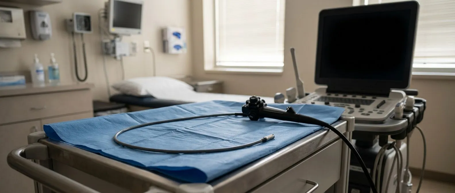
What the probe actually does once positioned: The TEE probe is about the thickness of a large piece of spaghetti and is marked with measurement lines so the operator knows exactly how far it has travelled. Once the tip reaches the correct position behind the heart, the cardiologist can rotate, angle, and tilt the transducer at the tip to capture images from multiple planes — essentially building a three-dimensional understanding of the heart from different vantage points. The whole imaging session usually takes between 15 and 30 minutes, depending on the complexity of what is being assessed.
During the scan, the echocardiographer may also use a technique called Doppler imaging, which measures the speed and direction of blood flow through the heart's chambers and valves. This is essential for grading how tight a narrowed valve has become or how much blood is leaking backward through a valve that should be closing fully. Together, the structural and flow information from a transoesophageal echocardiogram gives your cardiac surgeon a precise map of the terrain they will be working in. A landmark review published in The Lancet underscores how indispensable high-quality intracardiac imaging has become in modern cardiac surgery.
Transoesophageal Echocardiogram Preparation: Fasting and Medication Instructions
Proper transoesophageal echocardiogram preparation is the single most important thing you can do to ensure the procedure goes smoothly. The core requirement is fasting: you must not eat or drink anything for at least six hours before your appointment. This empty-stomach rule is not arbitrary — it exists to minimise the small but real risk of stomach contents coming back up during the procedure and entering your lungs while your gag reflex is suppressed by sedation. Your safety depends on following this instruction precisely.
What medications you should and should not take before TEE: If you take prescription medications, ask your cardiologist which ones to take on the morning of the procedure with a small sip of water and which to hold until afterward. Blood pressure medications are sometimes continued, but blood thinners such as Aspirin or anticoagulants may need specific guidance depending on the reason for your TEE. Diabetic patients need particular instructions about insulin or oral hypoglycaemics, since fasting changes your blood sugar response. Never stop a medication on your own — always confirm with your cardiac team.
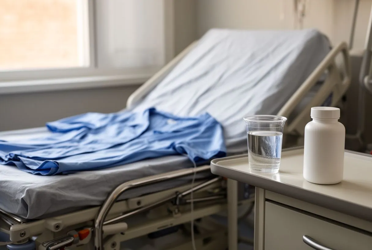
On the practical side, wear loose, comfortable clothing and leave jewellery and valuables at home. You will need to remove dentures, glasses, and any oral piercings before the probe is inserted. Arrange for a responsible adult to accompany you and drive you home afterward — the sedation used during the test will leave you drowsy and unsafe to drive for the rest of the day. If you are travelling from outside Guadalajara, Querétaro, or Hermosillo, plan to stay locally overnight rather than attempting a long journey immediately after the procedure.
What to Expect During the TEE Procedure Step by Step
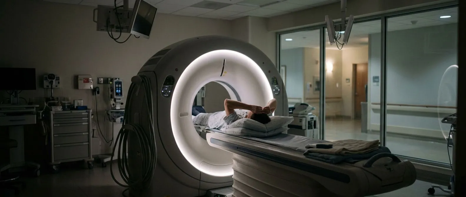
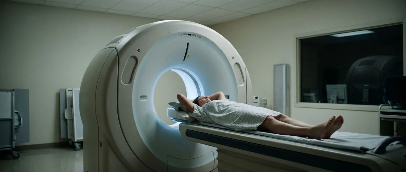
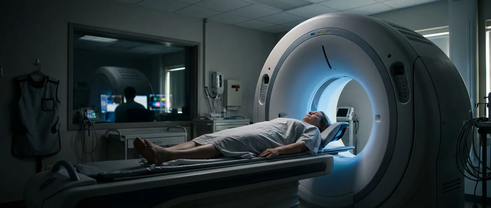
When you arrive for your transoesophageal echocardiogram, a nurse will confirm your fasting status, check your blood pressure and heart rhythm, and place a small intravenous line in your arm or hand. You will be asked to lie on your left side on a narrow examination table. A clip will be placed on your finger to monitor your oxygen levels continuously, and sticky electrodes on your chest will track your heart rate throughout the procedure. This monitoring is standard and nothing to worry about — it simply ensures your safety while you are sedated.
The moment of insertion and what you will feel: The doctor will spray a local anaesthetic at the back of your throat to numb it, which reduces the gag reflex. You may notice a bitter taste from the spray, but this passes quickly. The sedative is then given through your IV line, and within a minute or two you will feel deeply relaxed and drowsy — most patients remember very little of the actual procedure. The probe is gently guided into your mouth, and you will be asked to swallow as it passes into the oesophagus. Some people feel a brief pressure or mild discomfort in the throat, but significant pain is uncommon.
Once the probe is in position, the cardiologist will capture the necessary images, sometimes asking you to hold your breath for a few seconds at a time to improve picture quality. You may be aware of the probe moving slightly, but the sedation keeps most patients calm and comfortable throughout. When imaging is complete, the probe is gently withdrawn. The entire process, from numbing the throat to removing the probe, typically takes 30 to 45 minutes. After that, you will rest in a recovery area until the sedation wears off. Conditions such as atrial flutter or other rhythm disturbances can sometimes be assessed more precisely during this window.
TEE vs TTE: Understanding the Difference Between Heart Ultrasound Types
Patients are often confused about why they need a transoesophageal echocardiogram when they have already had a "regular" heart ultrasound. The distinction matters because the two tests serve different purposes and are used at different points in your diagnostic journey. A transthoracic echocardiogram (TTE) is the standard, non-invasive ultrasound performed through the chest wall — it is an excellent first-line tool and is completely painless. However, its image quality is limited by body habitus, lung tissue, and rib shadows, which can obscure fine cardiac structures.
When TTE is not enough and TEE becomes necessary: There are several clinical scenarios where your cardiologist will specifically request a TEE. These include detailed assessment of the mitral valve before repair surgery, detection of small blood clots in the left atrium or left atrial appendage (especially before procedures for atrial flutter), evaluation of suspected endocarditis when TTE is inconclusive, and intraoperative monitoring during cardiac surgery. TEE is also the modality of choice for checking whether a balloon-expandable valve has seated properly during transcatheter aortic valve replacement.
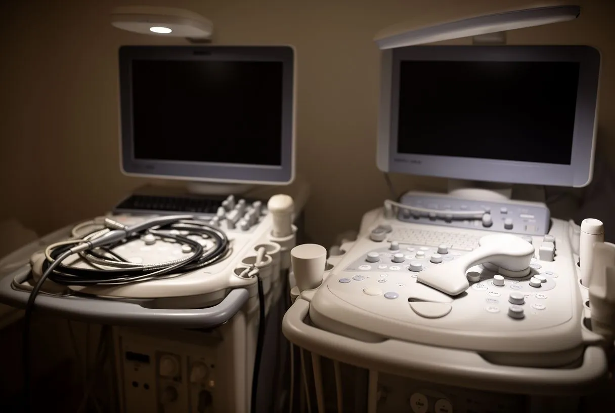
- TTE (Transthoracic): Non-invasive, done through the chest, no sedation, 20-30 minutes, lower image detail, best for routine screening.
- TEE (Transoesophageal): Probe through the oesophagus, mild sedation, higher image detail, best for surgical planning and complex valve assessment.
The decision is always clinical. Your cardiologist weighs the added information TEE provides against the small additional risk of sedation and probe insertion. In many cases, a TTE is perfectly adequate, and a TEE is only recommended when the clinical question truly demands the higher resolution.
Transoesophageal Echocardiogram Sedation: What You Should Know
Sedation is a key part of the transoesophageal echocardiogram experience, and understanding how it works can reduce anxiety considerably. Most TEE procedures use what is called moderate or "conscious" sedation — you are not fully asleep as you would be for a general anaesthetic, but you are deeply relaxed, drowsy, and largely unaware of what is happening. This level of sedation is intentionally chosen because it keeps you breathing on your own and able to follow simple instructions, such as swallowing when asked, while making the experience far more tolerable.
What sedatives are typically used and how they feel: The most common combination is a short-acting benzodiazepine such as midazolam, sometimes paired with a low dose of a pain reliever like fentanyl. You will feel the effects within one to two minutes of the injection: a warm, calm sensation, a sense of heaviness in your limbs, and gradually fading awareness. Most patients remember nothing of the procedure itself or have only vague, dream-like impressions. The medications used are short-acting and wear off relatively quickly, though you will still feel drowsy for several hours afterward.
Because sedation affects your reflexes and judgement for the remainder of the day, there are strict safety rules you must follow. You must not drive, operate machinery, sign legal documents, or drink alcohol for 24 hours after the procedure. A responsible adult must take you home and ideally stay with you for the evening. The monitoring team will track your heart rate, blood pressure, and oxygen levels throughout, and will not discharge you until they are confident you are recovering normally. If you have sleep apnoea or a history of reactions to sedation, mention this well in advance so the team can adjust their approach.
Transoesophageal Echocardiogram Risks and Contraindications
A transoesophageal echocardiogram is generally considered a safe procedure, but like any test that involves sedation and passing a probe into the oesophagus, it carries a small but measurable risk of complications. Understanding these risks honestly — without minimising them — is part of giving informed consent. The overall rate of major complications is very low, and most are minor and self-limiting, such as a sore throat or mild hoarseness that resolves within a day or two.
Who should not have a TEE or needs extra caution: Certain conditions make the procedure more risky or contraindicate it entirely. These include a history of oesophageal stricture (narrowing), previous oesophageal surgery, active oesophageal varices (dilated veins often related to liver disease), severe bleeding disorders, or significant cervical spine instability. Patients who have had radiation to the neck or chest may also need special evaluation. If you have ever had difficulty swallowing, experienced arm pain related to positioning, or been told you have a narrowed airway, share this information with your cardiologist before the day of the test. In complex scenarios, alternative imaging such as intracardiac echocardiography or CT may be recommended instead.
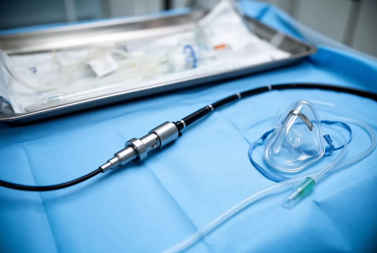
Among the rare but serious potential complications are dental or oral injury from the probe, oesophageal perforation (a tear in the wall of the oesophagus), aspiration of stomach contents, and adverse reactions to the sedation medications. Your cardiac team takes active steps to minimise each of these: the throat is numbed, the probe is inserted with care and imaging guidance, fasting is enforced, and your vitals are monitored continuously. Published clinical data, including case series examining adverse events during procedures such as those discussed in the study Wearable cardioverter‐defibrillator: friend or foe in suspected myocarditis?, reinforce that the risk-benefit balance favours proceeding when the clinical indication is strong.
TEE for Heart Valve Imaging: When Surgeons Need Detailed Pictures
One of the most important uses of the transoesophageal echocardiogram is detailed heart valve imaging. The heart has four valves — the aortic, mitral, pulmonary, and tricuspid — and each can develop problems that range from narrowing (stenosis) to leaking (regurgitation). When surgery or a transcatheter intervention is being planned, the surgeon needs to know the exact anatomy of the diseased valve, how severely it is affected, and what repair or replacement strategy offers the lowest risk for your specific situation. TEE provides this information with a level of detail that no surface echo can match.
Why the mitral valve especially benefits from TEE: The mitral valve sits deep in the heart, between the left atrium and the left ventricle, and is partially obscured by lung tissue when viewed from the chest wall. From the oesophagus, however, the view is direct and unobstructed. The cardiologist can see the individual leaflets of the valve, identify whether the problem is restricted movement, prolapse, or a tear, and measure the size of the opening and the volume of any leak. This information directly influences whether a surgeon can repair your valve rather than replace it — a decision with major implications for your long-term outcome. Studies of drug-induced cardiac toxicity, such as the report Lenalidomide-Induced Myocarditis, Rare But Possibly Fatal Toxicity of a Commonly Used Immunotherapy, further illustrate why precise cardiac imaging matters when diagnosing complex valve and myocardial conditions.
TEE is also indispensable during the procedure itself. In the operating room, the probe is positioned before the first incision and remains in place throughout the surgery, providing real-time feedback. The surgeon can confirm that a repaired valve is no longer leaking before closing the chest, or verify that a prosthetic valve is functioning properly immediately after implantation. For patients undergoing valve procedures involving the ascending aorta or requiring an autograft, this intraoperative imaging is a standard part of the surgical workflow, not an optional extra.
Transoesophageal Echo Recovery Time and What to Watch For Afterward
Recovery from a transoesophageal echocardiogram is typically quick and straightforward. Most patients spend 30 to 60 minutes in a recovery area after the procedure, resting while the sedation wears off. The nursing staff will check your blood pressure, heart rate, and oxygen levels at intervals and offer you a drink of water once your gag reflex has fully returned — they will confirm this before allowing you to eat or drink anything. When you feel alert and stable, and your vital signs are normal, you will be discharged into the care of the person accompanying you.
Normal after-effects versus warning signs to report: It is entirely normal to have a mild sore throat for a day or two, a slight hoarse quality to your voice, and a feeling of fatigue from the sedation. These effects resolve on their own without treatment. However, you should contact your cardiac team immediately if you experience any of the following warning signs: difficulty breathing, chest pain, fever, vomiting, coughing up blood, or severe or worsening throat pain that does not improve. These could indicate a complication that requires prompt evaluation.
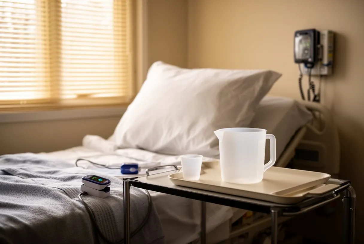
- Normal recovery: Mild sore throat, slight drowsiness, hoarse voice — resolves within 24 hours.
- Call your doctor if: Fever, chest pain, difficulty swallowing, vomiting, coughing blood, or breathing problems.
You should not eat or drink for at least one hour after the procedure, and when you do start, begin with cool water and soft foods. Avoid very hot liquids for the first few hours because your throat may still be partially numb and you could burn yourself without realising it. Resume your normal medications as directed by your cardiologist, and do not drive or make important decisions for 24 hours. Your doctor will discuss the results of the test with you, either on the same day or at a scheduled follow-up appointment, and will explain what the images mean for your treatment plan.
Booking a transoesophageal echocardiogram is a straightforward process, but it does require some advance planning because of the fasting and sedation involved. You will usually first see a cardiologist, who will review your history, examine you, and confirm that a TEE is the right test for your situation. This consultation is essential — no reputable cardiac practice will perform a TEE without first understanding why it is being requested and whether there are any contraindications specific to you.
What to expect regarding cost, insurance, and travel logistics: The cost of a transoesophageal echocardiogram in Mexico varies depending on whether the test is performed as a standalone diagnostic study or as part of a surgical procedure. It also depends on the facility, the anaesthesia support required, and whether the study is performed intraoperatively.
If you have private insurance, check with your provider whether TEE is covered under your plan and whether pre-authorisation is needed. Some policies cover diagnostic echocardiography but may have specific requirements for sedation-related procedures. The most important step is to discuss your individual circumstances with your cardiologist, who can recommend the most appropriate cardiac imaging approach for your needs. Do not self-diagnose or delay seeking care based on information you read online — your heart health deserves a personalised evaluation by a qualified specialist.
Booking a transoesophageal echocardiogram in Mexico and what it costs
Booking a transoesophageal echocardiogram at Cirugía de Corazón is a simple process, but it needs some advance planning because of the fasting and sedation involved. First you will have a consultation with a cardiologist, who will review your history, examine you and confirm that a TOE is the right study for your situation. That consultation is essential: no reputable cardiac centre will perform a TOE without first understanding why it is being requested and whether you have any specific contraindication.
What to expect on cost, insurance and travel logistics: The cost of a transoesophageal echocardiogram in Mexico varies depending on whether the study is done as a standalone diagnostic test or as part of a surgical procedure. It also depends on the centre, the anaesthetic support required and whether the study is done during the operation. If you are travelling from outside Mexico, we can help you coordinate appointments, schedule the blood tests needed before the procedure and guide you on the details of travel and accommodation in Guadalajara, Querétaro or Hermosillo.
If you have private insurance, check with your provider whether your plan covers a TOE and whether prior authorisation is needed. Some policies cover diagnostic echocardiograms but may have specific requirements for procedures that need sedation. The most important thing is to consult a cardiologist who can assess your particular circumstances and recommend the cardiac imaging method best suited to your needs. Do not self-diagnose or delay care on the basis of information you read online: your heart health deserves a personalised assessment by a qualified specialist.