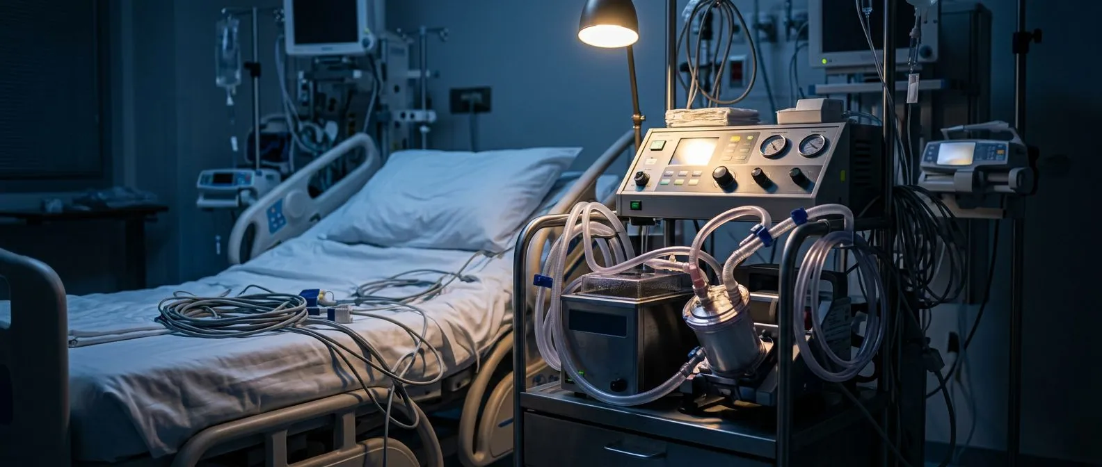
Veno-Venous ECMO for Severe Lung Failure
Veno-venous ECMO lung failure support gives damaged lungs time to heal.
Veno-venous ECMO for lung failure is a form of life support that takes over the work of lungs so damaged they can no longer keep the body alive on their own. If your loved one has just been told they need it, you are likely frightened and flooded with unfamiliar words. This guide explains, in plain language, what the machine does, who qualifies, what the risks are, and what recovery actually looks like — so you can ask better questions and make calmer decisions.
What Is Veno-Venous ECMO for Lung Failure?
Extracorporeal membrane oxygenation (ECMO) — the full name doctors use — means "oxygen added outside the body." Veno-venous ECMO for lung failure is the specific form used when the heart is pumping well but the lungs cannot exchange oxygen and carbon dioxide. Blood is drained from a vein, passed through an artificial membrane that adds oxygen and removes CO2, and returned to another vein. Your heart and circulation keep doing their own job; only the lungs are resting.
The word "veno-venous" simply describes the plumbing: both the drainage and the return cannulas sit in veins. Venous blood is naturally low in oxygen, so the machine's job is to enrich it before it returns to the right side of the heart and travels to the body. Nothing is opened inside the chest, and the heart itself is never touched by the circuit.
It helps to picture it as a temporary spare lung. It is not a treatment for the disease causing the lung failure — pneumonia, injury, or inflammation still need their own care. What it buys is time: days or weeks in which severely injured lung tissue can heal while the machine keeps oxygen levels safe.
How Does Veno-Venous ECMO Work?
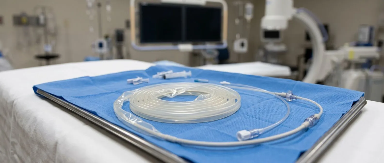
The circuit has three parts. First, a venous cannula — a soft plastic tube — drains blood from a large central vein, usually the femoral vein in the groin or the jugular vein in the neck. Second, a pump, most often a small centrifugal pump the size of a fist, moves the blood steadily. Third, an oxygenator membrane performs the gas exchange: oxygen flows on one side of the membrane, blood on the other, and carbon dioxide diffuses out while oxygen diffuses in.
The warmed, oxygen-rich blood then returns through a second cannula into a vein. Modern circuits are heparin-coated and monitored continuously for clots and for how much oxygen they deliver. The intensivist adjusts the blood flow rate and the oxygen sweep through the membrane depending on blood gases drawn every few hours.
At our team, we review every step of the circuit plan together before cannulation, because the smallest technical detail — cannula size, insertion site, flow target — shapes how well the system supports that particular patient. It is a team effort between surgeons, intensivists and perfusion specialists, and nothing is decided by one person alone.
Many patients are surprised to learn they can be conscious on ECMO. Because only the lungs are bypassed, sedation can often be kept lighter than on a full ventilator — one reason breathing tubes can sometimes come out earlier. This "awake" approach is increasingly used in specialised centres and is discussed in more detail below.
Who Needs VV ECMO Respiratory Failure Support?
ARDS — acute respiratory distress syndrome — is the most common reason. In ARDS the air sacs of the lung flood with inflammatory fluid, often after severe pneumonia, sepsis, aspiration or chest trauma. Standard care is a ventilator with careful pressure and oxygen settings. When even the best ventilation cannot keep oxygen levels safe — roughly, an oxygen index below about 80% despite optimised settings — ECMO candidacy is considered.
Severe pneumonia is the second classic scenario, including bacterial, viral and influenza-driven cases. During the COVID-19 pandemic, extracorporeal life support (ECLS) programmes worldwide cared for large numbers of patients whose lungs could not be supported any other way, and much of what we now know about safe ECMO practice was refined in those years.
Candidate selection criteria matter enormously, because ECMO is invasive and carries real risks. Typical considerations include:
- Reversible lung disease: the underlying cause must be expected to heal or be treatable, such as pneumonia, ARDS or post-operative lung injury.
- Reasonable overall health: advanced irreversible disease elsewhere, or incurable neurological injury, usually rules the therapy out.
- Timing: outcomes are better when ECMO starts before prolonged high-pressure ventilation has further damaged the lungs.
- Clear goals: the team and family agree on what success means and how long support will be offered before reassessing.
Age alone is not a disqualifier, but frailty and other organ problems influence the decision. Every major ECMO centre weighs these criteria individually — no checklist replaces clinical judgement.
Veno-Venous ECMO vs Veno-Arterial ECMO: Which One and Why?
The difference comes down to which organs are failing. In veno-venous ECMO, both cannulas sit in veins and only the lungs are supported; the heart must still pump the oxygenated blood around the body. In veno-arterial ECMO (VA ECMO), blood is drained from a vein but returned into an artery, so the circuit supports both the lungs and the heart's circulation. VA ECMO is used after cardiac arrest, in profound heart attack, and in patients whose heart cannot generate a pulse.
For pure lung failure, VV is almost always the right choice. It is technically simpler, avoids tying up an artery, and lets the heart keep working naturally, which preserves normal blood flow patterns. VA ECMO carries additional risks, including differences in blood pressure between the upper and lower body and a higher clot and stroke burden.
A sensible comparison:
- VV ECMO: lungs only; cannulas in veins; heart continues working; used for ARDS, severe pneumonia, lung transplant bridging.
- VA ECMO: lungs plus circulation; return cannula in an artery; used when the heart has also failed, for example after cardiac arrest or in cardiogenic shock.
Sometimes a patient starts on VV support and the heart weakens later; conversion to VA is possible. For families, the practical takeaway is simple: if the doctors say "veno-venous," the heart is still doing its job, and the machine is there purely as a lung substitute.
How Is ECMO Cannulation (Neck or Femoral) Done?
ECMO cannulation — inserting the cannulas — is usually done at the bedside in the intensive care unit under ultrasound guidance, with local anaesthetic and sedation. The two most common sites are the femoral vein in the groin and the internal jugular vein in the neck. Many centres drain from one femoral site and return through the right jugular vein, which sends oxygenated blood directly toward the heart and brain.
The procedure typically takes under an hour. The skin is cleaned thoroughly, wires guide each cannula into the correct position, and their placement is confirmed on ultrasound and X-ray before full flow begins. Sutures and secure dressings hold the tubes in place, and nurses check the insertion sites every shift.
In some patients — especially those needing support for many weeks or being evaluated for transplant — cannulas are placed centrally through a small chest incision, or a single dual-lumen cannula is used in the neck so only one puncture site is needed. The choice depends on anatomy, expected duration and whether the patient may need to sit up and walk while supported.
After insertion the patient is transferred to an ECMO-capable ICU. If a referring hospital cannot provide this, mobile ECMO retrieval teams can travel with the patient already cannulated, running a portable ECMO machine during transport — a service that has made this therapy reachable far beyond the few large centres.
What Are the Risks of ECMO Bleeding and Clots?
Bleeding and clotting are the two central risks. The circuit's surfaces activate the blood's clotting system, so patients receive a continuous anticoagulant — usually heparin — to keep blood from clotting inside the tubing. But the same medicine that prevents clots also makes surgical sites, cannula insertions and even the brain more prone to bleeding. Balancing these two risks is a daily task for the ICU team.
Clotting inside the circuit is the mirror problem. Small clots can form on the oxygenator or cannulas, reducing oxygen delivery or breaking loose into the bloodstream. Teams monitor clotting markers several times a day, watch the membrane for early signs of failure, and replace circuit components when needed.
Other recognised complications include infection around the cannula sites, limb ischaemia when a femoral cannula restricts arterial flow in the leg, and injury to the vessel during insertion. Kidney and liver function are watched closely, since prolonged critical illness affects every organ. Honest, evidence-based reviews of these risks are published in peer-reviewed literature, and any responsible centre will walk families through them before starting.
In our consultations we insist on explaining these risks in plain words — what the bleeding risk really is, what the clot risk really is — before anyone signs anything, and we answer every question the family brings, however basic it sounds. No patient or family should ever feel rushed into consenting to this therapy.
Awake ECMO and Life Inside the ICU
Awake ECMO is an approach in which the patient is not sedated and has no breathing tube. The cannulas sit in the neck or groin, the machine does the gas exchange, and the patient breathes on their own — talking, eating soft food, and in some programmes even doing physiotherapy. It is best suited to patients who are alert, cooperative, and stable enough not to need full ventilator support.
The benefits are real: avoiding a breathing tube reduces sedation-related complications, preserves muscle, and keeps the patient able to communicate with family. But awake ECMO demands a highly experienced nursing team and careful patient selection, because a sudden deterioration can require urgent re-intubation. Not every centre offers it, and not every patient qualifies.
For most patients on VV support, the ICU days follow a rhythm: blood gases every few hours, daily chest X-rays, ventilator settings reduced as the machine takes over, and careful watching for complications. Families usually find that the first 48 to 72 hours are the most intense, after which the routine becomes steadier. Asking the ICU team to explain the daily plan is always worthwhile — good teams expect and welcome that.
ECMO as a Bridge to Lung Transplant
ECMO lung transplant bridge support is a recognised use of this therapy for patients whose lungs will not recover on their own. For someone with end-stage lung disease awaiting a transplant, or with an acute deterioration who suddenly becomes a transplant candidate, VV ECMO can hold oxygen levels safe while the transplant team works through evaluation and organ matching. This is called "bridging" — the machine bridges the gap between the lungs failing and the new lungs arriving.
Awake bridging is a deliberate strategy in some transplant programmes: keeping the patient conscious, off the ventilator, and doing daily physiotherapy while waiting, because prolonged sedation before surgery is associated with worse outcomes. It requires the patient to be stable and the waiting period to be short enough to be practical.
It is important to be honest about the limits: ECMO cannot substitute for a transplant indefinitely, and the longer the bridge lasts, the harder the surgical course tends to be. Families should ask directly how long the team expects to bridge, and what happens if an organ does not come in time.
Our multidisciplinary team reviews every case like this against international practice guidelines before recommending anything, and the patient receives a clear proposal with the alternatives — the final decision is always theirs.
ECMO Weaning Success Rates and Lung Failure ECMO Survival
Weaning means gradually reducing support as the lungs recover. In practice, the team watches for the lung's own improvement: better oxygen levels at lower machine flows, less stiffness on chest X-ray, rising compliance. The flow is lowered in stages, the sweep gas reduced, and the patient observed for a trial period with the circuit off before the cannulas are removed. Weaning is a test, not a switch — the lungs have to prove they can manage.
Lung failure ECMO survival figures vary by cause and by how sick the patient was at the start. Published registry data show that a meaningful majority of patients with respiratory failure are successfully weaned, and that a substantial proportion survive to hospital discharge — figures that have improved steadily over the last two decades as selection, circuits and ICU care have matured. The honest framing is that ECMO gives a real chance to patients who would otherwise have very little, but it is not a guarantee, and the underlying disease matters more than the machine.
Factors that consistently predict better outcomes include starting ECMO before irreversible damage has occurred, a reversible underlying cause, younger age, absence of other organ failure, and the experience of the centre. Asking a centre about its own volumes and outcomes is entirely reasonable — high-volume centres generally achieve better results, and any confident team will answer.
ECMO Rehabilitation Recovery Timeline and When to Seek Help
Recovery after ECMO happens in stages. The first stage is in the ICU: cannulas removed, breathing tube out (if there was one), and gentle physiotherapy beginning. The second stage is the hospital ward, where the patient rebuilds strength — weeks of severe illness strip muscle even in young, fit people. The third is weeks to months of outpatient rehabilitation: breathing exercises, graded walking, and gradually returning to normal life.
The ecmo rehabilitation recovery timeline is different for everyone, but a realistic expectation is meaningful fatigue for several months. The lungs themselves often recover well after ARDS, though some patients are left with mild scarring and reduced exercise tolerance. Cognitive fuzziness after weeks in an ICU is common and usually improves with time and rehabilitation.
Watch for warning signs during recovery at home: new breathlessness out of proportion to activity, fever, chest pain, swelling or bleeding at the cannula site, or a leg that becomes cold or painful. Any of these deserves urgent medical review.
At our practice — consulting rooms in Guadalajara, Querétaro and Hermosillo — the first contact is a free video consultation: we get to know the patient, talk through the condition, and identify which studies are genuinely still needed so no unnecessary ones are ordered. If you or a loved one is facing severe lung failure or has been offered ECMO, a second opinion from a qualified cardiac and critical care team is a normal, sensible step — not a betrayal of your treating doctors. Book a consultation and bring your questions; we will take the time to answer them.
Choosing where to have this therapy matters as much as the therapy itself. Look for a centre with a dedicated ECMO team that includes cardiac surgeons, intensivists and perfusionists, a programme that runs regularly rather than occasionally, and a culture of explaining options rather than prescribing them. We actively encourage second opinions and treat seeking one as a normal part of deciding about heart and lung support surgery.
Finally, remember that ECMO is a tool, not an answer. The best outcomes come from the combination of timely decision-making, an experienced team, and a family who understands what is being proposed and why. Ask about the centre's experience, the weaning plan, and the realistic expectations for someone in your loved one's exact situation. No honest team will promise you a number; the best ones will tell you the truth.

In summary, veno-venous ECMO for lung failure gives badly injured lungs the time they need to heal while a machine quietly does their work. It is used for severe pneumonia and ARDS, as a bridge to transplantation, and in selected post-operative patients. It carries real risks — bleeding, clotting, infection — which are managed by experienced teams, and it works best when started early and in a centre that does it regularly. If you are facing this decision, discuss your own situation with a qualified cardiac team rather than self-diagnosing. The right first step is a conversation, not a scan.