Ventriculography: Measuring How Well the Heart Pumps
Ventriculography heart function testing measures how well your heart pumps.
When you or a loved one faces symptoms like unexplained shortness of breath or deep fatigue, understanding exactly what is happening inside the chest becomes the highest priority. The heart is a complex mechanical pump, and accurately measuring its performance is the first step toward creating a safe and effective treatment plan. This is where a specialized diagnostic tool comes into play. By measuring ventriculography heart function, your cardiology team can capture precise images of your heart's main pumping chamber, revealing exactly how much blood it pushes out with every beat and how well the muscle walls are moving. This guide walks you through the procedure, what to expect during recovery, and how these results inform critical decisions about your cardiac care.
What is ventriculography heart function testing?
At its core, ventriculography heart function testing is an imaging technique used to visualize the left ventricle, which is the heart's strongest chamber responsible for pumping oxygen-rich blood to the rest of the body. During the test, a contrast dye is injected directly into the ventricle through a thin, flexible tube called a catheter. As the dye fills the chamber, an X-ray camera records dynamic video images. This allows the cardiologist to watch the heart muscle contract and relax in real time, identifying areas that might be weak, stiff, or damaged by previous events like an Atypical Infarction where symptoms might have been vague and gone unrecognized initially.
Why the left ventricle matters most: The left ventricle does the heavy lifting for your circulatory system. If it cannot squeeze forcefully enough, blood backs up into the lungs, causing breathlessness. If it cannot relax properly to fill with blood, the body receives an inadequate supply. Ventriculography provides a direct, unobstructed view of this chamber's volume and pressure dynamics that surface tests simply cannot match.
The images produced are called a left ventriculogram. By tracing the outline of the dye at the end of a heartbeat and comparing it to the outline at the beginning of the next beat, doctors calculate the precise volume of blood ejected. This objective measurement replaces guesswork with hard data, ensuring that any subsequent treatment recommendations are based on your specific anatomy and actual mechanical performance rather than estimates alone.
How is ventriculography performed safely?
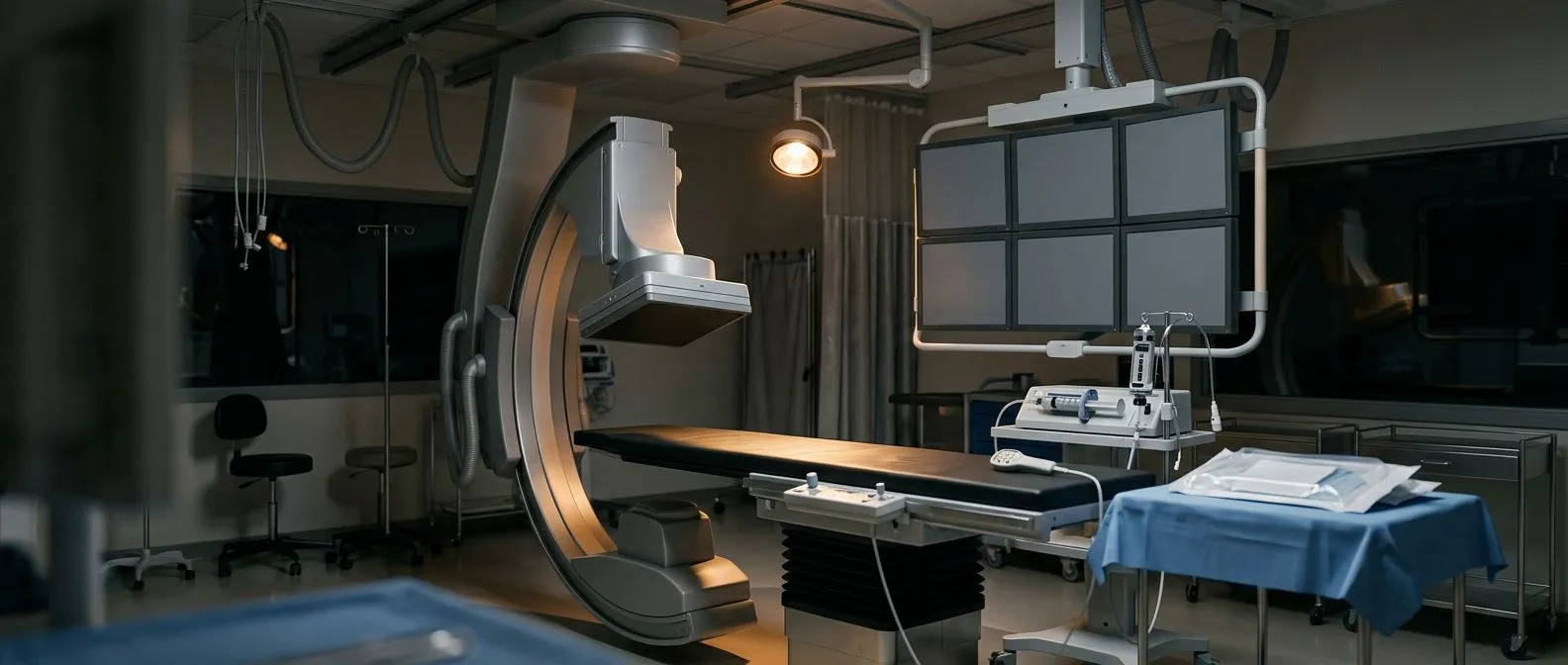
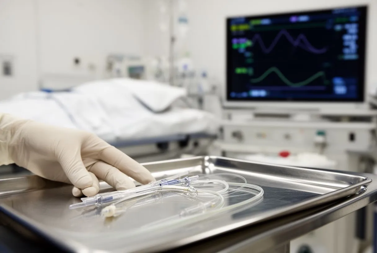
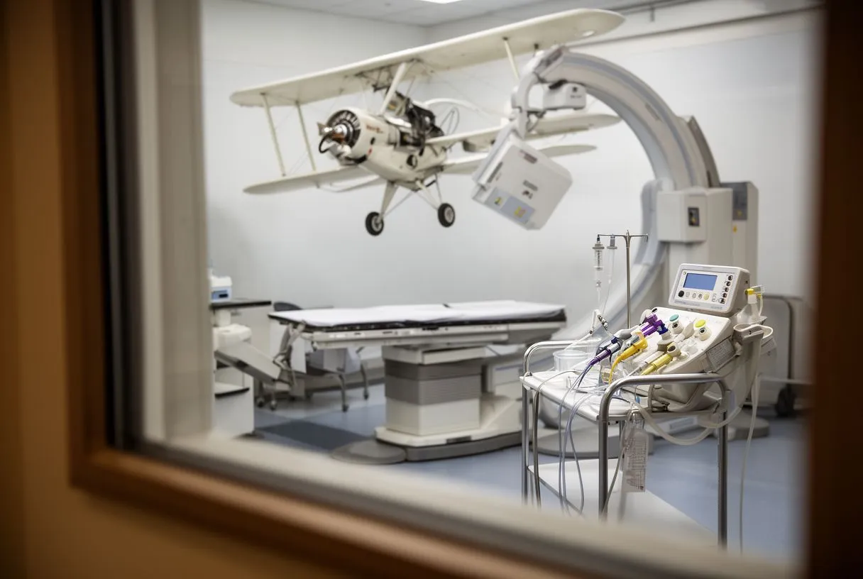
The procedure takes place in a specialized hospital room called a cardiac catheterization lab. You will typically be awake but given a mild sedative through an intravenous line to help you relax. The cardiologist cleans and numbs a small area of skin, usually in the wrist or the groin, before gently threading the catheter up through the blood vessels and into the heart. Continuous X-ray guidance ensures the tube follows the exact path needed without damaging the vessel walls.
What you will feel during the dye injection: When the contrast dye is released into your ventricle, it is completely normal to feel a sudden, warm flush spreading through your chest and body. Some patients describe a brief sensation of needing to urinate. This feeling passes in a matter of seconds. You will not feel the catheter moving inside your heart because there are no nerve endings for pain inside the heart chambers themselves, though you may feel mild pressure at the insertion site.
Throughout the procedure, your medical team closely monitors your vital signs. They track your heart rhythm and blood pressure continuously, ensuring your stability. According to the National Heart, Lung, and Blood Institute, catheterization procedures are common and generally safe when performed by experienced teams in controlled environments. The entire imaging portion of the test takes only a few minutes, though preparing the access site and guiding the catheter into position may take twenty to thirty minutes overall.
When is ventriculography needed for diagnosis?
Doctors typically recommend this test when less invasive evaluations leave unanswered questions. If a basic physical exam, including Auscultation listening to your heart with a stethoscope, reveals a new murmur, or if an electrocardiogram shows subtle changes, your doctor needs to know exactly what is causing those findings. Ventriculography is highly effective at identifying structural issues like leaky valves or structural damage from blocked arteries.
Key reasons your doctor may order the test:
- To evaluate the pumping strength after a suspected heart attack or severe viral illness.
- To check for valve regurgitation, where blood leaks backward through a valve that should stay closed.
- To assess congenital heart defects or abnormalities in the size and shape of the ventricular walls.
This test is also frequently performed when a patient is being evaluated for major cardiac surgery. If your surgical risk needs quantifying, knowing the precise strength of the heart muscle is essential. For instance, if an aneurysm is growing and the aortic diameter approaches guideline thresholds like greater than 5.5 cm, the surgical team must know if the ventricle can withstand the stress of a major operation. The data gathered here forms the foundation of your entire surgical plan.
Ventriculography vs echocardiogram: which is better?
Patients often wonder why a doctor would choose a ventriculogram when an echocardiogram, which uses harmless sound waves, is non-invasive and faster. An echocardiogram is indeed the standard first step for evaluating heart function. It provides excellent images of the valves and overall chamber size without any needles or radiation. However, sound waves can sometimes be blocked by body tissue, lung air, or bone, leading to suboptimal imaging in certain patients.
When invasive imaging wins: Ventriculography is typically reserved for situations where echo images are inadequate or when exact, high-resolution pressure and volume measurements are required. Research comparing diagnostic modalities, such as findings indexed in PubMed 39537272, shows that direct contrast imaging often provides superior geometric accuracy for the left ventricle in complex cases. It gives the physician direct visual confirmation of how the muscle behaves under real physiological conditions.
Ultimately, it is not a matter of one being better than the other, but rather using the right tool for the specific clinical question. In many cases, your cardiology team will use both. The echocardiogram establishes a baseline, and the ventriculogram, often performed simultaneously with a coronary angiogram to check for blockages, provides the definitive anatomic detail needed before deciding on interventions like stents or surgery.
Ventriculography preparation: what patients should know
Preparing for the test requires a few simple but important steps to ensure your safety during the procedure. You will usually be asked to avoid eating or drinking anything for six to eight hours beforehand. This reduces the risk of nausea while you are sedated and lying flat. Your medical team will provide specific instructions regarding your daily medications, particularly blood thinners like Aspirin, telling you whether to take them on the morning of the test or hold them temporarily.
Organizing your day: Arrange for a responsible family member or friend to drive you home afterward. You will not be allowed to drive for at least twenty-four hours because the sedatives affect your reaction time. Wear comfortable, loose-fitting clothing that is easy to change out of, and leave all valuables at home. You will typically change into a hospital gown for the procedure.
Before the test begins, the nursing staff will review your medical history, specifically asking about any allergies to iodine or contrast dye, and checking your kidney function through blood tests. The kidneys are responsible for filtering the dye out of your system. If you have chronic kidney disease, the team will take extra precautions, sometimes providing intravenous fluids before and after the test to help flush the contrast safely, or considering alternative imaging methods entirely.
Ventriculography risks and safety profile
Because ventriculography is an invasive procedure that involves entering the blood vessels and the heart, it carries certain risks. However, major complications are rare when performed by experienced cardiologists. The most common issues are minor and localized to the insertion site, such as minor bleeding, bruising, or a small hematoma where the catheter entered the skin. The medical team applies firm pressure to the site afterward to prevent excessive bleeding.
Understanding rare complications: More serious risks include an allergic reaction to the contrast dye, which can range from mild hives to severe anaphylaxis. In very rare instances, the catheter can irritate the heart muscle, causing arrhythmias like Atrial Flutter. The lab is fully equipped to handle these situations immediately. An Automated External Defibrillator and advanced resuscitation drugs are always on standby. In extreme, life-threatening cases of cardiac failure, extraordinary support systems like Awake ECMO might be discussed, though they are almost never needed for a diagnostic test.
To mitigate these risks, the team carefully screens patients beforehand. They calculate surgical risk scores, such as EuroSCORE II, to ensure the benefits of obtaining the diagnostic information outweigh the potential downsides. If you are at a higher risk due to advanced age or other health conditions, your doctor will discuss whether the test is truly necessary or if non-invasive alternatives can provide sufficient information to guide your treatment safely.
Ventriculography results interpretation explained
Once the images are captured, the cardiologist analyzes the video frame by frame. They are looking for several critical markers of heart health. The most important measurement is the ejection fraction, but they also look at wall motion abnormalities. If a specific segment of the heart muscle does not squeeze properly, it indicates that the blood supply to that area may be compromised, often due to severe Atheromatous Plaque buildup in the coronary arteries feeding that section of the heart.
Identifying mechanical problems: The test also reveals how well the valves are functioning. If the dye flows backward out of the aortic valve when the ventricle contracts, it indicates aortic regurgitation. If it flows backward into the left Atrium, it indicates mitral regurgitation. Quantifying these leaks accurately is vital, especially if a patient is being considered for advanced structural interventions, which are rigorously evaluated in clinical studies like the REPRISE II trial.
Your doctor will sit down with you to review these findings in plain language. A normal result means the heart muscle is strong, the walls move symmetrically, and the valves are competent. An abnormal result does not immediately mean disaster; rather, it provides a precise map of what is wrong. This map is exactly what your cardiac surgeon needs to plan the next steps, whether that means medication management, a targeted intervention, or a surgical repair.
Ventricular ejection fraction test details
The ventricular ejection fraction test is a critical component of the overall ventriculogram. The ejection fraction, often abbreviated as EF, represents the percentage of blood that is pumped out of the filled ventricle with each heartbeat. A normal EF is typically between fifty and seventy percent. This means that with every beat, the heart pushes out at least half of the blood it contains. It is the single most important number used to gauge the severity of heart failure.
What a low or high number means: An EF below forty percent indicates significant weakening of the heart muscle, often called systolic heart failure. This can leave patients feeling exhausted and short of breath because the organs are not receiving enough oxygenated blood. Conversely, an unusually high EF is not always a sign of an incredibly strong heart; it can indicate a thickened, stiff ventricle that cannot relax to fill properly, known as diastolic dysfunction.
Understanding your EF helps your medical team decide if you need aggressive treatment. If the pumping strength is severely reduced and blockages are present, your doctor might recommend coronary bypass surgery, utilizing an Arterial Graft to restore blood flow to the dying muscle. The goal is to preserve as much viable heart tissue as possible. If the EF is low but no blockages exist, the focus shifts to optimizing medications to help the heart pump more efficiently over time.
Heart catheterization ventriculography recovery time
Recovery from a ventriculography heart function test is usually straightforward and brief. Immediately after the catheter is removed, firm pressure is applied to the insertion site to stop the bleeding. If the access was through the groin, you will need to lie flat on your back for several hours to prevent the puncture from reopening. If the wrist was used, a tight pressure band is applied, and you can often sit up immediately, though you must avoid heavy lifting with that hand.
Managing post-procedure discomfort: It is normal to experience some soreness or mild Arm Pain if the radial artery in the wrist was used. The puncture site might look like a small bruise for a few days. You will be encouraged to drink plenty of water to help your kidneys flush the contrast dye out of your system. Before you are discharged, the nurses will ensure you can walk, eat, and use the restroom without difficulty.
Here are the standard recovery milestones to expect:
- First hour: Lying flat with pressure applied to the access site while nurses monitor vitals.
- Four to six hours: Gradually sitting up and walking with assistance before discharge.
- Next 24 hours: Resting at home, avoiding strenuous exercise, and keeping the site clean and dry.
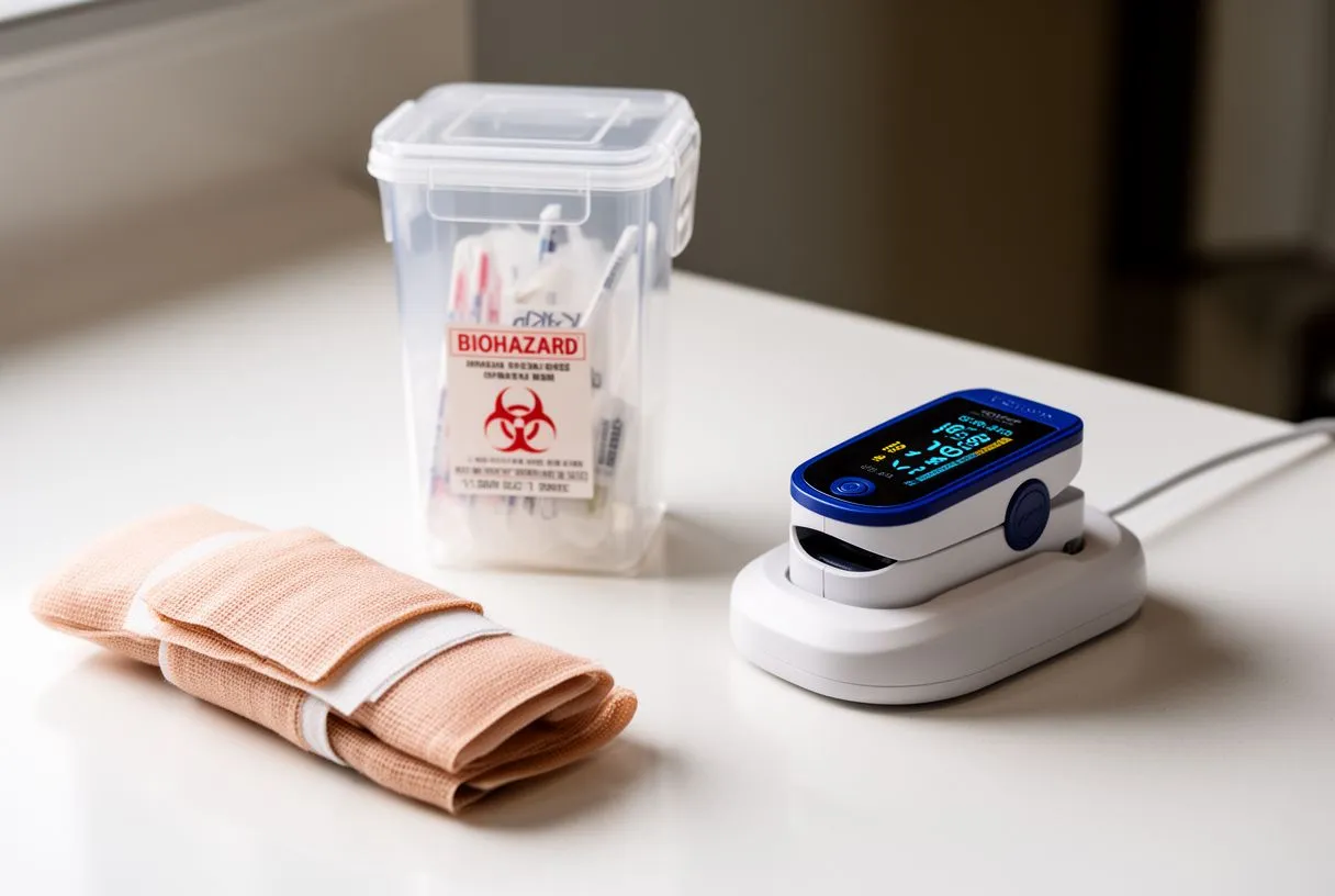
Most patients go home the same day. You should avoid heavy lifting, vigorous exercise, or driving for at least twenty-four hours. If you notice sudden swelling, severe pain, or continuous bleeding at the puncture site, or if you develop a fever, you should contact your medical team immediately. These are signs that warrant prompt professional evaluation to rule out complications.
Consulting your cardiac team in Mexico
Interpreting the results of a ventriculography heart function test requires deep clinical expertise. The numbers and images must be contextualized within your specific symptoms, medical history, and overall physical condition. This is why it is vital to have these conversations with a dedicated cardiac team rather than relying on internet searches. At Cirugía de Corazón, the practice of Dr Alejandro Molina in Guadalajara, Querétaro, and Hermosillo, we take the time to explain exactly what your imaging means for your daily life and long-term prognosis.

Making your next decisions: If your ventriculography reveals coronary blockages, we will discuss whether interventions like Balloon Angioplasty or bypass surgery represent the lowest risk for your situation. If your heart function is diminished but stable, the focus is usually on medical management strategies proven to improve quality of life. We provide realistic recovery timelines and honest discussions about expected outcomes, avoiding false reassurances while maintaining a calm, supportive environment for you and your family.
Your heart health is a deeply personal journey, and accurate diagnostics are the map that guides it. We encourage you to bring your questions, your fears, and your test results to a consultation. By sitting down with a qualified cardiac team, you can understand your condition fully and make informed, confident decisions about your care. Please book a consultation with our specialists to discuss your specific situation and determine the most appropriate path forward for your heart health.