Aortic Stenosis: Symptoms, Diagnosis and Treatment Options
Explore aortic stenosis treatment options, symptoms, and diagnosis.
Understanding cardiac health is crucial, especially when dealing with structural changes that affect blood flow. Aortic stenosis is one such condition, characterized by the narrowing of the heart's primary exit valve. This comprehensive guide explores the symptoms, diagnosis, and the most effective aortic stenosis treatment options available today, helping patients and caregivers make informed decisions about heart valve disease management.
What is aortic stenosis?
Defining valve obstruction: Aortic stenosis occurs when the aortic valve fails to open fully, restricting blood flow from the left ventricle to the aorta. The aortic valve is located at the Aortic Annulus, the structural foundation anchoring the valve leaflets. When these leaflets become thickened or calcified, they lose their flexibility. The outermost layer of the vessel wall, known as the Adventitia, provides structural support to the aorta, but the problem originates at the valve leaflets themselves.
Understanding the hemodynamic impact: As the valve narrows, the heart must generate significantly higher pressure to eject blood. Over time, this pressure overload causes the left ventricle to thicken, a condition known as concentric hypertrophy. While this initially helps the heart maintain adequate cardiac output, the thickened muscle requires more oxygen and eventually loses its ability to relax and fill properly, leading to diastolic dysfunction.
The gradual progression of this narrowing means that many patients remain asymptomatic for years. The valvular obstruction forces the myocardium to work harder against a fixed resistance, setting the stage for eventual heart failure if left untreated. Early detection and continuous monitoring are essential to intervene before irreversible myocardial damage occurs, ensuring the best possible outcomes for long-term cardiac health.
What causes aortic valve narrowing?
Age-related calcification: The most common cause of aortic stenosis is the buildup of calcium deposits on the valve leaflets over time. This degenerative process typically manifests in older adults as the valve endures decades of mechanical stress. The calcium deposits restrict leaflet motion, gradually narrowing the valve opening and restricting forward blood flow into the systemic circulation.
Congenital and rheumatic origins: Some individuals are born with a bicuspid aortic valve, which has only two leaflets instead of three. This structural anomaly accelerates wear and tear, leading to premature calcification and stenosis. Additionally, rheumatic fever, a complication of untreated strep throat, can cause scarring and fusion of the leaflets. Issues extending into the Aortic Arch can sometimes complicate the overall hemodynamic profile, though the primary narrowing remains at the valve itself.
In rare instances, patients may develop acute narrowing as part of a broader Acute Aortic Syndrome, though this is usually secondary to sudden structural failures rather than the slow, calcific process typical of the disease. Inflammatory conditions and radiation therapy to the chest can also contribute to valve fibrosis. Identifying the exact etiology helps clinicians predict disease progression and determine the optimal timing for surgical or transcatheter intervention.
What are common aortic stenosis symptoms?
Recognizing classic clinical signs: The hallmark symptoms of severe aortic stenosis include exertional chest pain (angina), unexplained syncope (fainting), and congestive heart failure symptoms like shortness of breath. Patients often experience fatigue and reduced exercise tolerance because the heart cannot increase cardiac output during physical activity. Sometimes, concurrent conditions like Anaemia can exacerbate these symptoms by further reducing oxygen delivery to the struggling myocardium.
Identifying late-stage manifestations: As the disease progresses and heart failure develops, patients may notice fluid retention. Ankle Swelling is a common physical finding, indicating right-sided heart congestion secondary to the left-sided obstruction. The heart's inability to pump efficiently causes blood to back up into the venous system, leading to peripheral edema. Managing these symptoms often requires careful fluid balance and may involve adjunctive medications like Amiodarone to control concurrent arrhythmias.

Because symptoms often develop insidiously, patients may unconsciously limit their physical activity to avoid discomfort, delaying clinical presentation. This is why a high index of suspicion is necessary for older adults presenting with new-onset exercise intolerance. Careful evaluation of any new heart murmur or unexplained dyspnea is critical, as timely diagnosis dramatically alters the clinical trajectory and overall survival rates.
How is aortic stenosis diagnosed?
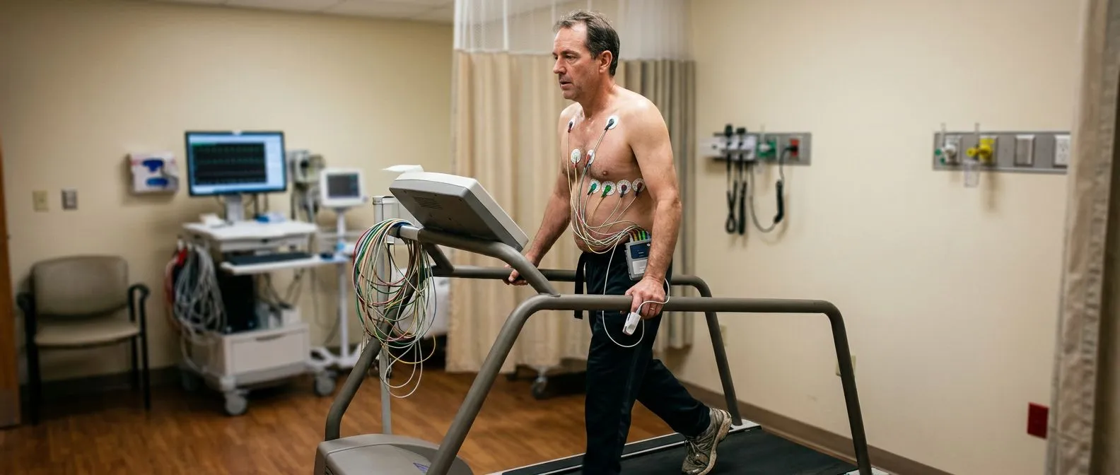
Utilizing non-invasive imaging: The primary diagnostic tool for aortic stenosis is transthoracic echocardiography. This ultrasound exam provides detailed visualization of the valve morphology, degree of calcification, and blood flow velocities. A critical measurement taken during an Annual Echocardiogram is the transvalvular gradient and the calculated aortic valve area. Annulus Sizing is also performed during this phase to prepare for potential future valve replacement procedures.
Assessing clinical severity: Beyond echocardiography, clinicians may use cardiac catheterization to directly measure the pressure gradient across the valve, particularly if non-invasive tests yield inconclusive results. According to the American College of Cardiology, comprehensive guidelines recommend a thorough evaluation that includes symptom assessment, as asymptomatic severe stenosis is managed differently than symptomatic disease. acc.org emphasizes the importance of standardized hemodynamic criteria to confirm the severity.
Additional imaging modalities like cardiac MRI or CT scans are increasingly used to provide cross-sectional views of the heart and great vessels. These advanced tools help assess ventricular function, myocardial fibrosis, and the extent of valvular calcification. Establishing an accurate diagnosis and severity grading is the foundation for developing an effective, patient-centered treatment strategy.
What are the stages of severe aortic stenosis?
Categorizing disease progression: The staging of aortic stenosis is based on valve anatomy, hemodynamics, and the presence of symptoms. Stage A includes patients with risk factors or mild valve thickening but no obstruction. Stage B involves mild to moderate obstruction, while Stage C represents severe obstruction that may still be asymptomatic. Progression to symptomatic disease drastically increases the risk of All-cause Mortality if left untreated.
Defining symptomatic thresholds: Stage D1 refers to classic symptomatic severe stenosis, where patients experience exertional dyspnea, angina, or syncope. Stage D2 involves low-flow, low-gradient stenosis with reduced ejection fraction, where the weakened heart muscle cannot generate high pressures across the valve. Managing these advanced stages often requires careful hemodynamic support, sometimes utilizing an Angiotensin Receptor Blocker to manage hypertension without causing excessive afterload reduction.

Understanding the distinction between these stages is vital because the treatment algorithm shifts dramatically once symptoms appear. Asymptomatic patients are typically monitored closely, whereas symptomatic patients require prompt intervention to relieve the mechanical obstruction. The transition from compensated to decompensated disease marks a critical turning point where medical therapy alone is insufficient, and valve replacement becomes medically necessary.
What are aortic stenosis treatment options?
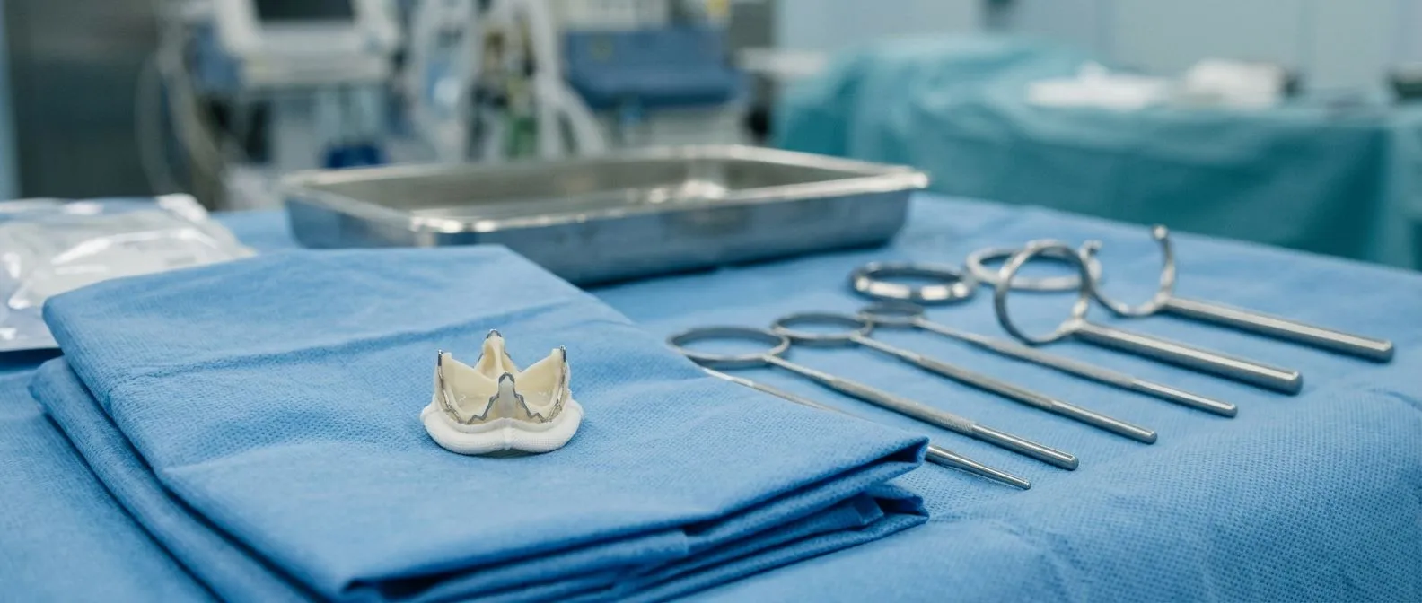
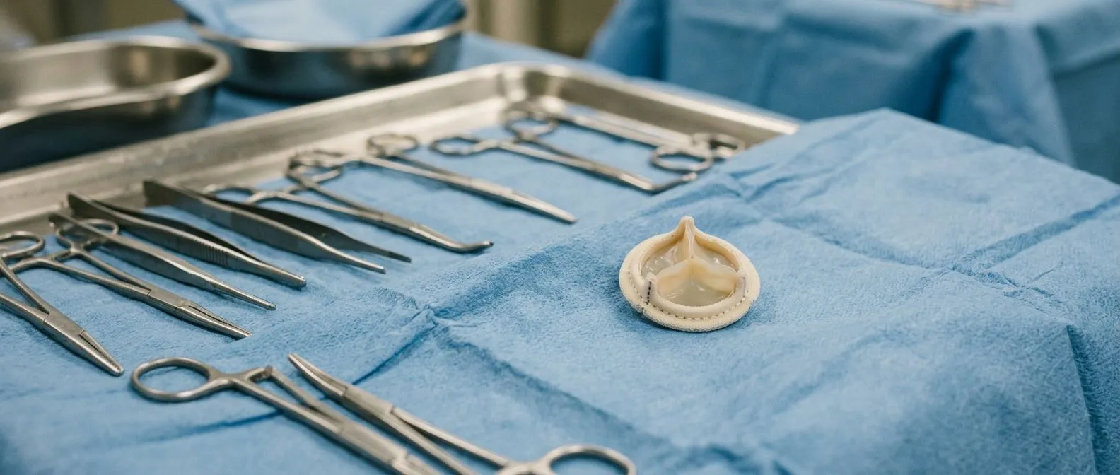
Evaluating surgical versus transcatheter approaches: The definitive treatment for severe symptomatic aortic stenosis is valve replacement. Options include surgical aortic valve replacement (SAVR) and transcatheter aortic valve replacement (TAVR). For patients with concurrent regurgitation, a repair procedure known as Annuloplasty may be considered, often utilizing an Annuloplasty Ring to stabilize the valve base. However, for true calcific stenosis, complete valve replacement is almost always required.
Selecting the appropriate intervention: The choice between SAVR and TAVR depends on surgical risk, patient anatomy, and life expectancy. Medical therapy remains crucial for symptom management and optimizing hemodynamics before intervention. Anticoagulation strategies are also vital, as highlighted in the COVID-19 and Thrombotic or Thromboembolic Disease: Implications for Prevention, Antithrombotic Therapy, and Follow-Up: JACC State-of-the-Art Review, which can be referenced at europepmc.org. Utilizing tools like home blood pressure monitors allows patients to track their cardiovascular stability effectively at home.
While no medication can reverse the mechanical obstruction of calcific stenosis, pharmacological management targets the secondary effects of the disease. Diuretics help relieve fluid overload, while beta-blockers and ACE inhibitors manage hypertension and reduce cardiac workload. Ultimately, the goal of medical therapy is to bridge the patient safely to a definitive mechanical solution, ensuring the heart is in the best possible condition to undergo the replacement procedure.
How does the TAVR procedure work?
Understanding the minimally invasive technique: The TAVR procedure offers a less invasive alternative to open-heart surgery. A biological valve mounted on a stent is compressed onto a catheter delivery system. This catheter is usually inserted through the femoral artery and advanced up to the heart. During this navigation, maintaining Antegrade Cerebral Perfusion is critical to prevent neurologic complications, particularly if adjunctive cerebral protection devices are deployed.
Detailing valve deployment: Once the catheter reaches the diseased aortic valve, the new valve is expanded, crushing the old calcified leaflets outward against the aortic wall. The new valve immediately begins functioning, restoring normal blood flow from the left ventricle to the aorta. Recovery is typically rapid, with patients often mobilizing within 24 hours. Patients recovering at home may find continuous cardiac monitoring wearables useful for tracking arrhythmias during the initial healing phase.
Transcatheter approaches have revolutionized the treatment landscape, making valve replacement accessible to patients previously deemed too high-risk for traditional surgery. Ongoing advancements in valve design and delivery systems continue to expand the indications for TAVR, pushing the therapy toward lower-risk populations. However, careful pre-procedural imaging and annular sizing remain the cornerstones of procedural success, ensuring the new valve is appropriately seated without paravalvular leakage.
What to expect from aortic stenosis surgery?
Preparing for open-heart surgery: Traditional surgical aortic valve replacement requires a median sternotomy and the use of cardiopulmonary bypass. The surgeon excises the diseased valve and sutures a mechanical or biological prosthesis in its place. In complex cases, this may be combined with an Aortic Aneurysm Repair to address dilatation of the ascending aorta. Preoperative preparation often includes an Antimicrobial Shower to reduce surgical site infection risk.
Navigating postoperative care: Following surgery, patients spend time in the intensive care unit for close hemodynamic monitoring. The use of Antibiotic Prophylaxis is standard protocol to prevent endocarditis, particularly in patients with prosthetic valves. Mechanical valves offer superior durability but require lifelong anticoagulation to prevent thrombosis, whereas biological valves have a limited lifespan but do not mandate permanent blood thinners. The choice depends on the patient's age and bleeding risk.

Cardiac rehabilitation is a critical component of the postoperative recovery journey. Structured exercise programs help patients rebuild their physical stamina and confidence. While the surgery addresses the mechanical obstruction, the heart requires time to reverse the ventricular hypertrophy caused by years of pressure overload. With appropriate postoperative care and surveillance, surgical valve replacement provides excellent long-term outcomes and restores near-normal life expectancy.
How to manage heart valve disease treatment?
Implementing lifelong monitoring protocols: Regardless of the treatment chosen, long-term management of heart valve disease requires vigilant surveillance. Regular echocardiograms are necessary to assess valve function, monitor for structural deterioration, and evaluate ventricular recovery. The landscape of antithrombotic therapy is complex, and historical trials like AFASAK have informed current guidelines regarding anticoagulation, often utilizing medications like warfarin alongside Antiplatelet Therapy in specific patient subsets.
Addressing comorbidities and lifestyle factors: Effective management extends beyond the valve itself; controlling systemic blood pressure, managing cholesterol, and maintaining a heart-healthy lifestyle are paramount. The interplay between various cardiovascular risk factors is intricate. As noted in the research paper Detection of widespread horizontal pleiotropy in causal relationships inferred from Mendelian randomization between complex traits and diseases, available at ncbi.nlm.nih.gov, genetic factors intricately link multiple cardiovascular traits. Some patients choose to integrate heart health recovery supplements into their regimen, though this should be guided by a physician.
Self-care strategies play an indispensable role in stabilizing the condition and improving quality of life. Patients must remain attuned to changes in their exercise tolerance, weight, and breathing. Adherence to prescribed medications, especially anticoagulants if a mechanical valve was placed, is non-negotiable. A collaborative approach between the patient, cardiologist, and primary care physician ensures that both the mechanical and medical aspects of the disease are optimally balanced.
- Regular Check-ups: Schedule routine visits to monitor prosthetic valve function and detect early signs of deterioration.
- Dietary Control: Maintain a low-sodium diet to prevent fluid overload and reduce afterload stress on the heart.
- Supervised Exercise: Engage in physician-guided cardiac rehabilitation to safely improve cardiovascular endurance.
What is the aortic stenosis prognosis?
Understanding survival metrics: The prognosis for aortic stenosis depends entirely on the stage of diagnosis and the timeliness of intervention. Asymptomatic patients have a relatively favorable prognosis, with a low rate of sudden cardiac death. However, once symptoms like severe dyspnea or angina develop, the median survival without treatment drops significantly, often to just a few years. Prompt valve replacement dramatically alters this trajectory, returning survival rates closer to that of the general age-matched population.
Evaluating long-term outcomes: Both surgical and transcatheter valve replacements boast excellent success rates and symptom relief. The choice of procedure and valve type dictates the long-term management plan, particularly regarding the need for anticoagulation. Biological valves eventually degrade, requiring a future reintervention, while mechanical valves last a lifetime but carry a persistent bleeding risk due to blood thinners. Patients must weigh these trade-offs carefully with their healthcare team.

In summary, aortic stenosis is a progressive disease that transitions from a silent structural anomaly to a life-threatening obstruction. The key to a favorable prognosis lies in proactive screening and timely intervention before irreversible myocardial damage sets in. By understanding the causes, recognizing the symptoms, and exploring the full spectrum of treatment options, patients can effectively navigate their diagnosis and maintain a high quality of life.
- Early Diagnosis: Catching the disease in asymptomatic stages allows for planned monitoring and timely intervention.
- Intervention Timing: Replacing the valve immediately upon symptom onset prevents irreversible heart failure.
- Quality of Life: Post-treatment, most patients experience a dramatic return to normal activity and symptom relief.

About the author
Dr Alejandro Molina is a cardiovascular surgeon practising in Guadalajara, Querétaro and Hermosillo. He trained in cardiothoracic surgery at the Universidad de Guadalajara, completed advanced training in cardiac surgery, minimally invasive surgery, robotic surgery and structural interventions at the University of Montreal, and a fellowship in structural cardiology and TAVR at CHU Bordeaux, France. He performed the first robotic mitral valve repair in the Ángeles Health System and the first robotic heart surgery in Guadalajara.
447