Blood Cultures for Endocarditis: How Infection Is Confirmed
Blood cultures endocarditis testing confirms heart valve infection.
What Are Blood Cultures for Endocarditis?
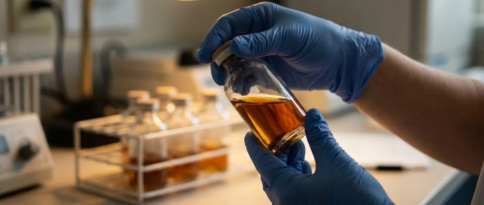
Blood cultures endocarditis testing is the cornerstone of diagnosing an infection of the heart's inner lining, particularly the valves. When bacteria or other microorganisms enter the bloodstream and settle on a heart valve, they can cause infective endocarditis, a condition that demands rapid identification of the responsible organism. A blood culture is essentially a laboratory test that takes a sample of your blood, places it in a nutrient-rich broth, and waits to see whether bacteria or fungi grow. This process allows your medical team to identify exactly which organism is causing the infection and which antibiotics will be most effective against it.
For patients and families hearing the word "endocarditis" for the first time, the anxiety is understandable. The heart valves are delicate structures — the aortic valve, the mitral valve, the tricuspid valve — and infection on these surfaces can damage them quickly. Blood cultures provide the evidence your cardiac team needs to move from suspicion to a concrete treatment plan. Without knowing which organism is present, doctors must rely on broad-spectrum antibiotics, which may not be the most targeted option for your specific infection.
Why blood cultures matter more than a single blood test: A routine blood test can show elevated inflammatory markers — such as C-reactive protein or white blood cell count — but it cannot tell you which organism is in your bloodstream. Blood cultures are the only test that grows and identifies the actual microbe. This distinction is critical because the choice of antibiotic, the duration of treatment, and the decision about whether surgery may be needed all hinge on that identification. In endocarditis, guessing is not good enough.
How Blood Cultures Endocarditis Testing Works
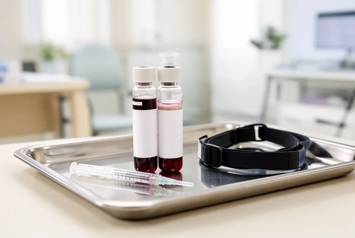
The process of drawing blood cultures is straightforward from the patient's perspective but highly structured behind the scenes. A nurse or phlebotomist cleans the skin thoroughly at the draw site — usually the arm — with an antiseptic solution to prevent contamination from skin bacteria. Then, using a sterile technique, they draw blood from two or three different vein sites, not just one. This multi-site approach is deliberate: if bacteria grow from only one bottle, it may be a contaminant from the skin, but if the same organism grows from multiple sites, it strongly suggests a true bloodstream infection.
Each blood draw fills two bottles: one designed to support aerobic organisms (those that need oxygen) and another for anaerobic organisms (those that do not). The bottles are sent to the laboratory and placed into an incubated system that monitors them continuously. Most clinically significant organisms grow within 24 to 48 hours, but some — particularly fastidious or slow-growing bacteria — may take five days or longer. The laboratory will hold the cultures for a minimum of five days before reporting them as negative.
What happens after bacteria grow: When an organism appears, the laboratory performs identification and susceptibility testing. Identification tells your team exactly what the bacteria or fungus is — for example, Streptococcus viridans, Staphylococcus aureus, or Enterococcus. Susceptibility testing, also called an antibiotic sensitivity panel, reveals which antibiotics kill that specific organism and which ones it resists. This information is what allows your cardiologist and infectious disease specialist to tailor your treatment precisely. According to The Lancet, timely and accurate blood culture results are central to reducing mortality in infective endocarditis, as delays in appropriate therapy worsen outcomes significantly.
When Doctors Order Blood Cultures for Endocarditis
Doctors order blood cultures endocarditis testing when a patient shows signs that suggest bacteria may have entered the bloodstream and settled on a heart valve. The classic scenario is a patient with a known heart valve condition — such as a bicuspid aortic valve, a prosthetic valve, or a history of rheumatic valve disease — who develops a persistent fever without another obvious cause. But endocarditis can also occur in patients with no prior valve problems, so the threshold for ordering cultures is deliberately low when clinical suspicion exists.
The symptoms that prompt blood cultures can be subtle or dramatic. A low-grade fever lasting days or weeks, night sweats, unexplained fatigue, and weight loss may be the only early signs. More advanced infection can produce new heart murmurs detected during auscultation — the clinical term for listening to the heart with a stethoscope — as well as small spots on the skin, the retina, or under the fingernails caused by tiny clumps of bacteria called vegetations breaking off from the valve. When a patient arrives with a prosthetic valve and a fever, the standard of care is to draw blood cultures before starting antibiotics, unless the patient is critically unstable.
Other situations that trigger blood cultures: Patients who have had recent dental procedures, intravenous drug use, or indwelling catheters are at higher risk. After cardiac surgery, a new fever in the first weeks may also prompt cultures to rule out infection of the surgical site or the valve itself. Your cardiac team will weigh the full clinical picture — your valve history, your symptoms, your blood test results — and decide whether the evidence supports drawing cultures immediately or monitoring first.
Endocarditis Diagnosis Criteria and Blood Culture Results
The diagnosis of infective endocarditis is not based on blood cultures alone. Cardiologists use a structured framework called the modified Duke criteria, which combines blood culture results with imaging findings and clinical evidence. This system awards points for major criteria and minor criteria, and the total determines whether the diagnosis is definite, possible, or rejected. Blood cultures play a central role: the presence of a typical endocarditis-causing organism in two separate blood cultures is a major criterion, as is evidence of endocardial involvement on echocardiography.
The Duke criteria matter because endocarditis can mimic other conditions. A patient with atrial flutter — an abnormal heart rhythm originating in the atrium, the upper chamber of the heart — might have palpitations and fatigue that overlap with early endocarditis symptoms. A patient with a bloodstream infection from another source, such as a urinary tract infection, could have positive blood cultures without heart valve involvement. The criteria force clinicians to consider the whole picture rather than a single test result.
How Duke criteria categorize your case: A "definite" diagnosis requires either two major criteria, one major and three minor criteria, or five minor criteria. A "possible" diagnosis meets the threshold for one major and one minor criterion, or three minor criteria. Blood culture results feed directly into both major criteria (typical organism from two cultures) and minor criteria (predisposing heart condition, fever above 38°C, vascular phenomena). Your cardiac team will explain where your case falls on this scale and what additional tests, such as echocardiography, are needed to solidify the diagnosis.
What Positive Blood Cultures Endocarditis Results Mean
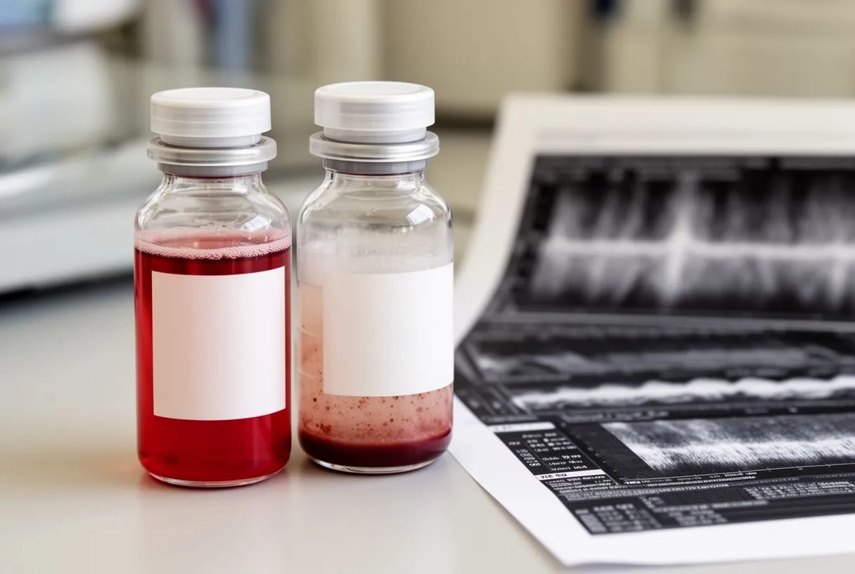
Positive blood cultures endocarditis results mean that bacteria or fungi have been found growing in your bloodstream. In the context of endocarditis, this finding is significant because it confirms that microorganisms are circulating in the blood — a condition called bacteremia when bacteria are involved, or fungemia when fungi are the cause. However, a single positive culture does not automatically mean you have endocarditis. Your team will look at the pattern: the same organism growing in multiple cultures drawn from different sites, over time, is far more meaningful than a single positive bottle.
The specific organism identified also shapes what happens next. Staphylococcus aureus, for example, is aggressive and has a high propensity to attach to heart valves; its presence in blood cultures strongly raises suspicion for endocarditis even before imaging is complete. Viridans group streptococci, which normally live in the mouth, are classic causes of endocarditis following dental procedures. Enterococci are associated with gastrointestinal and urinary tract sources. Each organism carries a different antibiotic resistance profile, a different expected duration of treatment, and a different likelihood that valve surgery will be needed.
Contaminant versus true infection — how your team tells the difference: Skin bacteria such as coagulase-negative staphylococci can contaminate a blood culture if the skin preparation is imperfect. When these appear in only one bottle or one draw site, they are often dismissed as contaminants. But in a patient with a prosthetic valve, even coagulase-negative staphylococci can be a true pathogen. Your team considers the organism, the number of positive cultures, the timing, and your clinical picture. As researchers noted in a study on wearable cardioverter-defibrillator use in suspected myocarditis, distinguishing true cardiac infection from other conditions requires integrating multiple diagnostic threads rather than relying on any single test.
Blood Culture Endocarditis Guidelines and Protocols
Blood culture endocarditis guidelines are designed to maximize the chance of catching the organism while minimizing false results. International consensus statements — from organizations such as the European Society of Cardiology and the American Heart Association — recommend drawing at least two sets of blood cultures from separate venipuncture sites before starting antibiotic therapy. Each set typically consists of one aerobic and one anaerobic bottle. The reasoning is statistical: drawing more cultures increases the probability of detecting low-level bacteremia, and using multiple sites helps distinguish true infection from skin contamination.
The timing of the draws also matters. Ideally, cultures are spaced at least 30 minutes apart, which helps demonstrate persistent bacteremia — a hallmark of endocarditis — rather than a transient shower of bacteria from a brief event. However, in a patient who is acutely septic or hemodynamically unstable, the team will draw cultures rapidly and start antibiotics immediately; the priority shifts from perfect timing to saving the patient's life. In subacute presentations, where the patient has been febrile for days or weeks, there is time to follow the full protocol.
- Minimum two sets: Drawn from two separate venipuncture sites before antibiotics whenever possible.
- Spaced timing: At least 30 minutes between sets to demonstrate continuous bacteremia typical of endocarditis.
- Two bottle types per set: One aerobic and one anaerobic bottle to capture organisms with different oxygen requirements.
- Five-day incubation: Cultures are held for a minimum of five days to catch slow-growing or fastidious organisms before reporting as negative.
The volume of blood drawn is another critical but often overlooked detail. Adult guidelines call for approximately 20 to 30 milliliters per culture set — typically two bottles of 8 to 10 milliliters each. Too little blood reduces the sensitivity of the test, potentially missing a low-grade infection. Pediatric volumes are adjusted by weight. Your cardiac team and the laboratory work together to ensure the protocol is followed precisely because every detail affects the reliability of the results you and your family will rely on for treatment decisions.
How to Confirm Endocarditis Beyond Blood Cultures
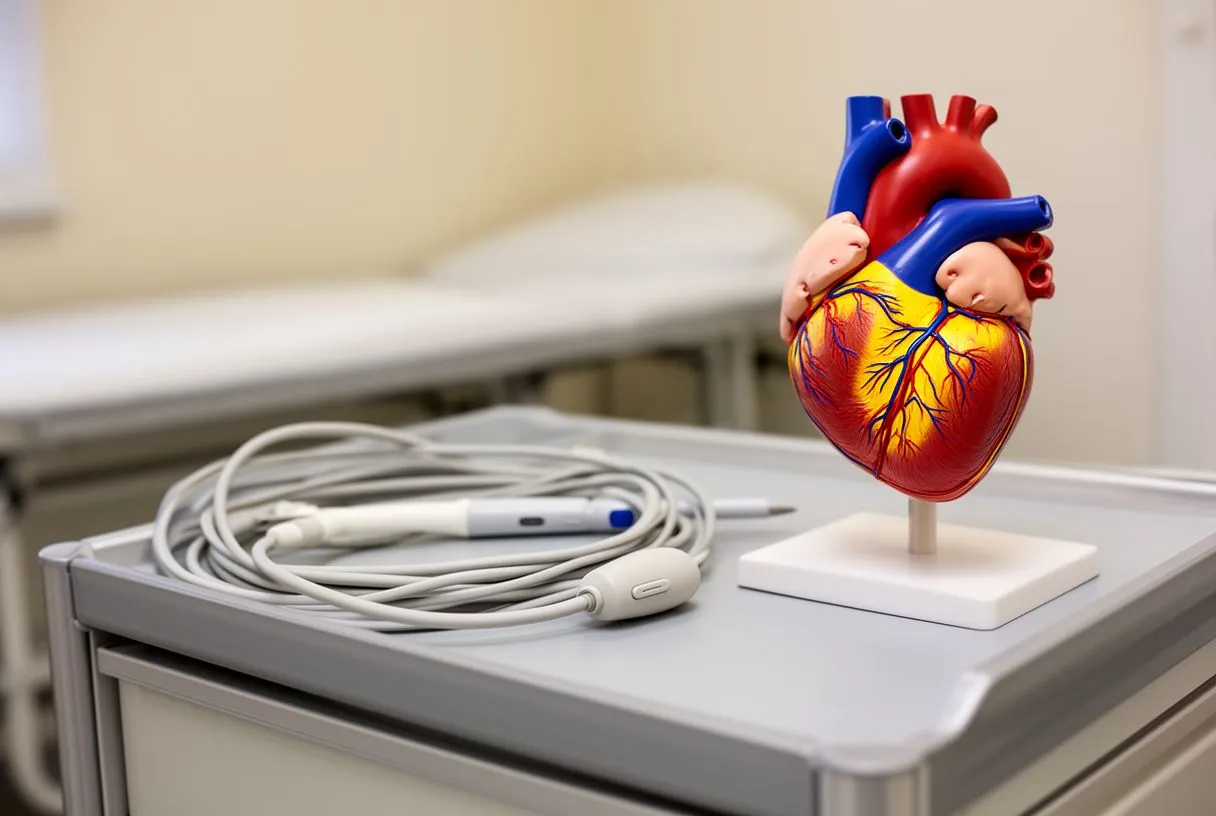
Blood cultures are essential, but they are one piece of a larger diagnostic mosaic. To confirm endocarditis, your cardiac team will also perform echocardiography — an ultrasound of the heart that can visualize the valves directly. A transthoracic echocardiogram, done by placing the probe on the chest wall, is the first-line imaging test. It is noninvasive and can often detect large vegetations — the clumps of bacteria and cellular debris attached to a valve. If the transthoracic study is inconclusive or if you have a prosthetic valve, the next step is a transesophageal echocardiogram, which provides much more detailed images by passing a probe down the esophagus to view the heart from behind.
Echocardiography looks for three specific findings: vegetations on the valve, abscesses (pockets of infection) in the surrounding tissue, and new partial dehiscence of a prosthetic valve — meaning the valve has loosened from its sewing ring. Any of these findings, combined with positive blood cultures, strengthens the diagnosis substantially. The ascending aorta, the first segment of the body's largest artery as it leaves the heart, and the aortic valve are common sites for infection-related complications, including abscesses that may require surgical drainage or valve replacement.
When imaging and cultures disagree: Sometimes blood cultures are negative but echocardiography shows clear vegetations. This scenario, called culture-negative endocarditis, can occur if antibiotics were started before the cultures were drawn, or if the organism is difficult to grow in standard laboratory media. In these cases, your team may order specialized molecular tests — such as polymerase chain reaction (PCR) assays — or serological tests that detect antibodies against specific organisms. The diagnosis does not collapse because one test is negative; it adapts.
Endocarditis Lab Tests That Complement Blood Cultures
Beyond blood cultures, several laboratory tests help your cardiac team assess the severity and consequences of endocarditis. A complete blood count may reveal an elevated white blood cell count, indicating active infection, or anemia, which can develop as a chronic inflammatory response. C-reactive protein and erythrocyte sedimentation rate are nonspecific inflammatory markers, but their trends over time are useful: falling levels suggest the antibiotic regimen is working, while persistently elevated levels may signal inadequate treatment or a complication such as an abscess.
Renal function tests are important because endocarditis can damage the kidneys through immune complex deposition — antibody clusters that lodge in the filtering units of the kidney. Liver function tests may also be affected, particularly in right-sided endocarditis involving the tricuspid valve. A urinalysis can reveal microscopic blood or protein, another sign of immune-mediated kidney injury. Electrolyte panels help monitor for disturbances caused by fever, poor oral intake, or antibiotic-related side effects.
- Complete blood count (CBC): Detects leukocytosis and anemia that accompany active endocarditis.
- CRP and ESR: Inflammatory markers tracked over time to gauge treatment response and detect complications.
- Renal and liver panels: Identify organ damage from immune complexes or embolic events during infection.
- Electrocardiogram (ECG): May show conduction abnormalities if an abscess extends near the heart's electrical pathways.
An electrocardiogram is a simple but important test in suspected endocarditis. If an aortic valve abscess extends into the tissue near the heart's electrical conduction system, it can produce a prolonged PR interval or even heart block — a dangerous slowing of the electrical signal between the atrium and the ventricles. Detecting these changes early can prompt urgent surgical intervention before the abscess causes complete heart block or structural catastrophe. As investigators discussed in a report on lenalidomide-induced myocarditis, cardiac infections and drug-induced cardiac inflammation share the principle that timely, multi-test diagnosis is essential for preventing irreversible damage.
Interpreting Endocarditis Blood Culture Results With Your Cardiac Team
Interpreting blood cultures endocarditis results is a collaborative process between you, your cardiologist, your infectious disease specialist, and sometimes your cardiac surgeon. The laboratory report will list the organism identified, the antibiotic susceptibility profile, and any resistance patterns. Your team will use this information to select the antibiotic — or combination of antibiotics — that will most effectively eradicate the organism from your bloodstream and from the vegetation on your valve. This choice is not always simple: some organisms are resistant to first-line drugs, and some require prolonged intravenous therapy lasting four to six weeks or more.
The duration of treatment is driven by the organism, the location of the infection, and whether the valve is native or prosthetic. Native valve endocarditis caused by highly susceptible streptococci may be treated with a four-week course, while staphylococcal prosthetic valve endocarditis often requires six weeks or longer, frequently with combination therapy. If you have a prosthetic valve and develop endocarditis, the treatment algorithm changes substantially because the foreign material of the valve is harder to sterilize than living tissue, and surgical replacement may be necessary if the infection does not respond promptly to antibiotics.
Questions to ask your cardiac team about your results: Ask which organism was identified and what its resistance profile means for your treatment. Ask how long the antibiotic course will be and whether it requires a peripherally inserted central catheter (PICC line) for home intravenous therapy. Ask whether imaging has shown vegetation size and whether the valve is leaking or obstructed. Ask what signs would prompt consideration of surgical intervention — such as new heart failure symptoms, large mobile vegetations, or persistent fever despite appropriate antibiotics. These conversations are the bridge between laboratory data and the decisions that shape your recovery.
Blood Cultures for Heart Infection: Next Steps After Diagnosis
Once blood cultures confirm endocarditis and antibiotic therapy is underway, the focus shifts to monitoring response and planning the full course of treatment. Your cardiac team will repeat blood cultures after starting antibiotics — typically within 48 to 72 hours — to confirm that the bacteremia has cleared. If cultures remain positive despite appropriate therapy, this is called persistent bacteremia, and it raises concern about inadequate source control, meaning the antibiotic cannot reach the organism effectively because it is sequestered in the vegetation or an abscess.
Throughout treatment, you will be monitored for complications. Embolic events — pieces of vegetation breaking off and traveling to the brain, lungs, spleen, or limbs — can occur even after antibiotics begin, though the risk falls significantly within the first two weeks of effective therapy. New neurological symptoms, sudden arm pain from an arterial embolus, or abdominal pain from a splenic infarct should be reported immediately. If you take aspirin for another cardiac condition, your team will review whether to continue it during active endocarditis, as bleeding risk may increase. Some patients may require a procedure such as balloon angioplasty for coronary complications, though this is less common in endocarditis than in atherosclerotic disease.
Looking ahead — recovery and long-term follow-up: After completing the antibiotic course, your team will arrange follow-up echocardiography to assess valve function and vegetation resolution. If the valve was damaged, you may need ongoing surveillance for progressive regurgitation or stenosis, and some patients will eventually require valve repair or replacement. If you had a blood culture endocarditis episode on a native valve, the risk of recurrence is real, and your team will discuss preventive strategies — including antibiotic prophylaxis before certain dental and medical procedures. The goal is not only to treat the current infection but to protect your heart for the decades ahead.
If you or a loved one is experiencing symptoms that could point to endocarditis — persistent fever, new heart murmur, unexplained fatigue, or night sweats — do not wait. Blood cultures are most informative when drawn before antibiotics are started, so timing matters. We encourage you to discuss your symptoms and your full cardiac history with a qualified cardiac team. At Cirugía de Corazón, Dr Alejandro Molina and his team in Guadalajara, Querétaro, and Hermosillo are available to evaluate your situation, order the appropriate tests, and guide you through every step of diagnosis and treatment. Book a consultation rather than self-diagnosing — endocarditis is a condition where early, expert intervention can make a decisive difference.