

Cardiac Ultrasound: What a Heart Scan Shows
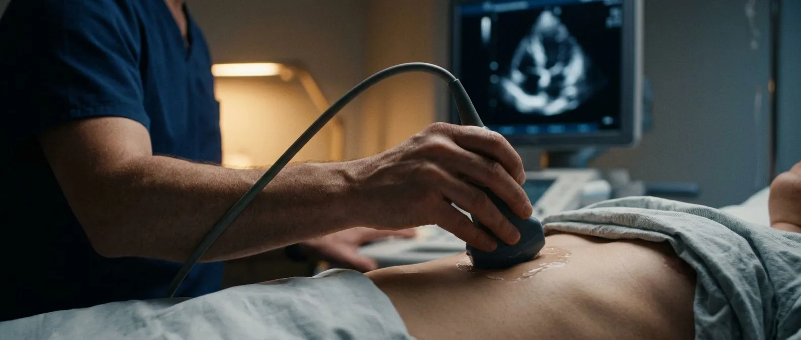
A cardiac ultrasound scan reveals valve function, blood flow, and heart muscle health in real-time.
A cardiac ultrasound serves as a vital window into the body's most important muscle, offering real-time images without using harmful radiation. For families facing the uncertainty of heart conditions, understanding exactly what this test reveals can bring immense clarity and peace of mind. This non-invasive imaging tool, also known as an echocardiogram, uses safe sound waves to capture moving pictures of your heart's chambers, valves, and blood flow. This guide walks you through precisely what to expect, how the technology works, and why it is a cornerstone of cardiac care.
What Is a Cardiac Ultrasound Scan and How Does It Work?
Understanding the basic technology: A cardiac ultrasound scan relies on high-frequency sound waves to create detailed, moving pictures of your beating heart. During a standard physical exam, your doctor might use auscultation—listening to your heart sounds through a stethoscope—to detect irregular rhythms or muffled valves. While this manual method is useful for initial screening, an ultrasound provides definitive visual proof of what is causing those sounds, allowing cardiologists to see the heart in action and diagnose structural issues with high precision.
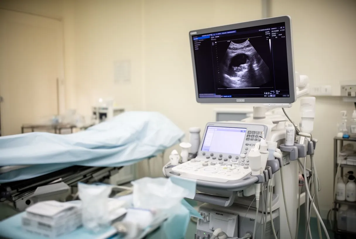
The role of the transducer and gel: A trained sonographer will apply a warm gel to your chest and glide a small device called a transducer over your skin. This probe emits sound waves that bounce off the dense tissues of your heart and return to the device, where a computer instantly translates these echoes into a live video feed. The gel is essential because it eliminates trapped air between the skin and the probe, ensuring the sound waves travel seamlessly into your body to produce clear, accurate images.
What the experience feels like: The procedure is entirely comfortable and non-invasive, typically lasting between thirty and forty-five minutes. You will lie on an examination table on your left side while the sonographer captures images from different angles around your chest. You will not feel the sound waves, though you may feel slight pressure from the probe as the technician presses firmly against your ribs to get a clearer view of the heart's lower wall.
What Does a Heart Scan Show About Valve Function?
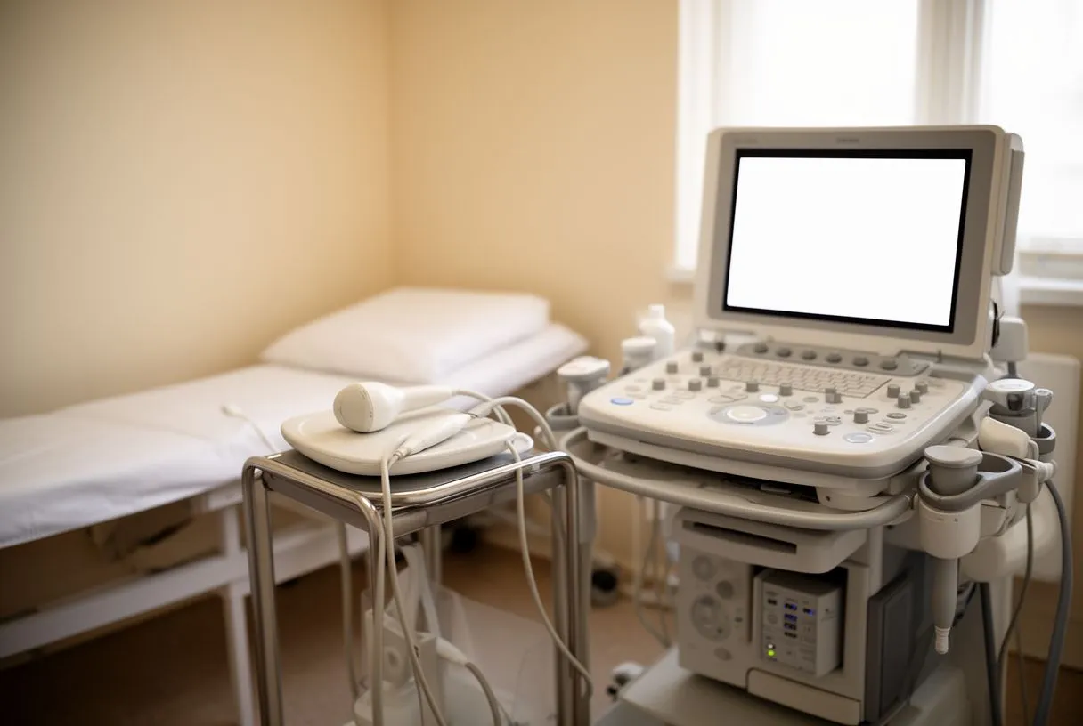
Identifying stenosis and regurgitation: One of the primary reasons a doctor orders a heart ultrasound is to evaluate the four main valves that control blood flow through the heart. The scan reveals if a valve has become stenotic, meaning it is stiff and narrowed, restricting blood from moving forward. It also shows regurgitation, where a valve does not close tightly, allowing blood to leak backward. Identifying these dysfunctions early is crucial to preventing long-term damage to the heart muscle.
Assessing the aortic valve and surrounding vessels: The ultrasound provides a clear view of the aortic valve and the Ascending Aorta, the major blood vessel carrying oxygen-rich blood to the body. If a patient has severe aortic stenosis, the scan helps determine if they might benefit from a Balloon Aortic Valvuloplasty, a procedure that temporarily widens a narrowed valve, or if they require a more permanent solution like a transcatheter aortic valve replacement (TAVR) using a Balloon-expandable Valve. According to ahajournals.org, accurate valve assessment is critical for planning these complex interventions.
Planning the next steps for valve disease: Once the ultrasound confirms the presence and severity of valve disease, your cardiac team can develop a tailored treatment plan. The images dictate whether a condition can simply be monitored over time, managed with medication, or requires surgical repair. By seeing exactly how the valves open and close with every heartbeat, surgeons can prepare for the specific anatomical challenges they will face in the operating room.
How a Heart Ultrasound Evaluates Heart Muscle Health
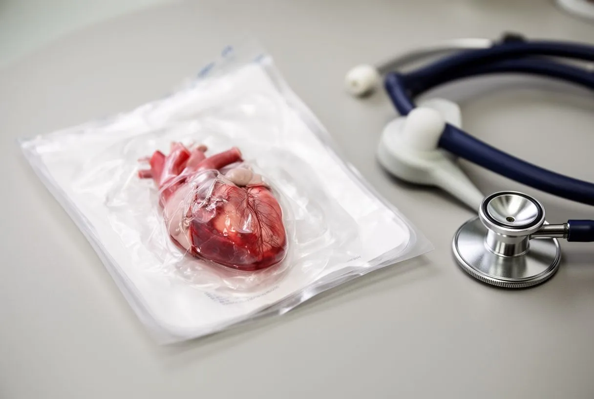
Measuring ejection fraction: Beyond the valves, a heart ultrasound is the gold standard for evaluating the pumping strength of the heart muscle. The scan calculates your ejection fraction, which is the percentage of blood that the left ventricle pumps out with each contraction. A normal ejection fraction typically ranges from fifty to seventy percent, and a reading below this threshold suggests that the heart muscle has weakened, a condition known as heart failure.
Detecting wall thickness and enlargement: The ultrasound measures the thickness of the heart's muscular walls. High blood pressure forces the heart to pump against greater resistance, causing the muscle to thicken abnormally, a condition called hypertrophy. The scan also captures the size of each Atrium—the upper chambers of the heart—and the ventricles. If a chamber is enlarged, it indicates that the heart has been stretching to accommodate excess blood volume or compensating for a leaky valve over a long period.
Evaluating damage from previous events: If a patient has suffered a heart attack in the past, the ultrasound will reveal areas of scar tissue on the heart muscle. These scarred areas do not contract normally, and identifying them helps the cardiologist understand the extent of the damage. This detailed mapping of muscle function is essential for determining a patient's prognosis and choosing the right combination of therapies to support the remaining healthy tissue.
Types of Cardiac Ultrasound: 2D, 3D, and Doppler Echo Tests
Standard 2D versus advanced 3D imaging: When exploring types of cardiac ultrasound, the 2D echocardiogram provides flat, slice-like images of the heart, which are highly effective for routine assessments. However, a 3D heart ultrasound captures a three-dimensional model, offering a surgeon a complete spatial understanding of complex structures before an operation. This advanced view is particularly useful when planning intricate valve repairs or when evaluating congenital defects where spatial relationships are distorted.
The power of Doppler technology: Doppler ultrasound is often integrated into standard echo tests to track the speed and direction of blood flow. This technology is invaluable for detecting turbulent blood flow, which appears as color patterns on the monitor. Doppler can definitively show the jet of blood leaking backward through a diseased valve and helps diagnose conditions like Atrial Flutter by visualizing how blood moves through the atria during rapid, irregular electrical signals.
Choosing the right test for your symptoms: The specific type of echo test ordered depends entirely on your symptoms and medical history. A resting echo might be sufficient for a general checkup, but if your symptoms only occur during exertion, a stress echo is required. Your cardiologist will select the combination of 2D, 3D, and Doppler techniques that provides the lowest risk for your situation while yielding the most actionable clinical data.
- 2D Echocardiogram: Provides flat, moving pictures of the heart used for basic structural and functional assessments.
- 3D Heart Ultrasound: Offers a detailed, three-dimensional view, helping surgeons plan complex structural interventions.
- Doppler Ultrasound: Tracks the speed and direction of blood flowing through the heart's chambers and valves.
What to Expect During a Transesophageal Echocardiogram
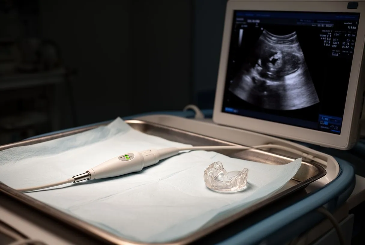
Why a closer view is sometimes needed: Sometimes, the chest wall, ribs, or lungs block the sound waves of a standard ultrasound, resulting in blurry images. In these cases, a transesophageal echocardiogram (TEE) is performed. Because the esophagus sits directly behind the heart, a specialized ultrasound probe passed down the throat provides remarkably clear, unobstructed images of the heart's posterior structures, making it the superior choice for detecting small blood clots or precise valve leaks.
The sedation and insertion process: Before the procedure, your throat is numbed with a spray to suppress your gag reflex, and a mild sedative is administered through an IV to help you relax. You will be conscious but drowsy as the cardiologist gently guides the thin, flexible tube containing the transducer down your esophagus. While the idea of swallowing a tube sounds uncomfortable, the sedation ensures the process is smooth, and most patients have no memory of the insertion afterward.
Recovery and immediate results: The actual imaging portion of a TEE takes about fifteen to twenty minutes, after which the probe is carefully removed. You will rest in a recovery area until the sedation wears off and the numbing in your throat subsides. Because your swallowing reflex will be temporarily impaired, you must avoid eating or drinking for an hour or two. The physician can often discuss the preliminary results with you before you leave the clinic.
Understanding the Stress Echo Test for Heart Disease
Combining exercise with imaging: A stress echo test is designed to evaluate how your heart performs under the increased demand of physical exertion. The test begins with a resting ultrasound to capture baseline images of your heart. Next, you will walk on a treadmill or pedal a stationary bicycle, increasing the intensity gradually. If you cannot exercise due to physical limitations, a medication is administered intravenously to safely simulate the effects of exercise on your heart.
Identifying hidden coronary artery disease: Immediately after your heart rate reaches its target peak, you will quickly lie down for a second ultrasound. By comparing the resting and stressed images, cardiologists can spot segments of the heart muscle that are not contracting properly. This lack of movement during stress often points to significant Atheromatous Plaque buildup in the coronary arteries, restricting blood flow to the muscle. If you experience symptoms like Arm Pain during the test, it further supports a diagnosis of ischemic heart disease.
From diagnosis to interventional planning: The results of a stress echo are critical for deciding whether you need further invasive testing or if your condition can be managed with medication. If the test shows severe blood flow restriction, your doctor may recommend a catheterization procedure like Balloon Angioplasty to open the blocked artery. Throughout this high-stress testing, medical teams are always prepared with an Automated External Defibrillator on standby to ensure your absolute safety in the rare event of a dangerous arrhythmia. When assessing patients for these procedures, clinical teams also reference broader systemic guidelines, such as the Global Strategy for the Diagnosis, Management, and Prevention of Chronic Obstructive Pulmonary Disease, to ensure the lungs can tolerate the cardiovascular stress of interventions.
Pediatric and Fetal Heart Ultrasound: What Parents Need to Know
Detecting congenital defects early: A pediatric echocardiogram is a specialized ultrasound used to diagnose congenital heart defects in infants and children. Because a child's heart is much smaller and beats faster than an adult's, specialized sonographers use probes designed for tiny chests. These scans can identify structural abnormalities, such as holes in the heart walls or abnormal blood vessel connections, allowing pediatric cardiologists to plan corrective surgeries long before the child experiences severe symptoms.
The timing and purpose of fetal imaging: A fetal heart ultrasound is typically performed between eighteen and twenty-two weeks of pregnancy. This specialized scan examines the four chambers of the developing heart, the outflow tracts, and the heart rhythm. If a mother has a family history of congenital heart disease, or if a routine anatomy scan shows an irregular heartbeat, this detailed fetal echo provides the crucial information needed to prepare for specialized care immediately upon delivery.
Keeping the child calm during the scan: Performing an ultrasound on a frightened child can be challenging, but the test is completely safe and painless. Parents are usually allowed to stay in the room and hold their child to provide comfort. In some cases, a mild sedative is given to very young infants to keep them still during the scan, as movement can blur the ultrasound images and make it difficult to measure the tiny structures accurately.
Pre-Surgery Heart Evaluation: Why Your Surgeon Needs an Echo
Establishing a pre-operative baseline: Before any major non-cardiac surgery, such as a joint replacement or abdominal procedure, your surgical team may require a pre-surgery heart evaluation. A cardiac ultrasound ensures that your heart is strong enough to withstand the stress of anesthesia and the surgical procedure itself. The scan checks your ejection fraction and ensures no hidden valve disease is present that could lead to complications like an Atypical Infarction while you are under anesthesia.
Optimizing medical management before the operating room: The results of this ultrasound help your anesthesiologist tailor their approach to keep your blood pressure and heart rate stable during the operation. It also guides decisions regarding your current medications. For example, if you are on blood thinners evaluated in studies like ARISTOTLE, or taking Aspirin for secondary prevention, the echo results—along with blood markers like Apolipoprotein B levels—help determine if these medications should be paused or continued before surgery to minimize bleeding risks without increasing clot risks.
Planning structural repairs and grafts: If the surgery is specifically a cardiac procedure, the ultrasound becomes the surgical blueprint. It shows the surgeon exactly where an Arterial Graft needs to be attached during bypass surgery or whether an Autograft (using the patient's own tissue) is viable for valve repair. Having these precise measurements and structural details mapped out beforehand reduces time spent in the operating room and improves surgical outcomes.
Are Cardiac Ultrasounds Safe and Does Insurance Cover Them?
The safety profile of ultrasound imaging: Unlike X-rays or CT scans, a cardiac ultrasound does not use ionizing radiation, making it entirely safe for repeated use, even for pregnant women and infants. The sound waves pose no known health risks. In complex cardiac care, where procedures might require an Arterial Line for continuous blood pressure monitoring or the use of Autologous Blood for transfusions, ultrasound is routinely used to guide the safe placement of an Arterial Cannula. It is even used to guide advanced life-support systems like Awake ECMO, ensuring vessels are accessed without damaging surrounding tissues.
Navigating insurance and evidence-based care: The clinical necessity of echocardiograms is universally recognized by medical guidelines. Clinicians rely on strict methodologies, such as those detailed in GRADE: an emerging consensus on rating quality of evidence and strength of recommendations, to ensure diagnostic tools meet the highest standards of safety and utility. Because of this strong evidence base, most health insurance plans, including international travel insurance, cover cardiac ultrasounds when ordered for valid clinical reasons, such as shortness of breath, chest pain, or pre-operative clearance.
Understanding the variables of coverage: While coverage is standard, the exact out-of-pocket cost depends heavily on your specific insurance policy and where the test is performed. Patients should always verify their benefits with their insurance provider before the appointment.
- Pre-authorization: Many international insurers require clinical documentation proving the test is medically necessary before they will approve coverage.
- Out-of-Pocket Costs: Self-pay rates are frequently available for international patients, offering clarity and predictability for medical travel budgets.
- Facility Fees: Total costs can vary depending on whether the scan is performed in an outpatient clinic or within a hospital setting.
Cardiac Ultrasound Near Me: Finding Expert Care in Mexico
Choosing a dedicated cardiac team: Finding a reliable facility for a cardiac ultrasound is the first step toward taking control of your heart health. At Cirugía de Corazón, the cardiac surgery practice of Dr Alejandro Molina, we pride ourselves on clear, calm explanations for our patients.
What to bring to your appointment: When you schedule your echo test, ask for clear instructions on how to prepare. Generally, you should wear comfortable, loose-fitting clothing that allows easy access to your chest, and you may be asked to avoid applying lotions or powders to your skin that day. It is also crucial to bring a complete list of your current medications, any previous heart records, and a referral note from your primary care physician if you have one, so the specialists can interpret your new scan in the context of your full medical history.
Take the next step for your heart: Understanding what a heart scan shows is empowering, but reading about it is only the beginning. If you or a loved one are experiencing symptoms like unexplained fatigue, chest pressure, or irregular heartbeats, do not wait for the situation to worsen. We encourage you to discuss your specific situation with our qualified cardiac team. Contact Cirugía de Corazón today to book a consultation and find the lowest risk path forward for your heart health.






