

Intramural Haematoma of the Aorta: Diagnosis and Treatment
Aortic intramural haematoma: learn symptoms, diagnosis & treatment from Guadalajara heart surgery specialists.
What is an aortic intramural haematoma?
You or a loved one may have recently heard the term aortic intramural haematoma during a medical evaluation, and the words alone can sound frightening. To understand what is happening, it helps to picture the aorta, the largest artery in your body, as a thick, layered hose that carries oxygen-rich blood away from the heart. An intramural haematoma is a localized collection of blood that has leaked into the wall of this major vessel, specifically within the middle muscular layer, even though the inner lining remains intact. This distinguishes it from a more typical aortic dissection where that inner lining tears open. Recognising this condition early allows your cardiac team to monitor the haematoma closely and intervene with medication or surgery if it begins to grow or show signs of instability.
Understanding the aortic wall layers: The aortic wall is composed of three distinct layers: the intima (the smooth inner lining), the media (the thick, muscular middle layer), and the adventitia (the tough outer wrapping). When a patient develops an aortic intramural haematoma, blood accumulates within the media layer. This accumulation creates a crescent-shaped thickening that can be seen on imaging scans. The blood usually arrives there through a tiny microscopic tear in the inner lining or from bleeding of the tiny blood vessels that supply the aortic wall itself, known as the vasa vasorum. The pressure of the trapped blood can still compromise blood flow to vital organs or weaken the aortic wall over time.
Why this condition requires urgent attention: Although the inner lining has not completely torn open, an intramural haematoma is still classified as an acute aortic syndrome. If the pressure builds too high, the wall can rupture outward, which is a life-threatening emergency. Because the clinical signs are nearly identical to other severe cardiac events, rapid evaluation in an emergency department is essential. According to the 2014 ESC Guidelines on the diagnosis and treatment of aortic diseases, prompt identification is essential to prevent progression to a full dissection or rupture, underscoring the need for specialized cardiac care.
How does an aortic intramural haematoma develop?
Spontaneous bleeding in the aortic wall: An aortic intramural haematoma typically develops in one of two ways. The most common mechanism involves a microscopic rupture of the vasa vasorum, the tiny network of vessels that feeds oxygen to the aortic wall itself. When these small vessels bleed directly into the media layer, the blood pools and expands the wall thickness. This process can happen without any large tear in the main aortic channel, which is why patients sometimes mistake the initial event for severe muscle strain or indigestion, as the primary symptom is abrupt pain without an obvious external cause.
Penetrating atherosclerotic ulcers: The second common pathway involves a penetrating atherosclerotic ulcer. Over time, plaque buildup—known as an atheromatous plaque—can erode the inner lining of the aorta. If this ulceration burrows deep enough, it can allow blood to seep into the aortic wall under high pressure. Patients with longstanding high blood pressure, severe atherosclerosis, or those with elevated levels of Apolipoprotein B are at a higher risk for this mechanism. The ulcer acts as a focal point where blood is driven into the wall with every heartbeat, gradually dissecting the layers apart from the inside.
Risk factors and anatomical vulnerability: Chronic high blood pressure remains the most significant risk factor, as the constant mechanical stress weakens the aortic wall over time. Aging also plays a critical role, as the aortic wall naturally loses some of its elastic properties and becomes more brittle. Understanding these mechanisms helps explain why your medical team focuses so heavily on strict blood pressure control immediately after diagnosis. By reducing the force of the blood against the compromised wall, the progression of the haematoma can often be halted, giving the body a chance to reabsorb the trapped blood naturally.
What are the symptoms of aortic intramural haematoma?
Sudden chest or back pain: The hallmark symptom of an aortic intramural haematoma is abrupt, severe pain. This pain is often described as tearing, ripping, or sharp, and it is usually located in the chest, upper back, or abdomen depending on which segment of the aorta is affected. Unlike a heart attack, which often causes diffuse chest pressure, aortic pain is frequently focal and intense from the moment it begins. The sudden, unrelenting nature of this pain is what drives most patients to seek emergency care, and it should never be ignored or attributed to simple muscle strain without imaging.
Radiating pain and neurological signs: Because the aorta supplies blood to the entire body, symptoms can vary if the haematoma restricts blood flow to specific branch vessels. If the arteries supplying the spinal cord are compromised, patients might experience numbness or weakness in the legs. If the arch vessels are affected, the pain might radiate, causing sudden arm pain or even focal neurological deficits. Auscultation, or listening to the chest with a stethoscope, might reveal altered blood flow sounds, though physical examination alone is rarely sufficient to diagnose an intramural haematoma definitively.
Atypical presentations: Some patients experience an atypical infarction, where the symptoms mimic other conditions like a pulmonary embolism or acute pericarditis. In older adults, the pain may be less pronounced, presenting as vague chest discomfort accompanied by shortness of breath or a sudden drop in blood pressure. If you or a family member experience sudden, unexplained, and severe chest or back pain that does not resolve with rest, it requires immediate emergency evaluation to rule out acute aortic syndromes. Time is critical, as delays in diagnosis increase the risk of the haematoma progressing to a rupture.
How is aortic intramural haematoma diagnosed?
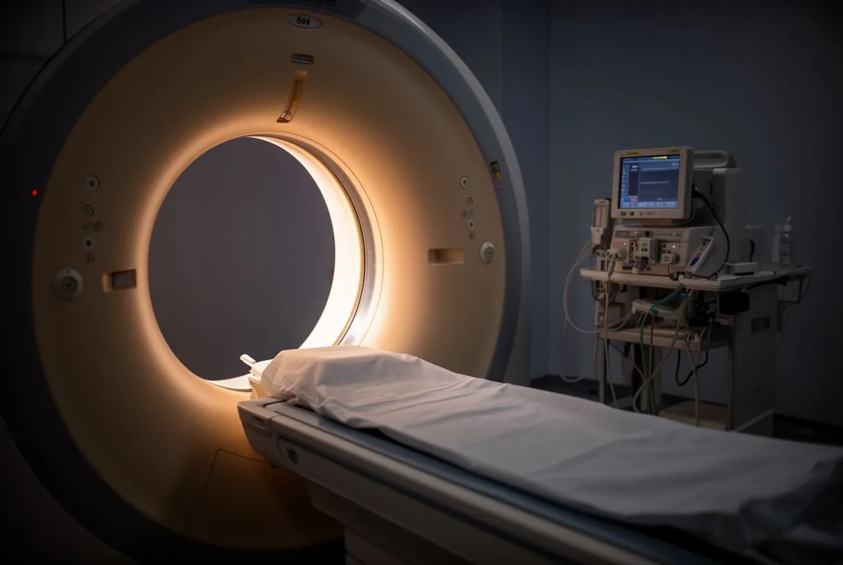
The role of advanced imaging: Diagnosing an aortic intramural haematoma requires high-resolution imaging, as clinical symptoms alone overlap heavily with other cardiac emergencies. The gold standard for diagnosis is a computed tomography (CT) scan with intravenous contrast. The intramural haematoma CT scan reveals the characteristic crescent-shaped thickening of the aortic wall, often measuring more than 5 millimeters in thickness. A transesophageal echocardiogram (TEE) may also be used, especially in unstable patients, to assess the heart valves and the proximal aorta in real-time without the need for radiation exposure.
Distinguishing the diagnosis: Because an intramural haematoma lacks the intimal flap that defines a classic dissection, radiologists look specifically for the localized wall thickening without a communicating channel. Magnetic resonance imaging (MRI) can also be utilized, offering excellent detail of the aortic wall and bleeding without exposing the patient to radiation, though it takes longer to perform. Research compiled by resources like europepmc.org highlights that accurate diagnosis heavily relies on the radiologist's ability to differentiate true intramural haematoma from severe atherosclerosis or a mural thrombus.
Assessing the extent of the condition: Once the diagnosis is confirmed, the cardiac team must determine if the haematoma involves the ascending aorta or is confined to the descending aorta. This distinction dictates the urgency of treatment. Blood tests, including D-dimer levels, may be drawn to support the clinical picture, though imaging remains the definitive proof. The rapid acquisition of these scans is critical, as delays in diagnosis increase the risk of the haematoma progressing to a rupture. Every minute counts in stabilizing the patient and formulating a definitive surgical or medical plan.
IMH vs aortic dissection: what is the difference?
Anatomical differences in the aorta: The primary difference between an aortic intramural haematoma and an aortic dissection lies in the innermost layer of the aorta. In a classic aortic dissection, the intima tears completely, allowing blood from the main channel to rush into the wall and create a false lumen, or a second channel for blood flow. In an intramural haematoma, the intimal layer remains intact; blood is trapped within the wall without an exit point back into the main aortic channel. This anatomical difference is crucial for the surgeon when planning the repair strategy.
Clinical progression and risks: While an intramural haematoma might sound less severe because there is no open tear, it carries significant risks. The trapped blood can expand outward, putting pressure on the adventitia and potentially causing the aorta to rupture outward. Alternatively, the pressure can build until the inner lining gives way, converting the intramural haematoma into a classic aortic dissection. Therefore, the management of both conditions is often similarly aggressive, particularly when the ascending aorta is involved and the patient's life is at immediate risk.
Implications for the patient: For the patient, the experience of an IMH vs aortic dissection feels identical—sudden, severe pain. However, the subtle anatomical distinction is vital for the surgeon. When planning aortic haematoma surgery, the surgical approach must account for the fragile, blood-soaked layers of the aortic wall. Replacing the damaged segment with a durable arterial graft requires meticulous technique to ensure the new suture lines hold securely in the compromised tissue, preventing postoperative bleeding and ensuring long-term stability of the repair.
Types of aortic intramural haematoma
Classifying the location: Aortic intramural haematomas are classified based on their location along the aorta, mirroring the Stanford classification system used for aortic dissections. This anatomical distinction is the most critical factor in determining whether the patient requires immediate surgery or can be managed with medication. The classification divides the condition into Type A and Type B, referring to the involvement of the ascending aorta versus the descending aorta, which dictates the treatment pathway and overall prognosis.
Type A intramural haematoma: A Type A intramural haematoma involves the ascending aorta, the section of the aorta that exits directly from the left ventricle and travels up toward the neck. This type is considered highly dangerous because the haematoma can easily extend backward into the heart, causing fluid to build up around the heart sac (pericardial effusion) or compromising the coronary arteries that supply the heart muscle. Due to the high risk of rupture or progression to dissection, Type A is typically treated as a surgical emergency requiring immediate intervention.
Type B intramural haematoma: A Type B intramural haematoma originates in the descending aorta, the portion that travels down the chest and into the abdomen. Because it does not directly threaten the heart or the aortic valve, Type B is usually managed medically in an intensive care setting. The goal is to lower the blood pressure to reduce the stress on the aortic wall, allowing the haematoma to stabilize and eventually be reabsorbed by the body over time. However, if complications arise, surgical or endovascular intervention may be necessary.
- Type A: Involves the ascending aorta, usually treated with emergency surgery to prevent rupture.
- Type B: Involves the descending aorta, typically managed with medication and strict blood pressure control.
- Complicated Type B: Requires intervention if it threatens organ blood flow or grows rapidly despite medical therapy.
When is surgery needed for aortic intramural haematoma?
Indicators for surgical intervention: While Type A intramural haematomas almost always require surgical intervention, Type B haematomas may also necessitate surgery if they exhibit specific high-risk features. If the aortic diameter exceeds 5.5 centimeters, if the wall thickness is greater than 10 millimeters, or if the patient experiences recurrent pain despite aggressive blood pressure control, the risk of rupture increases significantly. In these scenarios, the cardiac team will shift from a watch-and-wait approach to planning aortic haematoma surgery to prevent a catastrophic outcome.
Surgical approaches and complexity: Open surgical repair involves replacing the weakened section of the aorta with a synthetic arterial graft. This is a major operation requiring a heart-lung machine to maintain circulation, utilizing an arterial cannula to route the blood safely away from the heart while the surgeon works. In some complex cases, surgeons might use autologous blood salvaged during the procedure to minimize transfusion needs. The decision to proceed is carefully weighed against the patient’s overall health, often utilizing risk scoring systems like EuroSCORE II to estimate the potential mortality risk of the surgery itself.
Alternatives to open repair: For patients with descending aortic haematomas that require intervention but are not candidates for open surgery, endovascular repair offers a less invasive alternative. This involves threading a stent graft up through the femoral artery in the groin to reinforce the aortic wall from the inside. While this reduces the physiological stress on the patient, it requires specific anatomical characteristics to ensure the stent seals properly against the aortic wall. Both open and endovascular approaches aim to depressurize the haematoma and prevent a catastrophic rupture by redirecting blood flow away from the weakened area.
Medical management for descending aortic haematoma
Controlling blood pressure and heart rate: For patients with a stable Type B aortic intramural haematoma, medical management is the cornerstone of treatment. The immediate priority is to reduce the mechanical forces acting on the aortic wall. This is achieved through intravenous medications in an intensive care unit to maintain a strict heart rate and blood pressure target. By slowing the heart rate and lowering the systolic blood pressure, the sheer force of each heartbeat against the weakened wall is minimized, giving the haematoma the best chance to heal without progressing to a rupture.
Monitoring for complications: While in the hospital, the medical team will perform serial imaging to watch for any changes in the size or extent of the haematoma. During this time, patients are monitored for complications such as atrial flutter, a rapid heart rhythm that could compromise blood pressure control and require additional medication, as noted in the 2022 ESC Guidelines for the management of patients with ventricular arrhythmias and the prevention of sudden cardiac death. In rare emergencies where the heart stops, an automated external defibrillator might be used, though this is strictly a resuscitative measure, not a treatment for the haematoma.
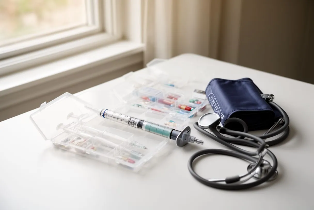
Long-term medication and lifestyle adjustments: Once the acute phase passes and the haematoma stabilizes, patients are transitioned to oral blood pressure medications, which they will likely need for the rest of their lives. Drugs like beta-blockers are heavily favored, often combined with other antihypertensives to keep the blood pressure in a safe range. Patients may also be advised to take aspirin for general cardiovascular protection, though this is independent of treating the haematoma directly. Strict adherence to these medications is non-negotiable to prevent future aortic events and maintain the structural integrity of the remaining aorta.
Recovery reality after aortic haematoma treatment
The hospital stay and early recovery: Recovery from an aortic intramural haematoma varies dramatically depending on whether the patient underwent surgery or was managed medically. Those treated with medication alone will spend several days in the intensive care unit followed by a step-down unit, focusing solely on pain control and strict blood pressure management. Surgical patients face a longer recovery, enduring the physical toll of a major chest incision, the use of an arterial line for continuous monitoring, and the gradual process of regaining strength. Both paths require patience and careful medical supervision.
Long-term follow-up imaging: Regardless of the treatment path, surviving an aortic intramural haematoma means entering a lifelong surveillance program. The aorta remains vulnerable, and new haematomas or dissections can form in different segments over time. Patients require regular CT scans or MRIs to monitor the aortic diameter. This commitment to ongoing imaging is not merely precautionary; it is the primary way your medical team catches silent changes before they become dangerous. Maintaining a close relationship with your cardiologist is essential for navigating this long-term phase.
Returning to daily life: Returning home brings a new set of adjustments. Patients must modify their lifestyle to avoid activities that spike blood pressure, such as heavy lifting or strenuous exercise. Managing the psychological impact is equally important, as surviving an acute aortic syndrome often leaves patients with anxiety about recurrent chest pain. Support from family, clear communication with the cardiac team, and a structured cardiac rehabilitation program can significantly ease the transition back to everyday life and rebuild physical confidence safely.
- Intensive Care: Initial stabilization requires continuous monitoring in a specialized cardiac unit.
- Medication adherence: Strict blood pressure control is vital to prevent recurrence and further aortic damage.
- Imaging schedule: Regular CT scans track aortic stability over the patient's lifetime to catch new changes early.
When to seek help for aorta complications
Recognizing warning signs at home: Once you are discharged after an aortic intramural haematoma, knowing when to return to the hospital is critical. Any return of the original tearing or sharp chest and back pain is a medical emergency. This pain could indicate that the haematoma has expanded, progressed into a classic dissection, or that a new area of the aorta has become compromised. Do not wait to see if the pain resolves; immediate emergency medical attention is required to prevent a fatal outcome.
Symptoms of compromised blood flow: Beyond pain, watch for signs that major organs are not receiving adequate blood supply. Sudden weakness, numbness, or paralysis suggests the spinal cord or brain is not getting enough blood. Fainting, severe dizziness, or a sudden inability to speak clearly are also red flags that require immediate evaluation. If you have been treated for an aortic condition and experience these symptoms, call emergency services immediately rather than attempting to drive yourself to the hospital.
Partnering with your cardiac team: Living with a history of aortic disease requires an active partnership with your specialists. If you are considering pregnancy, undergoing non-cardiac surgery, or experiencing side effects from your blood pressure medications, these discussions need to happen proactively with your surgeon. We encourage you to discuss your specific situation with a qualified cardiac team to determine the lowest risk approach for your ongoing care. To evaluate your personal risk factors or review your imaging, book a consultation with our practice to receive a tailored assessment.






