Janeway Lesions: A Skin Sign of Endocarditis
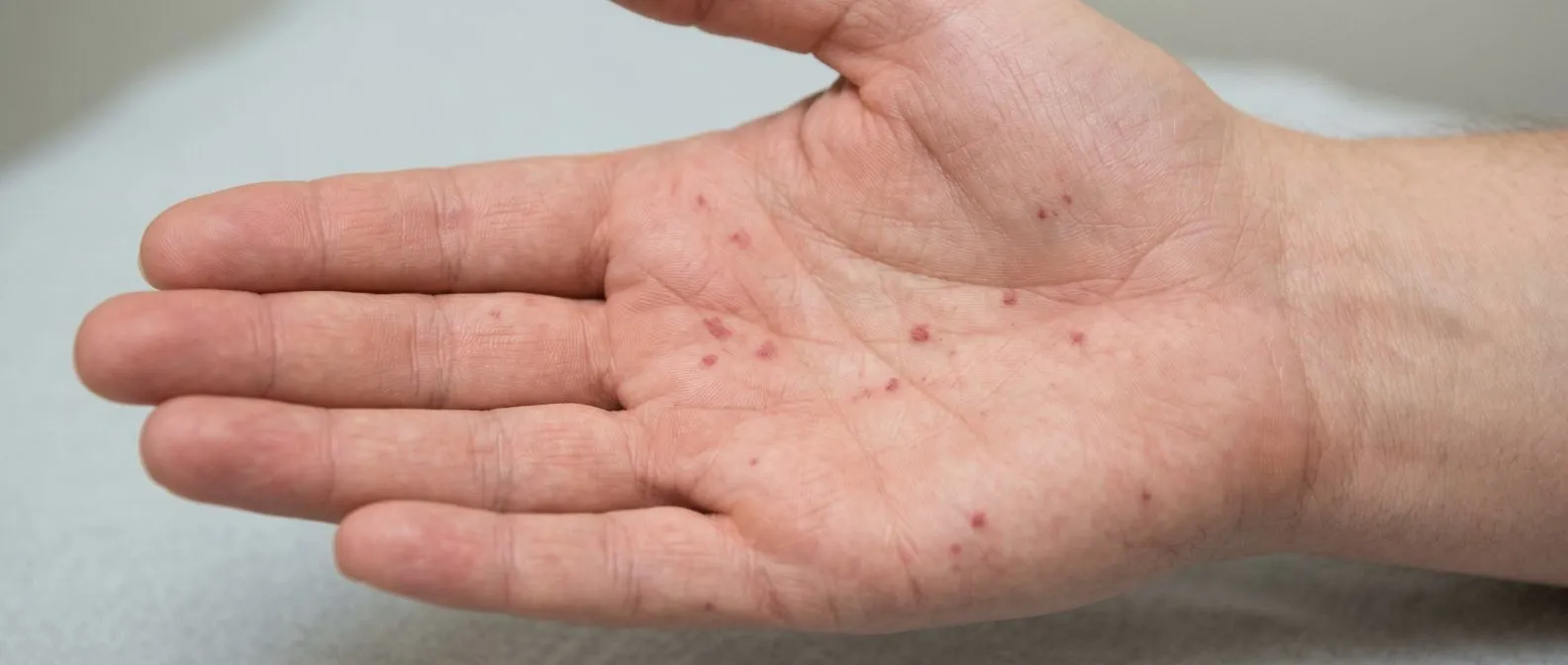
Janeway lesions in endocarditis can appear as painless red spots on the palms and soles.
Some medical signs carry an outsized message. Janeway lesions endocarditis — small, painless red spots on the hands and feet — are one of them. They are painless macules (flat, discolored patches) caused by septic emboli: tiny fragments of infected material that travel from an infected heart valve through the bloodstream and lodge in small vessels of the skin. For patients and families, recognizing what these spots mean can shorten the delay to diagnosis of a serious heart valve infection. This guide explains what Janeway lesions look like, what causes them, how they differ from Osler nodes, and what treatment — including heart surgery — may follow.
What Are Janeway Lesions?
Janeway lesions are painless red or purplish macules. They appear most often on the palms of the hands and soles of the feet, usually a few millimeters across, and they do not hurt when pressed. That painlessness is a defining feature — and it separates them from their better-known cousin, Osler nodes. Named after the physician Theodore Janeway, these lesions were described in the era before antibiotics, when infective endocarditis (an infection of the inner lining of the heart and its valves) was almost always fatal. Today they remain a classic physical finding, though antibiotics and better dental care have made them less common.
Why they matter to patients. A skin spot that doesn't hurt is easy to ignore. But in the right context — a fever that has lasted days, a new heart murmur, unexplained fatigue — Janeway lesions are a visible clue that infection may have seeded the heart valves. Dermatologic findings like these can help clinicians make an earlier diagnosis of infective endocarditis, which is critical because the condition can rapidly destroy heart valves if left untreated.
In our practice, when a patient mentions skin lesions together with fever or a new murmur, we treat that combination as a reason to investigate the heart promptly — never as a skin problem alone. In our consultations we explain what each study actually shows, in plain words, so patients understand why the workup is ordered.
What Causes Janeway Lesions in Endocarditis?
The mechanism is septic embolization. When bacteria settle on a heart valve, they form vegetations — friable, irregular masses of bacteria, fibrin, and platelets. Pieces of these vegetations can break off and travel through the arteries. When they lodge in the tiny vessels of the skin of the palms and soles, they cause a local area of infection and small-vessel blockage, producing the red, painless macules we call Janeway lesions. The palms and soles are affected because their small vessels are anatomically prone to this kind of lodging.
The bacteria involved are usually staphylococci. Staphylococcus aureus, in particular, is aggressive in producing vegetations and emboli, and is the organism most often associated with Janeway lesions and other peripheral signs of infective endocarditis. Research on the mechanisms of these lesions has been published in journals such as Frontiers in Cardiovascular Medicine, which examines how bacteria and immune responses interact in infective endocarditis.
Why does infective endocarditis cause Janeway lesions? Because the heart valve is a surface where the bloodstream deposits bacteria continuously, and the valve itself has no easy way to clear the infection. The immune system struggles to reach the vegetations, so the infection persists and sheds fragments into the circulation. Understanding the link between vascular findings and underlying infection helped shape the diagnostic criteria used today, and studies such as the one on in situ detection of Chlamydia pneumoniae and cytokines in cardiovascular patients show how deeply infection and vascular inflammation can be intertwined in cardiovascular disease.

Janeway Lesions vs Osler Nodes: Key Differences
Osler nodes are painful; Janeway lesions are not. This is the single most important distinction, and it reflects a different mechanism. Osler nodes are tender, raised lumps, typically on the pads of the fingers and toes, and they arise from an immune-complex reaction — the body's immune response depositing inflammatory complexes in the small vessels of the skin. Janeway lesions, by contrast, are flat, painless, and caused directly by septic emboli lodging in the vessels.
Where they appear also differs. Janeway lesions favor the palms and soles; Osler nodes favor the fleshy pads of the fingers and toes. Both are part of the classic peripheral signs of infective endocarditis, alongside splinter hemorrhages (tiny dark lines under the nails) and petechiae (small red or purple dots caused by tiny bleeds). Neither sign is specific on its own — but together they raise the level of suspicion considerably.
- Janeway lesions: flat, painless, red-purple macules on palms and soles; caused by septic emboli.
- Osler nodes: painful, raised, tender nodules on finger and toe pads; caused by immune-complex deposition.
- Shared meaning: both are peripheral signs of infective endocarditis and both warrant urgent evaluation.
Patients often ask whether Osler nodes and Janeway lesions are the same thing. They are not — but both point to the same underlying possibility: a heart valve infection that needs diagnosis and treatment.
First Signs of Infective Endocarditis
Most cases start with vague, flu-like symptoms. Fever, chills, night sweats, fatigue, and loss of appetite are common early complaints. A new or changing heart murmur — an abnormal sound the doctor hears through the stethoscope — may be the first clue that a valve is involved. Unexplained weight loss and aching joints also occur. Because these symptoms overlap with many common illnesses, diagnosis is often delayed, which is why skin signs like Janeway lesions can be so valuable.
Other signs to watch for. Beyond the skin, patients may notice blood in the urine, tiny hemorrhages in the retina of the eye, or a history of recent dental work or other procedures that could have introduced bacteria into the bloodstream. In people with prosthetic valves, pacemakers, or congenital heart disease, any unexplained fever should be taken especially seriously.
Red flags that demand urgent attention. Some symptoms suggest the infection is spreading or causing complications, and they require same-day evaluation:
- Neurological symptoms: weakness, numbness, slurred speech, or vision loss — possible embolic stroke.
- Severe breathlessness: the valve may be failing acutely.
- Spreading skin signs: new painful nodules, Janeway lesions, or splinter hemorrhages in a febrile patient.
How Doctors Diagnose Bacterial Endocarditis
Diagnosis rests on blood cultures and imaging. Blood cultures are drawn several times to identify the organism responsible, because treatment depends on knowing which bacteria are involved. The modified Duke criteria — the standard diagnostic framework — combine positive blood cultures, evidence of valve involvement on echocardiography, and clinical findings such as Janeway lesions, Osler nodes, and vascular phenomena.
Echocardiography is the cornerstone test. A transthoracic echocardiogram (ultrasound of the heart through the chest wall) is done first; if suspicion remains high, a transesophageal echocardiogram (a probe passed through the esophagus, giving a much closer view of the valves) is used. When structural complications are suspected, a coronary CT angiography or a Coronary Angiography may be added — especially if surgery is being considered, because the surgical team needs to know whether the coronary arteries need attention at the same time.
Additional tests are ordered selectively. A Coronary Calcium Score can clarify underlying Coronary Artery Disease in older patients before surgery. In our practice, we aim to order only the studies the patient genuinely needs — the first contact is a free video consultation, where we talk through the condition and identify which studies are still missing before anything is ordered.
Endocarditis Skin Manifestations Beyond Janeway Lesions
Janeway lesions are only one of several skin findings. Infective endocarditis skin manifestations also include splinter hemorrhages, petechiae, and Osler nodes, as described above. Rarer findings include Roth spots (hemorrhages in the retina with a pale center, seen on eye examination) and purpura. Some patients with sepsis from other causes can even develop Janeway-like lesions without valvular infection — one reason clinicians interpret these signs in context rather than in isolation.
Why the skin reflects the heart. The skin is the most visible organ affected by the small-vessel blockage and inflammation that endocarditis produces. Because the lesions are visible and often painless, they are frequently the first clue a patient or family member notices. This is why dermatologists and primary care physicians play a real role in early detection of infective endocarditis.
In our practice, we see the value of a careful physical exam that includes the hands and feet. Our team reviews every case against international practice guidelines before recommending any intervention, so a subtle sign like Janeway lesions is never overlooked in the wider picture.
Janeway Lesions Treatment
Janeway lesions do not need treatment of their own. They are a sign of the underlying infection, not a skin condition. Treatment is directed at the infective endocarditis itself: prolonged intravenous antibiotics, typically for four to six weeks, guided by the organism identified in blood cultures. As the infection clears, the skin lesions heal on their own.
Antibiotic choice matters. Empiric therapy — antibiotics started before the organism is identified — usually covers staphylococci and streptococci, then is refined once cultures return. The duration and route are set by the treating infectious disease team, often in coordination with cardiology. In patients with prosthetic valve endocarditis, treatment can be more complex and more often requires surgery.
Follow-up imaging is essential. Repeat echocardiography monitors how the valve is responding to treatment. If there is ongoing infection, worsening valve function, or evidence of uncontrolled embolization, the team will consider surgical intervention. In our practice, we review every case as a multidisciplinary team before proposing any treatment path, and we present the alternatives so the patient can make the final decision.
When Heart Surgery Is Needed for Endocarditis
Surgery is not required for every case — but it is often the safest route. Indications include heart failure caused by a leaking or destroyed valve, persistent infection despite adequate antibiotics, perivalvular abscess, embolic events that continue despite therapy, and infection with organisms that are difficult to eradicate. The goal is to remove infected tissue and reconstruct or replace the valve before the damage becomes irreversible.
Options range from conventional to transcatheter. Depending on the valve involved and the patient's condition, treatment may involve Coronary Artery Bypass Grafting if Coronary Artery Disease is also present, valve repair or replacement through conventional surgery, a minimally invasive or robotic approach, or in selected cases a transcatheter option such as TAVR (transcatheter aortic valve replacement) for the aortic valve. In our practice, we recommend the less invasive route only where the evidence supports it for that specific patient, and we are candid about when conventional surgery is the safer choice.
Recovery and realistic expectations. Hospital stays after valve surgery for endocarditis are typically longer than for elective surgery, because the intravenous antibiotics must continue and the patient needs close monitoring. Rehabilitation is gradual. Most patients return to a good quality of life, but outcomes depend on how early the infection is caught and whether complications have occurred. In our practice Dr Molina performed the first robotic mitral valve repair in the Ángeles Health System and the first robotic heart surgery in Guadalajara, and that experience shapes how we match each patient to the least invasive approach that is also safe for them.
Who Is at Risk of Heart Valve Infection?
Pre-existing heart conditions raise risk. People with a history of Rheumatic Heart Disease, congenital heart disease, a previous prosthetic valve, or prior infective endocarditis are at higher risk. Valvular heart disease — for example, a narrowed or leaking valve — provides an irregular surface where bacteria can more easily settle. Intravenous drug use, long-term dialysis, and chronic kidney disease also increase risk, as do recent dental procedures, particularly in patients with known valve disease.
Prevention is practical. Good oral hygiene, prompt treatment of skin and dental infections, and adherence to antibiotic prophylaxis guidelines where indicated all reduce risk. Patients with prosthetic valves or complex congenital heart disease should ask their cardiologist whether they need antibiotics before certain dental procedures.
- Brush and floss daily; dental bacteria are the most common source of bloodstream seeding for valves.
- Carry your valve history; any doctor or dentist who knows your valve status can make better prophylaxis decisions.
- Treat any fever seriously if you have a prosthetic valve, congenital heart disease, or a history of endocarditis.
When to Seek Help for Endocarditis Symptoms
Do not wait to "see if it goes away." If you have fever plus unexplained skin spots, a new murmur, night sweats, or breathlessness — and especially if you have a prosthetic valve, known valve disease, or a recent procedure — see a doctor promptly. Bring up any recent dental work, surgeries, or intravenous lines. Mentioning Janeway lesions and Osler nodes by name can help a clinician connect the dots faster.
What a proper evaluation looks like. Expect blood cultures, an echocardiogram, and a careful physical examination. In our practice, we encourage second opinions and treat seeking one as a normal part of deciding about heart surgery — a second opinion is never a sign of mistrust, but of a well-informed decision.
Getting help in Mexico. For patients in Guadalajara, Querétaro, Hermosillo, or traveling from abroad, the first step is simple: a consultation with a cardiac team that can evaluate the full picture. Our team of cardiac surgeons led by Dr Alejandro Molina — trained in cardiothoracic surgery at the Universidad de Guadalajara, in cardiac, minimally invasive, robotic and structural surgery at the University of Montreal, and in structural cardiology and TAVR at CHU Bordeaux — offers a free video consultation as a first step, so you can discuss your situation and decide what to do next. If you have noticed Janeway lesions, fever, or other symptoms that concern you, talk to a qualified cardiac team rather than self-diagnosing. Early diagnosis of infective endocarditis — and the right treatment at the right time — is the best way to protect your heart and your life.