


Marfan Syndrome and the Aorta: Monitoring and Surgery
Learn about marfan syndrome aorta surgery, monitoring, and expert treatment in Guadalajara.
What Is Marfan Syndrome and How Does It Affect the Aorta?
Marfan syndrome is a genetic condition that affects the body's connective tissue — the structural framework that holds cells, organs, and tissues together. Because connective tissue is found throughout the body, Marfan syndrome can affect many systems, but its most life-threatening impact is on the heart and blood vessels, especially the aorta. The aorta is the largest artery in the body, carrying oxygen-rich blood from the heart to the rest of the body. In people with Marfan syndrome, the aortic wall is weaker than normal, which makes it stretch and enlarge over time — a condition called aortic dilation or aortic aneurysm.
Why does the aorta matter most in Marfan syndrome? While Marfan syndrome can affect the skeleton, eyes, and lungs, cardiovascular complications remain the leading cause of serious health problems and early death in untreated patients. The weak connective tissue in the aortic wall cannot withstand the relentless pressure of blood pumped from the heart with each heartbeat. Over years, this pressure gradually widens the aorta, particularly at its root — the section where the aorta exits the heart. If this widening goes unchecked, the aortic wall can tear, creating what doctors call aortic dissection: a true cardiovascular emergency that requires immediate surgical intervention to prevent fatal consequences.
The encouraging news is that with careful monitoring and timely marfan syndrome aorta surgery, most patients can live long, fulfilling lives. Understanding how Marfan syndrome affects the aorta is the first step toward protecting your heart and making informed decisions about treatment. As Cleveland Clinic notes, early diagnosis and proactive management dramatically improve outcomes for people with this condition.
How Does Marfan Syndrome Cause Aortic Dilation?
Marfan syndrome is caused by mutations in a gene called FBN1, which provides instructions for making a protein called fibrillin-1. This protein is a critical building block of the elastic fibers in connective tissue. When fibrillin-1 is abnormal or deficient, the connective tissue throughout the body — including the layers of the aortic wall — becomes weaker and less resilient. The aorta normally expands and recoils with each heartbeat, but in Marfan syndrome the tissue loses its elasticity and gradually stretches out without springing back. Research into how genetic mutations affect tissue integrity has advanced significantly; for example, a study on Genetic compensation triggered by mutant mRNA degradation illustrates the complex molecular mechanisms at play.
What happens to the aortic wall over time? The aortic wall has three layers. In Marfan syndrome, the middle layer — which contains the elastic fibers — degenerates and thins. As this layer weakens, the constant pressure of blood leaving the heart causes the aorta to widen. This widening usually begins at the aortic root, the section closest to the heart. The aortic diameter is measured carefully during imaging tests; when it exceeds normal ranges, doctors diagnose aortic dilation. The larger the aorta grows, the higher the risk of a tear or rupture, because wall tension increases with diameter according to basic principles of fluid mechanics.
Not everyone with Marfan syndrome will develop severe aortic dilation at the same pace. The rate of enlargement varies from person to person and depends on factors such as blood pressure, age, and the specific genetic mutation involved. Some patients experience slow, stable enlargement over decades, while others see rapid growth in a matter of months. This variability is why regular monitoring is essential — it allows your cardiac team to detect changes early and recommend surgery before the aorta reaches a dangerous size.
What Are the Warning Signs of Aortic Dissection in Marfan Syndrome?
Aortic dissection is the most feared complication of Marfan syndrome. It occurs when the innermost layer of the aortic wall tears, allowing blood to surge between the layers and split them apart. This is an acute medical emergency that can be fatal if not treated immediately. Knowing the warning signs can save a life. The classic symptom is sudden, severe, tearing chest pain that may radiate to the back between the shoulder blades. This pain is often described as unlike anything the person has ever experienced — it comes on instantly and at maximum intensity.
What symptoms might accompany the chest pain? Depending on where the dissection occurs, patients may experience shortness of breath, fainting, a difference in blood pressure between the two arms, weakness or paralysis on one side of the body, difficulty speaking, or a sudden pulse deficit. If the dissection involves the Aortic Arch, it can disrupt blood flow to the brain, causing neurological symptoms. Some patients also report abdominal pain if the dissection extends down the descending aorta. Any of these symptoms in a person with Marfan syndrome should be treated as a life-threatening emergency requiring immediate hospital care.
It is important to understand that aortic dissection can sometimes occur even before the aorta has reached the threshold size normally recommended for surgery. This is why doctors focus not only on the aortic diameter but also on the rate of growth, family history of dissection, and other risk factors. If you or a loved one has Marfan syndrome and experiences sudden severe chest or back pain, do not wait — go to the nearest emergency department immediately and inform the staff about the Marfan diagnosis. Quick action can make the difference between life and death.

Marfan Syndrome Aorta Monitoring: What Tests Do You Need?
Regular monitoring is the cornerstone of managing Marfan syndrome. Because the aorta enlarges slowly and silently, imaging tests are the only reliable way to track its size and catch dangerous changes before they become emergencies. The foundation of monitoring is the Annual Echocardiogram, a painless ultrasound of the heart that lets your cardiologist measure the aortic root and watch how the heart valves are functioning. This test uses sound waves to create detailed images — no radiation, no needles — and is typically performed once a year, or more frequently if the aorta is growing quickly.
When is more advanced imaging needed? While echocardiograms are excellent for the aortic root, they may not provide clear images of the entire aorta. When your doctor needs a more complete picture, a computed tomography (CT) scan or magnetic resonance imaging (MRI) may be ordered. These scans can visualize the entire aorta from the heart to the abdomen and are especially useful when planning surgery or when the echocardiogram shows borderline measurements. MRI does not use radiation, making it a good choice for younger patients who will need repeated scans over many years.
In addition to imaging, your cardiac team will monitor your blood pressure closely, since high blood pressure increases stress on the aortic wall and accelerates enlargement. Medications such as beta-blockers are commonly prescribed to lower blood pressure and reduce the force of each heartbeat, which can slow aortic growth. Your doctor may also discuss lifestyle modifications — avoiding contact sports, heavy lifting, and strenuous competitive athletics — all of which help reduce sudden spikes in blood pressure that could stress the aorta. Monitoring is not a one-time event; it is a lifelong commitment that adapts as your body changes.
When Is Aortic Aneurysm Surgery Recommended for Marfan Patients?
The decision to recommend marfan syndrome aorta surgery is not made lightly. Surgeons weigh multiple factors, but the single most important measurement is the aortic diameter. For adults with Marfan syndrome, most guidelines recommend prophylactic aortic surgery — meaning surgery done before a complication occurs — when the aortic root reaches approximately 5 centimeters in diameter. However, this threshold may be lowered to 4.5 centimeters if there are additional risk factors, such as a rapid rate of growth (more than 0.5 centimeters per year), a family history of aortic dissection at a small size, or the presence of Aortic Regurgitation, where the aortic valve leaks blood back into the heart.
How do doctors decide the timing? The goal is to operate before the aorta dissects or ruptures, but not so early that the patient faces unnecessary surgical risk. This balancing act depends on careful measurements taken over time. If the aorta is growing steadily but slowly, your surgeon may recommend continued monitoring. If growth accelerates or the aortic valve begins to leak significantly, the conversation shifts toward scheduling surgery. The rate of aortic growth and evidence of valve dysfunction are as influential as the absolute diameter in timing the operation.
Other considerations include the patient's overall health, age, and readiness for a major operation. Some patients have additional cardiac features related to Marfan syndrome — such as Aortic Insufficiency, where the valve does not close properly — that may move the surgical timeline forward. The surgical plan is always individualized. Your cardiac team in Guadalajara, Querétaro, or Hermosillo will discuss the specific thresholds that apply to your situation and help you understand the risks of waiting versus the risks of proceeding. The aim is always to intervene at the optimal moment — late enough that the surgery is clearly necessary, but early enough to prevent a catastrophe.

What Are the Surgical Options for Marfan Syndrome Aorta Repair?
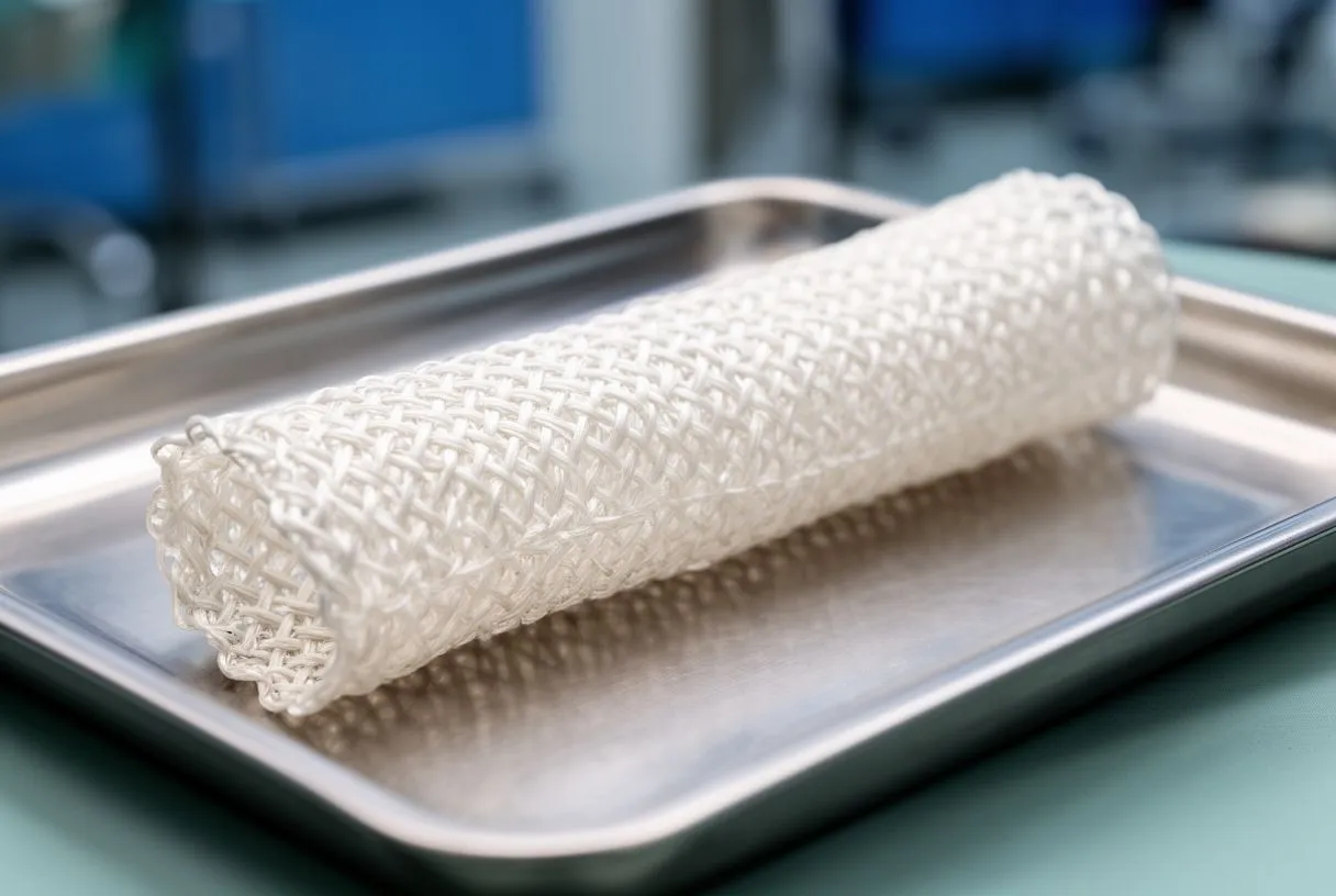
Several surgical procedures are available for Marfan patients, and the choice depends on which part of the aorta is affected and whether the aortic valve is also damaged. The most common operation is aortic root replacement, which addresses the section of the aorta where it meets the heart. There are two main approaches to this operation. The first is the Bentall procedure, in which the surgeon replaces both the dilated aortic root and the aortic valve with a mechanical or biological valve attached to a synthetic tube called a graft. This is a well-established operation with decades of proven results.
Can the natural valve be preserved? The second approach is valve-sparing aortic root replacement, often called the David procedure. In this operation, the surgeon replaces the dilated aortic tissue with a synthetic graft but reimplants the patient's own aortic valve inside the graft. This avoids the need for lifelong blood-thinning medication that mechanical valves require. Valve-sparing surgery is technically more complex and not suitable for every patient — if the valve itself is severely damaged or malformed, replacement may be the better option. Your surgeon will evaluate the valve's condition during preoperative imaging and sometimes confirm it visually during the operation itself.
If the aneurysm extends beyond the root into the ascending aorta or the arch, additional sections may need to be replaced during the same operation or in a separate stage. Aortic Arch Replacement is a more complex procedure that may require circulatory arrest — cooling the body to a very low temperature to protect the brain while circulation is temporarily stopped. Antegrade Cerebral Perfusion, a technique that maintains blood flow to the brain during arch surgery, has improved safety significantly in recent years. The surgical plan is tailored to the exact anatomy of each patient's aorta, and your surgeon will explain which approach is recommended and why.
- Bentall procedure: replaces the aortic root and valve with a graft containing a mechanical or biological valve — durable and well-tested.
- Valve-sparing (David) procedure: replaces the aortic root with a graft while keeping your own natural aortic valve — avoids lifelong blood thinners.
- Arch and descending repair: extends the graft beyond the root when the aneurysm involves the aortic arch or descending aorta, sometimes requiring circulatory arrest.
Aortic Root Replacement in Marfan Syndrome: What to Expect
Understanding what happens during aortic root replacement can ease much of the anxiety that naturally comes with facing heart surgery. The operation is performed under general anesthesia, meaning you are completely asleep and feel no pain. The surgeon makes an incision down the center of the chest to access the heart. A heart-lung bypass machine takes over the work of pumping blood and oxygenating it, allowing the surgeon to operate on a still, bloodless aorta. The Aortic Cross-clamp is applied to isolate the section being worked on, and the damaged aortic tissue is carefully removed and replaced with a synthetic graft.
How long does the surgery take, and what happens right after? Aortic root replacement typically takes between four and six hours, depending on complexity. After the operation, you will spend the first night in the intensive care unit, where your heart rhythm, blood pressure, and breathing are continuously monitored. Most patients remain in the hospital for about a week. During this time, the care team manages pain, watches for any signs of bleeding or infection, and begins early mobilization — sitting up, standing, and taking short walks — to promote healing and reduce complications. Annulus Sizing, the process of measuring the valve opening to select the correct graft and valve size, is performed precisely during surgery to ensure a proper fit.
Families often find the waiting period during surgery to be the hardest part. Knowing the typical timeline can help: your surgical team will provide updates as milestones are reached. Once the operation is complete and you are stable in the ICU, your surgeon will meet with your family to explain what was done and what to expect in the coming days. Recovery is a gradual process — the first few days are the most intense, and steady improvement follows. Ask your surgeon beforehand about their specific protocols so there are no surprises on the day of surgery.
Are There Alternatives to Open Heart Surgery for Marfan Patients?
Many patients ask whether less invasive options exist, especially given the size and recovery associated with open-heart surgery. Transcatheter aortic valve replacement (TAVR) is a minimally invasive technique in which a replacement valve is delivered through a catheter, usually inserted through the groin, and positioned inside the diseased valve without opening the chest. TAVR has become a well-established option for older patients with severe aortic stenosis who are not good candidates for open surgery. However, TAVR is generally not recommended as the primary treatment for young Marfan syndrome patients with aortic root aneurysms.
Why is TAVR usually not the first choice for Marfan patients? Marfan syndrome weakens the entire aortic wall, not just the valve. Simply placing a new valve inside the aorta does not address the underlying structural problem — the dilated aortic tissue remains and continues to be at risk for dissection or rupture. The goal of surgery in Marfan syndrome is to remove the weakened aortic tissue and replace it with a strong synthetic graft, something that a catheter-based valve cannot accomplish. For this reason, open surgical Aortic Aneurysm Repair remains the gold standard for Marfan patients with significant aortic dilation.
That said, catheter-based techniques can sometimes play a supporting role. In certain complex or emergency situations, endovascular stent grafts may be used to reinforce sections of the descending aorta, especially if a dissection has already occurred. These approaches are highly individualized and are not substitutes for elective root replacement in patients who meet surgical criteria. If you have read about TAVR or other minimally invasive approaches and wonder whether they apply to you, the best step is to discuss your specific anatomy and surgical history with a cardiac surgeon who specializes in aortic disease in Marfan syndrome.
Recovery After Marfan Syndrome Aorta Surgery: What Patients Should Know
Recovery from marfan syndrome aorta surgery is a marathon, not a sprint. The first six to eight weeks at home are focused on healing, building strength, and gradually returning to daily activities. You will be encouraged to walk daily, increasing distance as tolerated, but to avoid lifting anything heavier than a few kilograms, pushing, pulling, or any activity that strains the chest. Most patients can return to desk work in about six to eight weeks, while more physical occupations may require three months or more. Your surgeon will give you a personalized timeline based on the specific procedure performed and your overall progress.
What medications will you need afterward? If you received a mechanical valve, you will need lifelong anticoagulation therapy — commonly with a medication called warfarin. Regular blood tests are necessary to ensure the dose keeps your blood in the correct thinness range. If your own valve was preserved, you may instead need Antiplatelet Therapy for a period, and possibly medications like Amiodarone if you experience irregular heart rhythms after surgery. Your care team will also prescribe blood pressure medications to protect the remaining aortic tissue. Antibiotic Prophylaxis — taking antibiotics before certain dental or medical procedures — may be recommended to prevent infections from settling on the valve or graft.

Complications are uncommon but important to recognize. Some patients develop Anaemia (a low red blood cell count) after surgery, which can cause fatigue and is usually temporary. Others may experience Ankle Swelling as the body adjusts to changes in circulation, which can often be managed with compression stockings and elevation. If you experience fever, increasing chest pain, shortness of breath, or any wound redness or drainage, contact your surgical team immediately. Attending all follow-up appointments is critical — these visits allow your team to confirm that the graft is healing well and that the rest of your aorta remains stable.
- Anticoagulation: mechanical valve recipients need lifelong blood thinners with regular INR monitoring.
- Wound care: keep incisions clean and dry; report any redness, drainage, or opening promptly.
- Activity: walk daily, avoid heavy lifting and strenuous exercise for at least six to eight weeks.
- Follow-up: attend all scheduled echocardiograms and surgeon visits — your aorta needs lifelong check-ups.
Living Well After Marfan Syndrome Aortic Surgery: Long-Term Care
Surgery is not the end of the Marfan journey — it is a major milestone in an ongoing care plan. Even after a successful aortic root replacement, the remaining portions of the aorta still contain Marfan-affected tissue and must be monitored indefinitely. Your cardiologist will continue annual or biennial imaging to check the ascending aorta, arch, and descending aorta. If any new area begins to dilate, further surgery or intervention may be needed in the future. The good news is that with modern surgical techniques and vigilant follow-up, most Marfan patients who undergo elective aortic surgery can expect an excellent long-term outcome.
What lifestyle changes help protect your aorta long-term? Maintaining controlled blood pressure is the single most important lifestyle factor. Your doctor will help you find the right combination of medication and daily habits to keep your blood pressure within a safe range. Avoid contact sports, heavy weightlifting, and activities that involve sudden, intense physical exertion. Moderate, low-impact exercise such as brisk walking, swimming, or cycling at a conversational pace is generally encouraged and beneficial. Always check with your cardiologist before starting any new exercise program. Avoiding smoking is essential, as smoking damages blood vessels and accelerates aortic deterioration. Research has shown that Aortic Stiffness Is an Independent Predictor of All-Cause and Cardiovascular Mortality in Hypertensive Patients, underscoring the importance of blood pressure control and arterial health.
For women with Marfan syndrome, pregnancy requires special planning. The cardiovascular changes of pregnancy — increased blood volume and heart rate — put additional stress on the aorta. If the aortic root is dilated, pregnancy carries a significant risk of dissection. Your cardiac team should evaluate your aortic size and overall heart health before you conceive, and some women may be advised to have aortic surgery before pregnancy to make it safer. With careful planning and a collaborative team that includes your cardiologist and obstetrician, many women with Marfan syndrome can have successful pregnancies. Every person's situation is unique, and decisions about surgery, lifestyle, and family planning should be made in close partnership with a qualified cardiac team. If you or a loved one has Marfan syndrome, we encourage you to schedule a consultation with Dr Alejandro Molina and the team at Cirugía de Corazón in Guadalajara, Querétaro, or Hermosillo to discuss your aortic health and create a personalized care plan.






