Peripheral Oedema and Heart Failure: Swelling Explained
Peripheral oedema heart failure signals poor cardiac pumping.
Peripheral oedema is one of the most visible signs that the heart is struggling to keep up with the body's demands. When the pumping chambers lose efficiency — whether after a heart attack, due to a stiffened valve, or from long-standing high blood pressure — fluid begins to back up in the tissues, often collecting first in the feet, ankles, and lower legs. This guide explains what that swelling means, how it links to heart failure staging, what diagnostic clues your cardiologist looks for, and what treatment paths exist. The goal is simple: to turn a frightening symptom into something you understand well enough to discuss confidently with a qualified cardiac team.
What Is Peripheral Oedema in Heart Failure?
Peripheral oedema refers to the accumulation of excess fluid in the soft tissues of the limbs, most commonly the lower legs and feet. In the context of heart failure, this swelling is not a localised problem with the skin or veins — it is a downstream consequence of the heart's inability to maintain adequate forward blood flow. When the right side of the heart pumps inefficiently, blood returning from the body via the vena cava backs up, raising pressure in the venous system and forcing fluid out of capillaries into surrounding tissue.
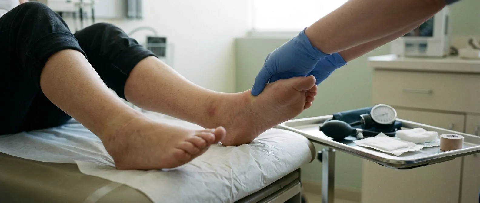
What the patient actually sees and feels: The swelling usually begins around the ankles and the lowest part of the leg because gravity pushes fluid downward during the day. You may notice that socks leave deep indentations, shoes feel tighter by evening, or the skin over the shin looks stretched and shiny. Pressing a thumb into the swollen area may leave a visible depression that takes several seconds to rebound — a hallmark called pitting that your clinician will assess during a physical examination.
It is worth noting that not all leg swelling comes from the heart. Venous insufficiency, kidney disease, liver cirrhosis, certain medications, and prolonged immobility can all produce similar signs. This is why a cardiologist never diagnoses heart failure from swelling alone — the symptom is one piece of a clinical puzzle that includes auscultation (listening to the heart and lungs with a stethoscope), blood tests, and imaging. Open-access research indexed by DOAJ provides numerous peer-reviewed studies on the differential diagnosis of lower-limb oedema that clinicians consult when narrowing the cause.
How Does Heart Failure Cause Leg Swelling?
To understand the mechanism, picture the heart as two parallel pumps. The left side pushes oxygen-rich blood into the ascending aorta and onward to the body. The right side receives oxygen-depleted blood from the veins and sends it to the lungs. When the left side weakens — often because of damage from an atheromatous plaque rupturing in a coronary artery — pressure backs up through the lungs into the right heart, and eventually into the systemic veins. That backward pressure is what drives fluid into your legs.
Why the body retains sodium and water: Heart failure triggers a cascade of neurohormonal responses. The kidneys perceive reduced blood flow and respond by activating the renin-angiotensin-aldosterone system, which holds onto sodium and water in an attempt to boost circulating volume. Unfortunately, the failing heart cannot handle that extra volume, so the excess fluid leaks into tissues. This is why a low-sodium diet is a cornerstone of management — it directly targets the mechanism that worsens the swelling.
Several structural cardiac problems set the stage for this fluid retention. A stiff or narrowed aortic valve forces the left ventricle to work harder over years. An enlarged left atrium — sometimes caused by chronic atrial flutter, a rapid irregular rhythm in the upper chambers — prevents effective filling and emptying. When these conditions reduce cardiac output over time, the body's compensatory mechanisms paradoxically accelerate fluid accumulation, creating a vicious cycle that medication and, in selected cases, procedural interventions aim to break.
Early Signs of Swollen Legs Heart Failure
Many patients dismiss early swelling as a normal consequence of standing all day, aging, or weight gain. Recognising the pattern early can lead to treatment before the heart sustains further damage. The earliest signs tend to be subtle: a ring that no longer fits, a watch strap that leaves a mark, or the need to loosen shoelaces by mid-afternoon. These changes reflect fluid that has not yet become clinically obvious.
Accompanying symptoms that should prompt evaluation: Swelling rarely occurs in isolation when the heart is the cause. You may notice increased breathlessness when lying flat — a condition called orthopnoea — or the need to sleep on extra pillows. Some people wake at night gasping for air, a phenomenon known as paroxysmal nocturnal dyspnoea. Others describe a persistent dry cough, fatigue that is disproportionate to activity, or arm pain that appears with exertion and eases with rest, which can suggest ongoing coronary artery disease contributing to the heart's decline.

It is also important to understand that some heart attacks present atypically, especially in women, older adults, and people with diabetes. An atypical infarction may produce nausea, jaw discomfort, or profound fatigue rather than the classic crushing chest pain. If the heart muscle is damaged silently, the first obvious sign might be gradual fluid retention weeks or months later. This delayed presentation underscores why unexplained or progressive leg swelling should never be ignored or self-attributed to lifestyle alone.
Pitting Oedema Diagnosis and What It Means
Pitting oedema is assessed by pressing a finger firmly against the swollen area — typically over the shin or just above the ankle — for several seconds and then releasing. If a visible indentation remains and slowly fills back in, the swelling is described as pitting. The depth and duration of the pit are graded on a scale from 1+ (slight, 2 mm) to 4+ (deep, 8 mm or more). This grading gives the clinician a quick bedside indicator of how much fluid has accumulated and helps track whether treatment is working over time.
What different grades tell your doctor: A 1+ or 2+ score may reflect early or moderate fluid retention that could respond to dietary changes and a single oral diuretic. A 3+ or 4+ score suggests significant volume overload and may require more aggressive medical management, adjustment of existing heart failure therapy, or investigation for an acute decompensation trigger such as an arrhythmia, infection, or missed medication. Non-pitting swelling, by contrast, often points to non-cardiac causes such as lymphatic obstruction or thyroid disease and changes the diagnostic path.
Your cardiologist will combine the pitting exam with other findings. Listening for crackles in the lungs, checking for an enlarged liver, measuring jugular venous pressure, and reviewing recent weight trends all help determine whether the oedema is purely cardiac in origin. Blood tests — including markers of kidney function and a peptide called NT-proBNP, which rises when the heart is under strain — add objective data. Together these elements move the assessment well beyond the simple observation of swollen ankles.
Heart Failure Stage Classification and Oedema
Heart failure is classified using the ACC/AHA staging system, which progresses from Stage A (risk factors present, no symptoms) through Stage D (advanced symptoms despite maximal therapy). Peripheral oedema typically appears at Stage C, when structural heart disease is established and symptoms have become clinically apparent. Understanding where a patient sits on this spectrum helps guide both urgency and intensity of treatment.

How staging affects treatment decisions: At Stage A or B, the focus is on controlling blood pressure, managing cholesterol, and addressing structural problems before symptoms emerge. A patient with a severely narrowed aortic valve but no fluid retention might be monitored or scheduled for a balloon aortic valvuloplasty as a bridge to definitive valve replacement. Once oedema appears, the conversation shifts toward diuretics, sodium restriction, and possible dose optimisation of medications that reduce the heart's workload over the long term.
At Stage D, oedema may become resistant to standard oral therapy. Patients in this group often require intravenous diuretics, specialised heart failure services, or evaluation for advanced therapies such as mechanical circulatory support. The staging system is not merely academic — it maps directly onto guideline-driven thresholds that cardiologists use to decide when to escalate care, when to refer to a surgeon, and when to have honest conversations with patients and families about prognosis and goals.
Pulmonary vs Peripheral Oedema: Key Differences
Both pulmonary and peripheral oedema stem from the same underlying problem — the heart's inability to circulate blood effectively — but the manifestations differ because of where the fluid accumulates. Peripheral oedema collects in dependent tissues: the legs when standing, the sacrum when bed-bound. Pulmonary oedema fills the air sacs of the lungs, producing acute breathlessness, a feeling of drowning, and sometimes frothy pink sputum. Left-sided heart failure tends to produce pulmonary oedema first; right-sided failure tends to produce peripheral swelling first.
Why some patients develop both: As heart failure progresses, the two sides of the heart rarely fail in complete isolation. A patient with long-standing left-sided failure may eventually develop right-sided dysfunction, leading to a mixed picture where leg swelling and breathlessness coexist. Similarly, an acute event — a blocked stent, a sudden arrhythmia, or a silent infarction — can rapidly tip a compensated patient into bilateral fluid overload. Recognising this interplay is essential because treatment must address both the volume excess and the underlying trigger. A Brazilian case report documented terminal ischaemic cardiomyopathy following a stent complication, illustrating how quickly fluid overload can escalate after a procedural setback (Cardiomiopatia isquêmica terminal associada à complicação do uso de stent no tratamento de infarto agudo do miocárdio).
Distinguishing the two in practice also matters for patient safety. Pulmonary oedema is a medical emergency requiring immediate attention, sometimes oxygen therapy, and occasionally non-invasive ventilation. Peripheral oedema, while a serious sign, rarely requires the same urgency unless it is accompanied by rapid weight gain, breathlessness, or signs of a clot. If you ever experience sudden severe breathlessness alongside leg swelling, treat it as an emergency and seek immediate medical care rather than waiting for a scheduled appointment.
Diuretic Therapy for Oedema Management
Diuretics — often called water pills — are the primary pharmacological tool for relieving peripheral oedema. They work by prompting the kidneys to excrete excess sodium and water, reducing the volume that the failing heart must handle. Loop diuretics such as furosemide are most commonly used for heart failure because they act powerfully and quickly. The goal is not to eliminate all swelling overnight but to bring fluid levels down to a point where breathing is comfortable, weight is stable, and daily activity is manageable.
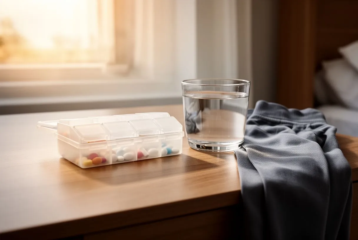
Balancing effectiveness with safety: Diuretic dosing requires regular monitoring. Too little and the oedema persists; too much and the patient risks dehydration, low blood pressure, and electrolyte disturbances that can trigger dangerous arrhythmias. Potassium levels, kidney function, and blood pressure are checked periodically, and doses are adjusted accordingly. Some patients require a combination of diuretics that work at different sites in the kidney to achieve the right balance. Long-term outcomes depend not just on fluid removal but on addressing the coronary disease that contributed to the heart failure — a topic explored in a study comparing outcomes for in-stent restenosis chronic total occlusions against de novo lesions (Procedural Results and Long-Term Outcomes of Percutaneous Coronary Intervention for in-Stent Restenosis Chronic Total Occlusion Compared with de novo Chronic Total Occlusion).
Patients sometimes ask whether natural remedies or herbal diuretics can substitute for prescription medication. While a low-sodium diet, elevation of the legs, and compression stockings can support medical therapy, they cannot replace diuretics when the heart is the root cause. Self-medicating with over-the-counter products can interact with heart medications, alter potassium levels, and mask worsening symptoms. Any change to diuretic strategy should be discussed with the cardiologist managing your overall heart failure care plan.
When Ankle Swelling Signals Cardiac Problems
Not every swollen ankle warrants a cardiology referral, but certain patterns elevate the concern. Bilateral swelling that progresses over days or weeks, swelling accompanied by unexplained weight gain, and swelling that coexists with breathlessness, fatigue, or palpitations all point more strongly toward a cardiac origin. Unilateral swelling, especially if it is sudden, painful, and associated with redness or warmth, raises a different concern — deep vein thrombosis — which itself can be provoked by reduced cardiac output and immobility.
Red-flag features that need urgent attention: Seek prompt medical evaluation if swelling is accompanied by chest pain, fainting, a rapid irregular heartbeat, or breathlessness at rest. These combinations may indicate an acute cardiac event, a significant arrhythmia, or a pulmonary embolism. Patients with known coronary disease who are on aspirin and other preventative medications should not assume that current therapy protects them from decompensation — any new or worsening fluid retention warrants reassessment by their treating team.
Other cardiac procedures and devices also intersect with swelling management. A patient who has had balloon angioplasty to open a narrowed coronary artery may still develop oedema if the underlying heart muscle is damaged. Someone with an automated external defibrillator implanted for life-threatening arrhythmias may experience fluid retention if their heart function subsequently declines. Each clinical context is unique, which is why general online checklists can only go so far — a face-to-face assessment with a cardiologist remains essential for accurate diagnosis and safe treatment planning.
Sudden Weight Gain and Fluid Retention in Heart Failure
Daily weight monitoring is one of the simplest and most powerful self-management tools for heart failure patients. A weight gain of two kilograms or more over two to three days, without any change in diet, is a strong indicator of fluid retention — even before visible swelling worsens. Many heart failure programmes ask patients to weigh themselves each morning under consistent conditions: same time, same clothing, after using the bathroom, before eating or drinking.
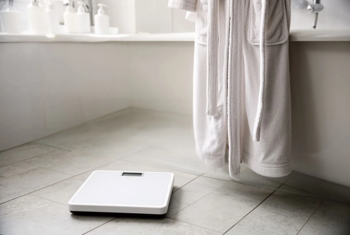
What the weight trend tells your care team: A steady upward trend signals that the current diuretic dose may no longer be adequate, that sodium intake has crept up, or that a new trigger — an arrhythmia, a missed medication, a respiratory infection — is placing extra strain on the heart. Catching the trend early often allows for a simple outpatient adjustment rather than a hospital admission. This proactive approach is a core principle of guideline-directed medical therapy and is supported by long-term outcome data from peer-reviewed open-access journals catalogued by DOAJ.
Families sometimes ask whether a weight gain of a kilogram or two over a week is equally concerning. A slower trend may reflect dietary lapses or reduced activity and is worth mentioning at the next clinic visit, but it does not usually require urgent action. The key distinction is speed. Rapid gain equals rapid fluid accumulation, and rapid fluid accumulation in a compromised heart can progress to pulmonary oedema with little warning. Keeping a written or app-based weight log that you can share with your cardiologist makes these patterns far easier to interpret during a consultation.
Getting Your Swelling Assessed
If you or a family member are experiencing persistent or worsening leg swelling, a structured cardiac assessment is the most reliable way to determine whether the heart is involved. A thorough evaluation typically includes a clinical history, physical examination, an electrocardiogram to detect rhythm problems or signs of past heart attacks, an echocardiogram to assess valve function and pumping strength, and blood tests to check kidney function and heart failure markers. Each piece builds a picture that no single test can provide alone.
What to expect from a consultation with Dr Alejandro Molina: Patients attending the Guadalajara, Querétaro, or Hermosillo clinics receive a calm, detailed discussion of findings rather than a rushed prescription. If heart failure is confirmed, the conversation covers staging, medication options, lifestyle adjustments, and — where relevant — procedural or surgical pathways. The approach is always individualised: what works for one patient's anatomy and circumstances may not suit another, even with the same diagnosis. The emphasis is on evidence-based recommendations and honest discussion of expected outcomes and recovery realities.
- Symptom review: When did the swelling start, how fast has it progressed, and what other symptoms accompany it?
- Cardiac imaging: Echocardiography and sometimes additional scans reveal the heart's structure and pumping efficiency.
- Treatment roadmap: A clear plan covering medications, follow-up intervals, warning signs to watch for, and when to escalate.
For international patients and families travelling to Mexico for cardiac care, the practice provides guidance on logistics, documentation, and continuity of care with referring physicians at home. The goal is not simply a single appointment but an ongoing relationship that supports long-term heart health. If swelling is causing you concern, the most important step is to have it evaluated rather than to guess at the cause — book a consultation with a qualified cardiac team and bring your questions, your medication list, and your weight record with you.