Veno-Arterial ECMO: How Cannulas Support the Heart
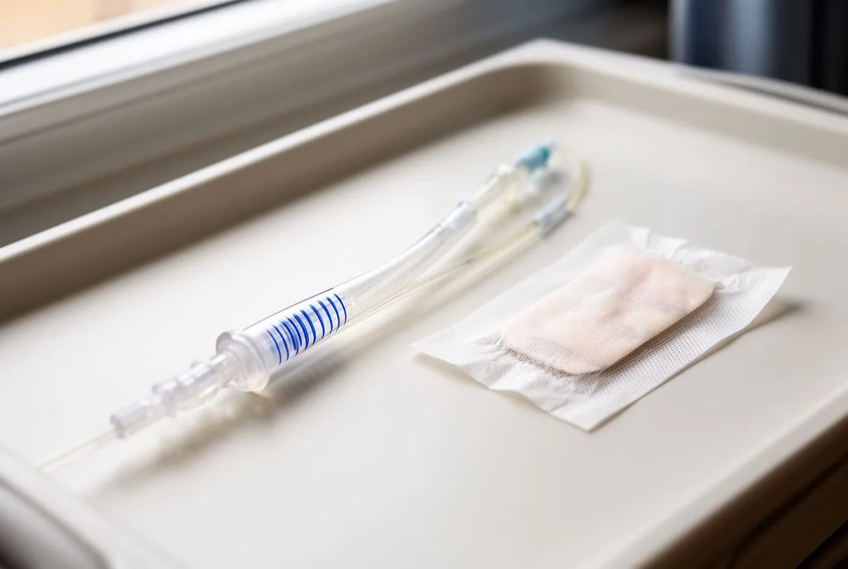
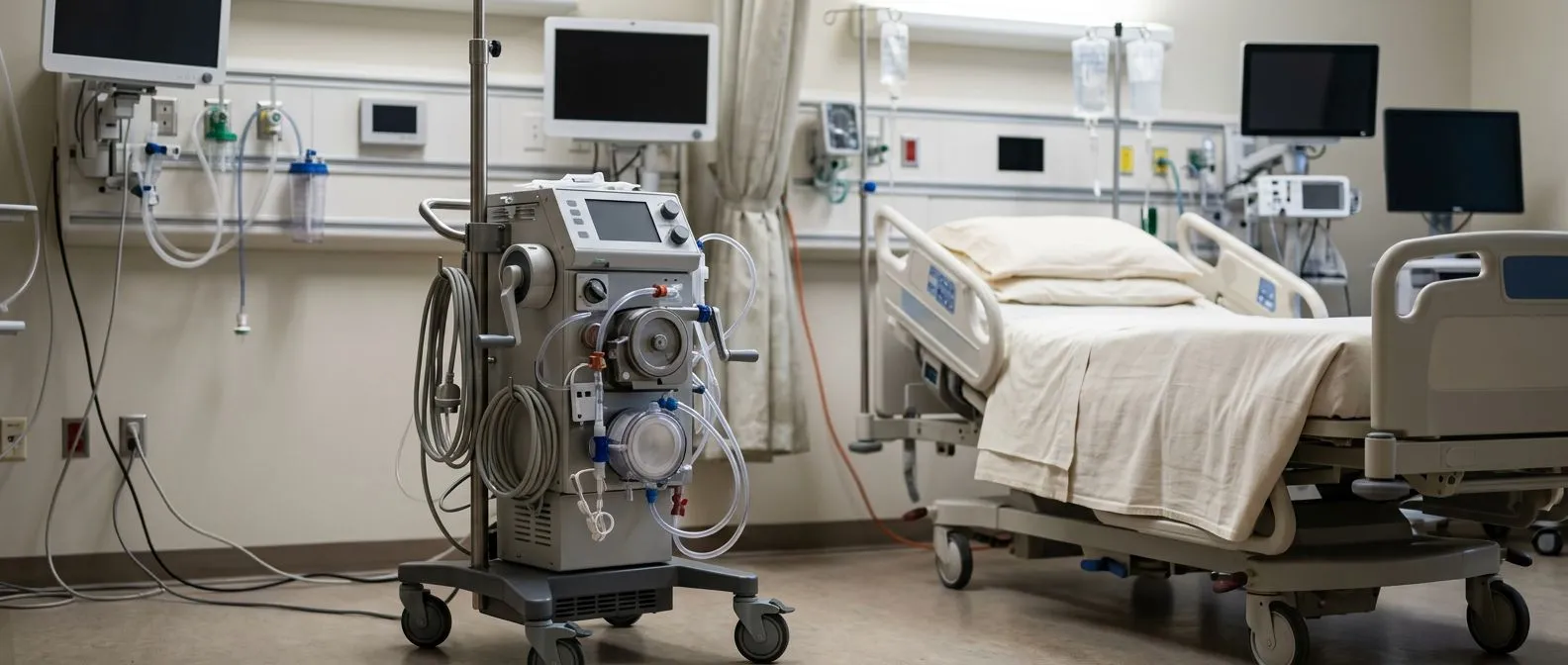
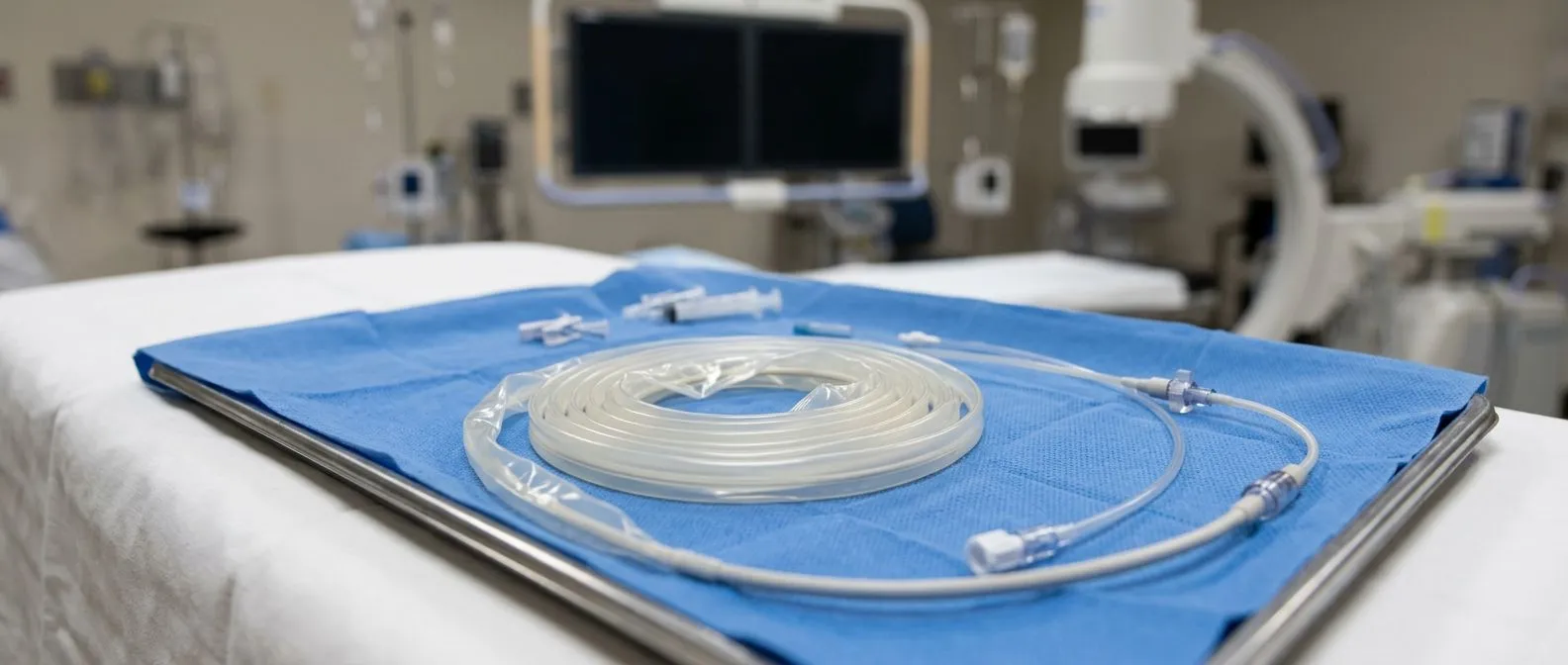
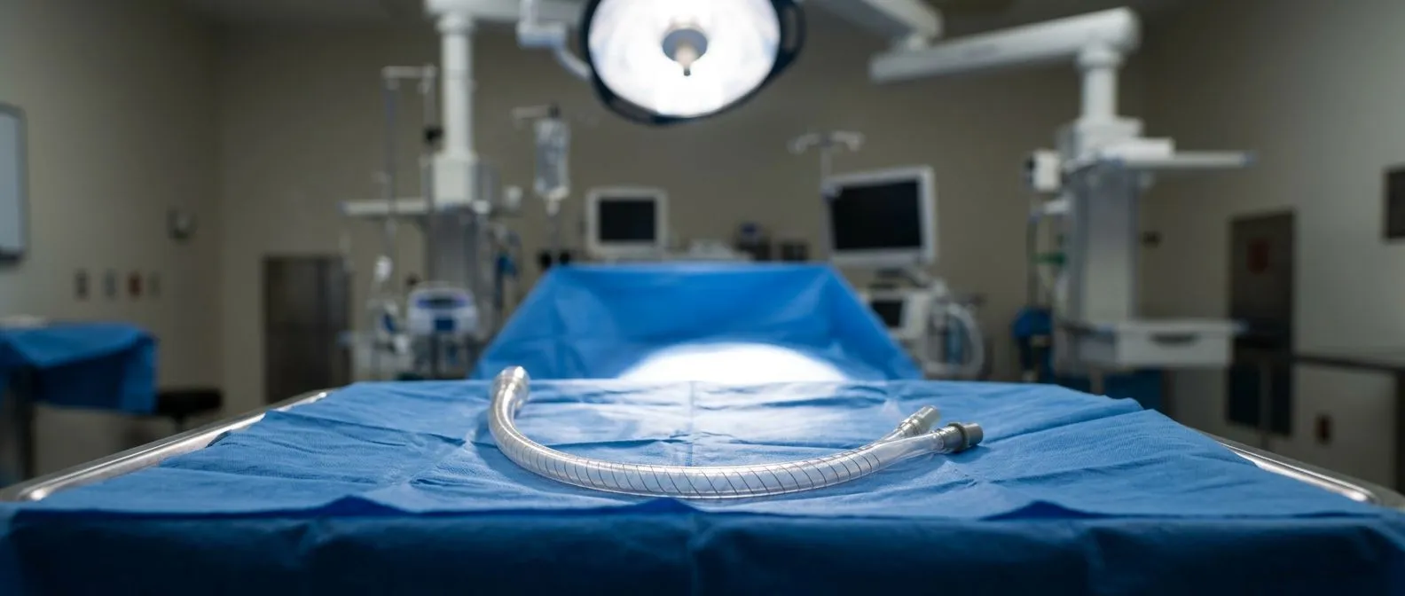
Veno-arterial ECMO cannulas connect the patient to the circulatory support machine.
When a heart is too weak to sustain the body on its own, medicine has a way to give it time to recover: extracorporeal membrane oxygenation, or ECMO, a machine that takes over the work of the heart and lungs. Veno-arterial ECMO cannulas are the tubes that connect the patient's circulation to that machine — a venous cannula drains blood out, an arterial cannula returns it under pressure, and the machine in between oxygenates the blood before it re-enters the body. For patients and families, understanding what these tubes are, where they are placed, and what they do is one of the best ways to feel less afraid of an intimidating machine. This guide walks through each part of the system in plain language.
What Is Veno-Arterial ECMO?
Veno-arterial ECMO, often abbreviated VA ECMO, is a form of mechanical circulatory support in which blood is drained from a vein, passed through a membrane oxygenator that adds oxygen and removes carbon dioxide, and then returned to an artery. Because the machine is connected to the arterial side, the oxygenated blood it returns carries part of the load of the failing heart — the machine is effectively pumping for the patient.
How it differs from the lung-support version. The other main configuration, veno-venous ECMO, supports the lungs only: blood is drawn from and returned to veins, so it oxygenates the blood but does not help the heart move it. That is why veno-arterial ECMO is the configuration chosen when the problem is the heart itself — a massive heart attack, cardiogenic shock (when the heart cannot pump enough blood despite adequate filling), or a heart that is stunned after cardiac surgery.
Common reasons a cardiac team chooses veno-arterial ECMO include severe cardiogenic shock after a myocardial infarction, fulminant myocarditis (inflammation of the heart muscle), failed resuscitation from cardiac arrest, and postcardiotomy support — the situation in which a patient cannot come off the heart-lung machine after open heart surgery. In each of these cases, the goal is the same: give the heart time to rest and recover, or to bridge to a durable solution such as a ventricular assist device or transplantation.
How Do Veno-Arterial ECMO Cannulas Work?
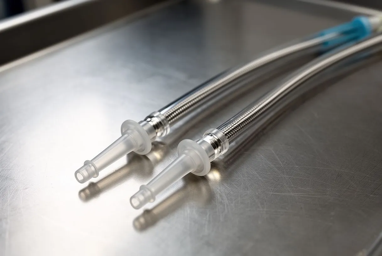
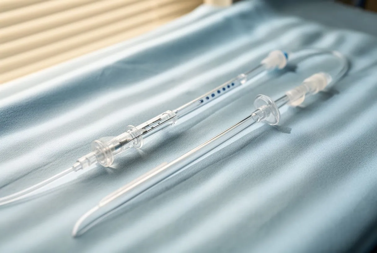
Every ECMO circuit has three parts: the tubing that takes blood away (the drainage side), the pump-oxygenator unit that treats the blood, and the tubing that brings it back (the return side). The cannulas are the short, reinforced tubes at the ends of that circuit — the points of contact with the patient. Their design matters enormously because they determine how much blood can be moved, and how much damage the blood suffers on the way.
Why the design matters so much. ECMO cannulas are made of biocompatible, non-thrombogenic (clot-resistant) materials, usually with a wire-reinforced wall so they cannot kink when the patient moves. Their tips are shaped to sit harmlessly in the vessel — a tapered tip for the arterial side, a multi-stage or flexible tip for the venous side. Because blood moves through them at hundreds of milliliters per minute, the diameter of each cannula is the single biggest determinant of maximum achievable flow.
The difference between drainage and return. The venous cannula works by suction: the pump pulls blood out of the venous system. The arterial cannula works by pressure: blood is pushed back into the arterial circulation. This asymmetry is why venous cannulas tend to be larger in diameter than arterial ones of the same circuit — the veins must give up blood at low pressure, while the arteries accept it at a higher one. Getting the geometry of the pair right is what makes the circuit behave properly, and it is a large part of why cannulation technique matters.
Venous Cannula ECMO: Where the Blood Is Removed
The venous cannula is placed in a large vein carrying blood back toward the heart. In peripheral veno-arterial ECMO, the commonest site is the femoral vein in the groin — the vein is large, easy to reach, and can be cannulated without surgery at the bedside. The tip of a femoral venous cannula is advanced up the inferior vena cava until it reaches the right atrium, the chamber of the heart that collects blood from the body. If the vein in the groin is not usable, the internal jugular vein in the neck is the usual alternative.
What the team is aiming for. The correct tip position is checked by ultrasound, X-ray, or transesophageal echocardiography before the circuit runs at full speed. A cannula that is too shallow will not drain enough blood; one that has advanced too far can press against the wall of the heart. Adjusting position at the start is far easier than discovering problems later.
Drainage capacity. The amount of blood the venous side can deliver depends on the vein's diameter, the patient's circulating volume, and the cannula's inner diameter. The team confirms it can be done before the circuit is committed; that is precisely what our team checks in the multidisciplinary review before recommending veno-arterial ECMO — whether the patient's vessels can support a drainage and return arrangement that will meet the target flow.
Arterial Cannula ECMO: Where the Blood Returns
The arterial cannula is the tube that returns oxygenated blood to the body, and in veno-arterial ECMO it is where some of the most consequential decisions are made. The standard peripheral site is the common femoral artery in the groin. Oxygenated blood is then pumped retrograde — backward — up the aorta, the body's main artery, assisting the heart's own output from below.
The retrograde perfusion problem. When blood is returned through the femoral artery, it travels up the aorta in the opposite direction to the heart's own output. If the heart is recovering and starts ejecting again, the two flows can meet inside the aorta and compete, which reduces the area the machine supports. This is why teams monitor the balance between native heart output and ECMO flow carefully, often adjusting the circuit as the heart recovers.
The Harlequin phenomenon. A well-known complication of femoral arterial cannulation is differential hypoxia: the patient's native heart ejects poorly oxygenated blood (if the lungs are also failing), which flows up the arch and supplies the head, while the ECMO returns oxygenated blood that flows down the descending aorta. The upper body can become cyanotic while the lower body is well oxygenated — an appearance teams call the Harlequin, or North-South, syndrome. If this occurs, it can often be managed by adding a second oxygenator connection or by switching to a central cannulation arrangement.
Peripheral vs Central ECMO Cannulation Sites
Cannulation sites come in two broad families, and the choice between them shapes the entire course of support. Peripheral cannulation uses the groin and neck vessels; central cannulation requires opening the chest and connecting directly to the great vessels.
Peripheral ECMO cannulation. Peripheral cannulation is fast, can be performed at the bedside, and is the usual choice for resuscitation and for most patients in shock. The drainage cannula typically sits in the femoral or internal jugular vein; the return cannula in the femoral artery. The downside is that groin vessels have finite limits — the artery cannot always accept a large enough cannula, and retrograde flow carries the risks described above.
- Femoral vein: the workhorse drainage site; large, accessible, and cannulated percutaneously (through the skin, no surgical cut).
- Internal jugular vein: the neck alternative; particularly useful if the femoral veins are unusable or the patient needs a longer drainage length.
- Common femoral artery: the standard peripheral return site; often accompanied by a small distal perfusion cannula to keep the leg supplied.
Central ECMO cannulation. Central cannulation means the surgeon opens the chest and places the drainage cannula directly in the right atrium and the return cannula in the ascending aorta. This is the standard arrangement in postcardiotomy patients — those who cannot come off the heart-lung machine after surgery — and it is chosen when central flows are needed, when the femoral vessels are inadequate, or when retrograde flow risks complications. It requires the chest to remain open or surgically closed, with a longer recovery, but it delivers superior flow characteristics and avoids the differential hypoxia problem entirely.
How the team decides. In our practice, the choice between peripheral and central cannulation is reviewed by the multidisciplinary team against international practice guidelines before a recommendation is given, and the patient receives a clear proposal with the alternatives laid out — the final decision is always theirs.
How ECMO Cannula Size Selection Is Decided
Cannula sizes are measured in French units (Fr), a scale that describes the outer diameter of the tube — higher numbers mean larger tubes. Venous cannulas in veno-arterial ECMO commonly range from 21 to 25 Fr, and arterial cannulas from 15 to 19 Fr, though the exact numbers depend on the manufacturer and on the patient's own anatomy. The size selection is a genuine engineering trade-off: a larger cannula allows more flow, but a larger cannula needs a larger vessel and carries a higher risk of vessel injury.
Matching the cannula to the patient. The team measures the target vessel with ultrasound before cannulating — a femoral artery smaller than about 7 mm in diameter is generally considered borderline for a large arterial cannula, and many teams then plan to place a smaller arterial cannula, or to use a side-graft (sewing a small tube onto the artery surgically so the cannula enters through the graft rather than through the artery wall). This approach protects the vessel while still permitting large flows.
What the flow numbers depend on. Maximum achievable flow is governed by the pressure gradient across the cannula and the cannula's internal resistance — longer, narrower cannulas resist flow more. The published ECMO literature, including reviews on the technique published by major academic societies, consistently emphasizes that undersizing the cannulas is one of the most common reasons a circuit fails to deliver the target flow. Planning the cannula sizes before the patient is cannulated is therefore a planning exercise in the true sense, not a formality.
What Happens During the ECMO Cannula Insertion Procedure?
Peripheral cannulation is usually performed in the intensive care unit, the catheterization lab, or the operating room, with local or general anesthesia depending on the patient's condition. The insertion itself takes several minutes to a few hours, depending on how many cannulas are placed and whether any complications arise.
The sequence of steps. A typical percutaneous insertion proceeds as follows:
- Ultrasound mapping: the vessel is located and its diameter measured so cannula size can be chosen confidently.
- Wire access: a fine guidewire is passed into the vessel under ultrasound guidance, then the tract is dilated stepwise to accommodate the cannula.
- Cannula insertion: the cannula is advanced over the wire to the planned depth, and its position is confirmed by imaging or echocardiography.
- Securing and connection: the cannula is sutured to the skin, the wire is removed, and the circuit is connected, air is purged, and support is initiated.
What the patient feels and sees afterward. Once the cannulas are in place and secured, the patient is connected to the ECMO circuit, and the pump begins circulating blood immediately. A patient on peripheral veno-arterial ECMO lies still with the cannulas coming out of the groin and often the neck, connected to the ECMO machine by the circuit tubing. The patient is on a ventilator (breathing machine) and sedated in most cases, and the team monitors the circuit continuously — flow, pressure, oxygenation, and the patient's own cardiac function.
What we do before the patient arrives. In our practice, the first contact is a free video consultation, where we get to know the patient, talk through the condition, and say which studies are still needed so no unnecessary ones are ordered; a face-to-face visit follows when the patient decides to go ahead. When the patient is already in the hospital, the same sequence is adapted to the urgency of the situation.
ECMO Cannula Flow Rates and What They Mean
Flow rates are the numbers the team watches most closely, and they are measured in liters of blood per minute. Adult veno-arterial ECMO typically supports a flow of 3 to 5 liters per minute, which is enough to take over the majority of the cardiac output when the cannulas are sized and positioned well.
Why more flow is not automatically better. Higher flows are limited by the venous drainage — if the pump tries to draw more blood than the vein can supply, the circuit will collapse the vein, and the drainage will falter. This "circuit collapse" is avoided by balancing the drainage cannula size, the patient's fluid volume, and the pump's speed. The team sets the pump to a speed that produces the target flow without over-drawing the vein.
What the flow tells the family. In practice, a flow of 4 liters per minute with a good heart means the heart has been substantially relieved of its workload. The team will often reduce the pump speed gradually over the following days if the heart is recovering, using the flow reduction as a test of the heart's function. Weaning — the process of reducing ECMO support until the heart can stand alone — is done step by step, with the patient's own cardiac function measured by echocardiography at each stage.
ECMO Cannula Complications to Know About
Cannulas carry real risks, and the best teams are candid about them rather than reassuring. The complications fall into a few families: vascular injury, bleeding, clotting, limb ischemia, and infection.
Leg ischemia. When the arterial cannula is placed in the femoral artery, the blood supply to the leg below the cannula can be compromised. The standard solution is a distal perfusion cannula — a small tube placed in the artery below the main cannula, sometimes called a "back-bleed," which keeps the leg supplied. The leg is monitored continuously for signs of poor circulation, and if the limb is at risk, the team can change the cannulation strategy.
Bleeding and clotting. Patients on ECMO receive anticoagulation (medication to prevent blood clotting), usually heparin, and the team monitors clotting with a range of blood tests. This medication prevents clotting in the circuit but increases bleeding risk at the cannulation sites, which is why bleeding is one of the commonest complications of prolonged ECMO. Clotting in the circuit itself can also occur, and if it does, the cannula or the circuit components may need to be replaced.
Infection and vascular injury. The cannulation sites are a constant route of infection, and they are kept clean and monitored. Pseudoaneurysm (a bulge in the artery wall at the cannulation site) and vessel injury are also documented risks. To reduce the risk of neurological complications from blood clots, teams monitor for signs of stroke and act promptly if they occur.
When Do Veno-arterial ECMO Cannulas Get Removed?
The cannulas come out when the patient's heart has recovered enough to sustain the body on its own — or when a decision is made that the circuit is no longer serving the patient. The removal process is a deliberate one, and understanding it helps families follow the plan.
Weaning, not switching off. Before removal, the team reduces the pump speed gradually while the heart's own function is tested, using echocardiography to assess how well the heart is ejecting. A patient who tolerates a low flow without deterioration is a candidate for decannulation. This process can take days, and it is the point at which the cannulas and the circuit are no longer needed.
Decannulation. The removal procedure itself is performed in the intensive care unit or the operating room, under local or general anesthesia. The femoral arterial cannula is usually removed with the artery closed by direct pressure or a small suture, often with a closure device (a small device that seals the hole in the artery) to reduce the risk of bleeding or pseudoaneurysm. The venous cannula is usually removed with simple pressure. If the cannula was placed surgically, the surgical sites are closed and a dressing is applied.
Recovery and follow-up. After the cannulas are removed, the patient is monitored in the intensive care unit for complications — bleeding, infection, clotting — before being stepped down. The recovery period is longer than most patients expect: weeks, not days, and this is true for every option the team recommends. When the patient recovers, the team encourages a second opinion at any time, as a normal part of deciding about heart surgery — a practice that supports a sound choice for the patient, and one that our team encourages as a routine part of the process.
What to expect long term. Many patients who survive veno-arterial ECMO go on to a full life, especially if the underlying condition was reversible. Some patients, however, require long-term follow-up for the condition that led to the support — the heart muscle that was damaged, the valves that were not working, or the arteries that were affected. The team follows the patient through that process, and the plan is discussed in advance rather than being discovered after the fact.
If you or a family member is facing a decision about heart surgery, mechanical circulatory support, or any cardiac procedure, the most valuable next step is to speak with a qualified cardiac team. We encourage every patient and family to discuss their own situation with a specialist rather than to self-diagnose or to decide alone — and to book a consultation with a cardiac surgeon who can review the imaging and the studies, explain the findings in plain words, and lay out the options with the alternatives. In our own practice, the first contact is a free video consultation, and we take the time to explain what the studies actually show, in plain words, and to answer every question before any decision.