

Ventilation-Perfusion Scan for Pulmonary Embolism
Ventilation-perfusion scan embolism detection explained: how the V/Q test diagnoses pulmonary clots, what results mean, and how to prepare.
A sudden shortage of breath, chest pain that worsens when you breathe deeply, or an unexplained racing heartbeat can all point to one serious condition: a pulmonary embolism, which is a blood clot that has travelled to the arteries of the lungs. One of the classic tests used to confirm or rule out this diagnosis is a ventilation-perfusion scan embolism evaluation, often shortened to a V/Q scan. This article explains, in plain language, what the test involves, when doctors choose it over a CT scan, how the results are read, and what you should expect before, during and after the appointment.
What Is a Ventilation-Perfusion Scan?
A ventilation-perfusion scan is a two-part nuclear medicine study of the lungs. "Ventilation" refers to the air you breathe in and out; "perfusion" refers to blood flow through the lungs. The test compares the two, because a pulmonary embolism typically blocks blood flow in an area of the lung that is still receiving air normally. That mismatch between breathing and circulation is the signature the scan looks for.
How the two parts work: in the ventilation phase, you inhale a small amount of a mildly radioactive gas or aerosol, and a special camera records how air distributes through your lungs. In the perfusion phase, a tiny quantity of a radioactive tracer is injected into a vein, and the camera maps where blood is flowing. Neither substance is a dye that affects the kidneys, and the radiation dose is small.
The formal name you may see on a report is a nuclear perfusion scan or a ventilation-perfusion scan; some older reports call it a lung scan for embolism. All of these describe the same principle: looking at air and blood side by side to find where they disagree.
How a V/Q Scan Detects Pulmonary Embolism
The logic behind the test is elegant. Lung tissue is unusual because it receives blood from two circuits: the pulmonary arteries, which carry oxygen-poor blood from the heart to the lungs, and the bronchial circulation, which supplies the lung tissue itself. When a clot lodges in a pulmonary artery, the blocked area still gets air and still gets some bronchial blood, so the tissue usually stays alive. What changes is the perfusion pattern, and that is exactly what the tracer reveals.
What the camera sees: on the perfusion images, a clot shows up as one or more "cold" areas — segments of lung where the tracer cannot reach because the feeding artery is blocked. On the ventilation images, those same areas usually light up normally, because air still flows there. This combination of absent perfusion with preserved ventilation is called a "mismatch," and it is the finding that raises suspicion for pulmonary embolism.
It is worth understanding the anatomy involved. The pulmonary arteries and the pulmonary veins are separate structures: clots lodge in the arteries, while the veins return oxygenated blood to the heart. A large clot, or several clots, can raise the pulmonary artery pressure because the right side of the heart has to push against a blockage. This is one reason an embolism can strain the heart and not just the lungs.
When Doctors Order a Ventilation-Perfusion Scan for Blood Clots
Doctors do not send every patient with chest pain straight to a nuclear scanner. The usual first step is a D-dimer blood test, which detects breakdown products of clots. If that is negative and your pre-test risk is low, pulmonary embolism is usually ruled out without imaging. Imaging comes next when suspicion remains.
Common reasons a V/Q scan is chosen:
- Kidney concerns: the CT alternative requires iodine contrast dye, which can stress the kidneys; a V/Q scan avoids contrast entirely.
- Pregnancy: the radiation dose to the unborn child is lower with a V/Q scan, so it is often preferred for expectant mothers.
- Known clot history: patients with pre-existing lung disease or previous embolism sometimes benefit from the functional information a nuclear scan provides.
- Contrast allergy: anyone who has reacted badly to iodinated contrast can be scanned safely with the nuclear technique.
In our team's experience reviewing cases across Guadalajara, Querétaro and Hermosillo, the test is most useful when it is chosen deliberately — after the multidisciplinary review weighs the patient's kidney function, pregnancy status and prior imaging — rather than as a default first test for every patient with breathlessness.
V/Q Scan vs CT Pulmonary Angiography
The most common competitor to the V/Q scan is CT pulmonary angiography, a CT scan performed after injecting contrast dye into a vein to light up the pulmonary arteries directly. In many hospitals it has become the default test because it is fast and shows the clot itself, not just its consequences.
Where CT wins: a CT pulmonary angiogram gives an anatomical picture. It shows the size and location of a clot, can reveal alternative diagnoses such as a dissection or pneumonia, and can be completed in minutes. Modern dual energy CT techniques also add functional information, narrowing the gap between the two modalities.
Where the V/Q scan wins: it avoids iodine contrast and its radiation dose to the breast tissue is lower, which matters for younger women and pregnant patients. It is also the preferred tool when doctors suspect chronic thromboembolic pulmonary hypertension, a long-term complication in which old clot material narrows the pulmonary arteries and raises pulmonary artery pressure permanently. For that condition, the perfusion pattern on a nuclear scan can be more informative than a CT.
In our practice we review every imaging decision as a team before recommending one test or the other, and we explain to the patient in plain words why a particular route is proposed — and what the alternatives would be. The final choice always rests with the patient.
How to Prepare for a Lung Ventilation Scan Procedure
Preparation for a V/Q scan is minimal, which is one of its quiet advantages. You can eat and drink normally beforehand, and you do not need to fast or stop most medications.
Practical steps before the appointment:
- Tell the nuclear medicine team if you are pregnant or breastfeeding, so the dose can be adjusted or the test reconsidered.
- Mention blood thinners and any recent nuclear scans, because residual tracer from another study can interfere with the images.
- Bring recent Pulmonary Function Tests or imaging if you have them; they help the reading physician interpret areas of pre-existing lung disease.
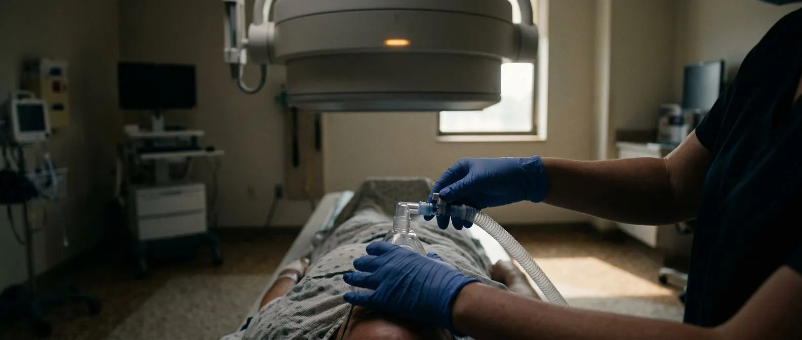
During the scan itself: the ventilation part usually involves breathing the tracer gas through a mouthpiece or mask for a few minutes, followed by a set of images. The perfusion part involves a small injection into an arm vein, then several sets of images taken from different angles while you lie still on the camera table. The whole appointment typically takes between thirty minutes and an hour, and the camera never touches you. In our consulting rooms we always take the time beforehand to explain exactly what the study shows and to answer every question, so patients arrive for the scan knowing what to expect.
Ventilation-Perfusion Scan Results Interpretation
Understanding your report helps you take part in the decision that follows. Nuclear medicine physicians classify V/Q results into probability categories, based on how many mismatched defects appear and what shape they are.
How findings are graded: a "normal" scan shows even perfusion matching ventilation, which effectively rules out pulmonary embolism. "Low probability" studies show small or non-segmental defects that could come from many other causes. "High probability" studies show several large, wedge-shaped mismatched defects — the classic embolism pattern. Intermediate results fall in between and usually trigger further testing.
What happens after an abnormal result: a high-probability scan usually leads straight to anticoagulant treatment, the blood-thinning medication that stops clots growing and prevents new ones. A large central clot with signs of heart strain may prompt consideration of a Ventilation-perfusion Scan-guided pathway toward more aggressive options, including catheter-based clot removal. In historical practice, an inferior vena cava filter — a small basket placed in the large abdominal vein to catch clots travelling from the legs — was used for patients who could not take anticoagulants, a device described in detail in a review of the permanent inferior vena cava filter for prevention of pulmonary artery embolism (creation of the permanent inferior vena cava filter for prevention of pulmonary artery embolism). Modern practice favours retrievable filters used only when anticoagulation is truly impossible.
Sometimes the scan is not the whole story. An embolism can occasionally be the first sign of an underlying disease, including cancer; there are published case reports of recurrent venous thromboembolism as the initial clinical presentation of gastric cancer (Recurrent Venous Thromboembolism as the Initial Clinical Presentation of Gastric Cancer: A Case Report). When clots recur without an obvious cause, a responsible team looks for the reason rather than simply treating each episode.
How Accurate Is a V/Q Scan?
Accuracy depends heavily on the patient. In someone with healthy lungs and a clear chest X-ray, a normal perfusion scan makes pulmonary embolism extremely unlikely, and a high-probability pattern makes it very likely. Those two ends of the scale are where the test performs best.
Where accuracy drops: patients with asthma, chronic bronchitis, emphysema or previous lung surgery often have areas where both ventilation and perfusion are reduced together — a "matched" defect. Matched defects can be caused by lung disease alone, which makes the scan harder to read and pushes results into the intermediate category. In those patients, CT is usually preferred.
The landmark PIOPED study, which compared V/Q scans against pulmonary angiography in large numbers of patients, established the probability categories still used today and showed why reading the scan alongside the patient's clinical picture matters more than reading it alone (Europe PMC). In our own case reviews we never act on a scan in isolation; every imaging result is discussed against international practice guidelines before a treatment recommendation is made.
Ventilation-Perfusion Scan Risks and Limitations
The V/Q scan is one of the lowest-risk imaging studies in medicine. There is no needle in an artery, no contrast dye and no sedation. The main consideration is radiation, and it is modest — comparable to or lower than a CT of the chest, and delivered through two very small tracer doses that the body clears naturally within hours to a day.
Known limitations to discuss with your doctor:
- Intermediate results: a meaningful share of scans land in the middle categories and do not give a definite answer on their own.
- Underlying lung disease: emphysema and similar conditions create matched defects that blur the picture.
- Availability and speed: the test takes longer than CT and requires a nuclear medicine department with specific tracers in stock.
- No alternative diagnoses: unlike CT, a V/Q scan cannot show what else might be causing your symptoms if embolism is ruled out.
Serious allergic reactions to the tracers are rare. The injection site may bruise slightly, and some patients notice a metallic taste from the ventilation gas for a few minutes. In our team's experience, patients tolerate the study comfortably; the greater difficulty is usually the waiting, which is why we explain the timeline and the next steps clearly from the first video consultation onwards.
V/Q Scan in Pregnancy and Special Situations
Pregnancy deserves its own paragraph because it changes the calculus. Suspected pulmonary embolism in an expectant mother cannot simply be ignored, since untreated embolism is dangerous for both mother and baby — but every imaging choice must balance diagnostic certainty against radiation exposure to the fetus.
Why the V/Q scan is often first choice in pregnancy: the perfusion component alone frequently provides the answer, and it delivers a lower dose to breast tissue and to the fetus than CT pulmonary angiography. Guidelines from international thoracic imaging societies support a perfusion-only scan in many pregnant patients: if that study is normal, embolism is excluded and no ventilation phase is needed at all.
Other special situations include patients with severe kidney impairment, where avoiding contrast is the priority; patients with a history of reaction to iodinated contrast; and patients being evaluated for chronic thromboembolic pulmonary hypertension, where the perfusion pattern helps plan treatment, sometimes involving procedures such as antegrade cerebral perfusion during complex surgery for associated conditions like total anomalous pulmonary venous drainage. In those complex cardiac cases the imaging picture must be read alongside other studies, including a Stress Test on a treadmill or a treadmill test with imaging, and measurement of Pulmonary Artery Pressure through echocardiography or catheterisation, to build a complete picture before any operation.
For international patients considering care in Guadalajara or elsewhere in Mexico, cost is a fair question — but it should never be the only one. Nuclear medicine lung scans are widely available in major Mexican private hospitals, and prices vary considerably between facilities. Rather than quoting figures that may not match your situation, we encourage patients to ask any hospital for a written quote that includes the radiopharmaceutical, the reading fee and any follow-up imaging that an intermediate result might require.
What to look for when choosing where to be tested:
- A nuclear medicine department that performs V/Q studies regularly, not occasionally.
- A team that reads the scan together with your full clinical history, not in isolation.
- A clear pathway for what happens if the result is intermediate — the plan should exist before you lie on the camera table.
In our practice, the first contact is a free video consultation: we get to know the patient, talk through the condition, and identify exactly which studies are still needed so no unnecessary ones are ordered. When the patient decides to move forward, a face-to-face visit follows at our consulting rooms in Guadalajara, Querétaro or Hermosillo. Dr Molina trained in cardiothoracic surgery at the Universidad de Guadalajara and in cardiac, minimally invasive, robotic and structural surgery at the University of Montreal, and the team's experience spans conventional, minimally invasive, robotic and transcatheter procedures — we recommend the less invasive route only where the evidence supports it for that specific patient. We also encourage second opinions, and we treat seeking one as a normal, healthy part of deciding about heart care.
A ventilation-perfusion scan remains a valuable, low-risk tool for diagnosing pulmonary embolism, particularly for pregnant patients, those with kidney disease and those who cannot receive iodine contrast. Its greatest strength is what a normal result tells you: when perfusion is even and matches ventilation, a clot in the lungs is effectively excluded. Its honest limitation is the intermediate result, which demands a team willing to think through the next step with you. If you have been experiencing unexplained breathlessness, chest pain on breathing, or symptoms after a long flight or surgery, do not self-diagnose — discuss your situation with a qualified cardiac team, ask what a ventilation-perfusion scan embolism workup would involve in your case, and book a consultation rather than waiting to see if the symptoms pass.






