Annual Echocardiogram After Valve Surgery: What It Checks
Annual echocardiogram after valve surgery checks valve function, heart pumping, and leak symptoms.
An annual echocardiogram after valve surgery is the single most useful test for keeping track of how your repaired or replaced valve is doing. It is a painless ultrasound scan of the heart, and it takes pictures of the valve in motion while your heart beats. Whether you had a Stented Valve, a bioprosthesis, or a repair of your own tissue, the yearly scan gives your cardiology team an early warning long before you would feel any change. This guide explains what the test checks, what to expect on the day, and how the results shape your long-term care.
Why Do I Need an Annual Echocardiogram After Valve Surgery?
Your valve changes over time, quietly. A replaced or repaired valve does not stay frozen at the moment of surgery. Tissue valves slowly stiffen, tiny leaks can develop, and the heart muscle itself adapts to its new plumbing. An annual echocardiogram after valve surgery catches these shifts while they are still easy to manage, often years before symptoms would appear.
Guidelines call for lifelong monitoring. International practice guidelines recommend regular surveillance imaging for every surgical or catheter-based valve, including Transcatheter Aortic Valve Implantation patients and people who have had a Valve-sparing Root Replacement. The schedule is usually a baseline study soon after surgery, then periodic checks — commonly yearly once things are stable. This heart valve replacement monitoring test is not optional; it is part of the treatment itself.
Peace of mind has real value. Many patients tell us they live more freely knowing their valve is checked every year. Follow-up echos are interpreted against international guidelines, and you should receive a clear explanation of what changed — or what did not — and what, if anything, needs to happen next.
What Does an Echocardiogram Check After Valve Surgery?
The short answer is: the valve, and everything around it. A post valve surgery cardiac ultrasound measures how well the valve opens, how well it closes, and how the heart is coping with it. The sonographer and cardiologist look at several things in every study, and each one answers a different question about your recovery and your future.
- Valve function: whether the valve leaflets or cusps open fully and seal completely with each heartbeat, and whether any stenosis (narrowing) or regurgitation (leaking backward) has appeared.
- Heart pumping strength: the ejection fraction, a percentage that shows how forcefully the left ventricle pushes blood out each beat.
- Chamber size and pressures: whether the heart's chambers are enlarging or the lung pressures are rising, which can signal a valve problem before you feel it.
- Structures around the valve: the Valve Annulus (the ring the valve sits in), the aorta, and — after repair — the tissue the surgeon worked on.
Comparing this year to last year is the real power of the test. A single echo gives a snapshot, but an echocardiogram after heart valve replacement becomes truly informative when measured against your previous studies. Small changes in valve gradients, leak severity, or heart size tell a story over time. That trend is what guides decisions, not any one number on its own.
How Long Does an Echocardiogram Last and What Happens During It?
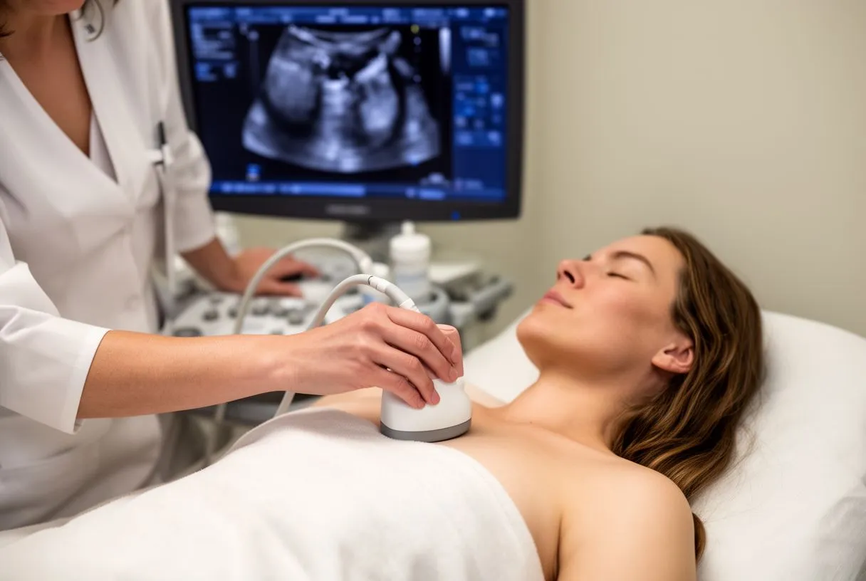
The standard test takes about 30 to 45 minutes. You lie on an exam table, a gel is applied to your chest, and a technician moves a small transducer — a smooth handheld probe — across it. The probe sends harmless sound waves into the chest and records the echoes that bounce back, building a live moving image of your heart. There is no radiation, no needle, and no pain.
You will hear the sounds of your own heart. Part of the study uses Doppler, which turns blood flow into audible sound and colored images. The clicks of a mechanical valve or the gentle flow through a biological valve are normal findings, not warnings. Expect to change position a few times and to hold your breath briefly for some measurements.
Preparation is minimal. You eat, drink, and take your medications as usual for a standard scan. In the consultation the team explains what the studies show, in plain words, and answers every question before any decision.
Can an Echocardiogram Detect All Heart Problems?
No — and it is worth being honest about that. An echo is superb at seeing valves, heart muscle, chamber sizes, and pumping function. It is far less useful for the coronary arteries themselves, which are too small and move too much to image directly. A blocked artery that caused no valve problem will not show up on your yearly valve scan.
Some questions need other tests. Rhythm disturbances are better seen on an electrocardiogram or a Holter monitor, and a Stress Echocardiogram is used when your team wants to see how the heart performs under exertion rather than at rest. For the coronary arteries, your cardiologist may order a CT angiogram or, rarely, a catheterization. Think of the echo as one essential instrument in an orchestra, not the whole band.
This is why a yearly visit includes more than the scan. At a proper follow-up appointment, your symptoms, medications, blood pressure, and imaging are all reviewed together. Our team reviews every case against international practice guidelines before making any recommendation, so a normal echo is read in the context of the whole person, not in isolation.
What Is a TEE Test and When Is It Needed?
TEE stands for transesophageal echocardiogram. Instead of pressing a probe against the chest, the team gently passes a slim ultrasound probe down the food pipe (esophagus), which sits directly behind the heart. From there the pictures are dramatically clearer, because there is no rib and no lung in the way. The transesophageal echocardiogram vs transthoracic valve question comes up often, and the answer is that they are partners, not rivals.
A TEE is not part of routine yearly follow-up. Your yearly heart echo after valve repair is almost always the simple chest scan. A TEE is reserved for specific situations: suspected Valve Thrombosis (a clot on the valve), infection of the valve (endocarditis), very small leaks the chest scan cannot fully measure, or planning before a complex procedure. It requires fasting and usually light sedation, and you will need someone to drive you home.
When a TEE is needed, it usually changes the plan for the better. In our experience, a well-timed TEE answers questions a chest echo genuinely cannot — for example, precisely sizing a paravalvular leak before deciding on treatment. If your team recommends one, ask exactly what question it is meant to answer; a good team will be able to tell you.
Do You Get Echocardiogram Results Immediately?
You usually get a preliminary answer the same day. The cardiologist who supervises the study can look at the moving images live and tell you the broad picture: the valve is working, the heart is pumping well, or something needs a closer look. That conversation alone often eases the anxiety people carry into the exam room.
The formal report takes a little longer. The sonographer's images must be measured, reviewed, and written up, which typically takes a few days. Serious or urgent findings are flagged and communicated right away — teams do not sit on bad news. Everything else is discussed at your scheduled follow-up, ideally with your previous scans side by side for comparison.
Ask for the discussion, not just the paper. A report full of numbers means little without explanation. Ask your doctor to walk you through what the study shows, what has changed since last year, and what — if anything — needs to be done. You should leave that conversation knowing exactly where you stand.
What Shouldn't You Do Before an Echocardiogram?
For a standard chest echo, there is very little to avoid. You do not need to fast, and you should take your normal medications unless your doctor says otherwise. A few practical points help the study go smoothly: wear a two-piece top that is easy to remove, skip heavy body lotion on the chest (it makes the probe slip), and arrive a few minutes early so the images are not rushed.
The exceptions apply to the special versions of the test. A transesophageal echo requires fasting for several hours beforehand, and a stress echo may require holding certain medications or avoiding caffeine on the day, depending on your doctor's instructions. Always follow the specific instructions your team gives you for the specific test you are having — not a generic list.
Bring what makes the comparison possible. If you are having your follow-up at a new center, bring a copy of your surgical report and, if you can, your previous echo reports. Knowing whether you received a Porcine Valve, a mechanical valve, or a transcatheter valve matters enormously to the people reading your scan, because each prosthesis has its own expected measurements and its own quirks.
Heart Valve Deterioration Signs the Echo Watches For
Tissue valves wear gradually, and the echo sees it first. Structural Valve Degeneration — the slow stiffening or tearing of a biological valve — is the main reason a biological valve yearly checkup matters so much. On the scan, the first hints are rising pressure gradients across the valve, subtle thickening of the leaflets, or a new leak. Stenosis progression monitoring is measured in precise millimeters of mercury, compared against your own baseline, year after year.
Symptoms, when they appear, follow a recognizable pattern. The echo detects trouble long before you would, but it helps to know what eventually to watch for between scans:
- New or worsening breathlessness: especially with activities that used to be easy, such as climbing the same flight of stairs.
- Swelling in the ankles or legs: fluid retention can signal the heart is struggling to keep up with a failing valve.
- Dizziness or fainting spells: a narrowed valve can limit blood flow to the brain during exertion.
- A new or changing heart murmur: a heart murmur follow up echo is often the very test that explains it, and many murmurs after surgery are entirely innocent.
Detection early means options stay open. When an echo check for valve regurgitation or stenosis catches a change early, treatment can often be planned calmly — sometimes a medication adjustment, sometimes a scheduled intervention such as a Valve-in-valve procedure inside the existing prosthesis. Caught late, the same problem can arrive as an emergency. This is the clearest argument for keeping every yearly appointment. In our practice, we have seen precisely this pattern many times: an asymptomatic change on a routine scan that became a planned, low-risk procedure instead of an urgent one.
Mechanical Valve or Biological Valve: Does Follow-Up Differ?
Both valve types need the same imaging, for different reasons. An annual echo for mechanical valve patients focuses on valve function, heart performance, and — critically — making sure the blood thinner is doing its job without causing problems. Mechanical valves are extremely durable and rarely fail structurally, but they require lifelong anticoagulation, and the yearly scan checks the heart is thriving on that regimen.
Biological valves trade durability for freedom from blood thinners. A Porcine Valve or other tissue prosthesis usually spares most patients lifelong warfarin, which many people value. The price is a limited lifespan: tissue valves slowly degenerate, which is why the stenosis progression monitoring described above is taken especially seriously in these patients. Neither choice is universally better — the right one depends on your age, lifestyle, and other conditions.
Your follow-up should be tailored, not templated. Valve surgery long term care also depends on what was done: a Mitral Valve Prolapse repair, a Supra-annular Valve placement, a Valve-sparing Root Replacement, or a catheter-based procedure all have different surveillance priorities. Every case in our practice is reviewed by the multidisciplinary team against international guidelines before any recommendation is made, and the final decision always rests with the patient.
How Often Echocardiogram After Valve Replacement? Choosing Your Team
Most patients settle into a yearly rhythm. A typical schedule after an uncomplicated valve replacement looks like this: an early post-operative study, a check at six to twelve months, and then an echocardiogram roughly once a year for life, with more frequent scans if a change appears. Your own cardiologist appointment after valve surgery should confirm the exact interval for you, because factors such as valve type, residual leaks, and heart function all shift the schedule. This is the practical answer to how often echocardiogram after valve replacement: annually for most, more often for some, and never less than what your team's guidelines require.
What happens during a catheter-based procedure matters for follow-up too. Patients who received a valve through TAVR — Transcatheter Aortic Valve Implantation — go through the same annual surveillance, and it helps to understand what the procedure involved. The valve is delivered through a vessel, usually in the groin, and that access point is closed carefully at the end; published techniques describe dedicated closure devices used to achieve hemostasis in challenging anatomy, including reoperative patients (a published case report on access-site closure). Likewise, open valve replacement relies on briefly stopping the heart with a cardioplegia solution; studies of single-dose lignocaine-based cardioplegia in valve replacement patients show how protection of the heart muscle during surgery continues to be refined (Brazilian Journal of Cardiovascular Surgery). Knowing these details helps you ask sharper questions at follow-up.
Choose a team that will tell you the truth, every year. If you are looking for the best heart valve clinic Guadalajara has to offer, or a second opinion closer to home, look for three things: a multidisciplinary team that reviews your imaging together, a willingness to explain results in plain words, and a clear statement of all your options — including doing nothing yet, when that is genuinely reasonable. We encourage second opinions and treat seeking one as a normal part of deciding about heart surgery. Dr Molina trained in cardiothoracic surgery at the Universidad de Guadalajara, in cardiac, minimally invasive, robotic and structural surgery at the University of Montreal, and in structural cardiology and TAVR at CHU Bordeaux, and the first contact with our team is a free video consultation where we talk through your condition and say which studies are truly needed.
The takeaway is simple. An annual echocardiogram after valve surgery is your early-warning system: it tracks your valve, your heart muscle, and your overall cardiac health with no discomfort and no radiation. Keep every yearly appointment, bring your previous reports, ask questions until the answers make sense, and discuss your own situation with a qualified cardiac team — a consultation with our practice or a trusted local team is the right next step, never self-diagnosis. The Centers for Disease Control and Prevention notes that regular follow-up care is an essential part of living well with heart disease (CDC), and your yearly echo is the cornerstone of that care.